Review
. 2018 Jun 19;48(6):1104-1117.
doi: 10.1016/j.immuni.2018.05.013.

Innate Lymphoid Cells: Diversity, Plasticity, and Unique Functions in Immunity

Affiliations
Review

Innate Lymphoid Cells: Diversity, Plasticity, and Unique Functions in Immunity

Marco Colonna. Immunity. .

Abstract

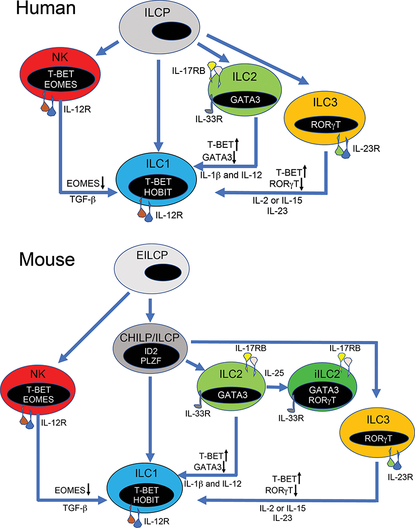
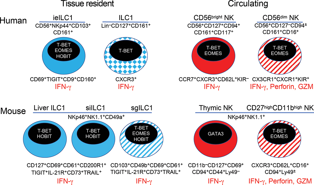
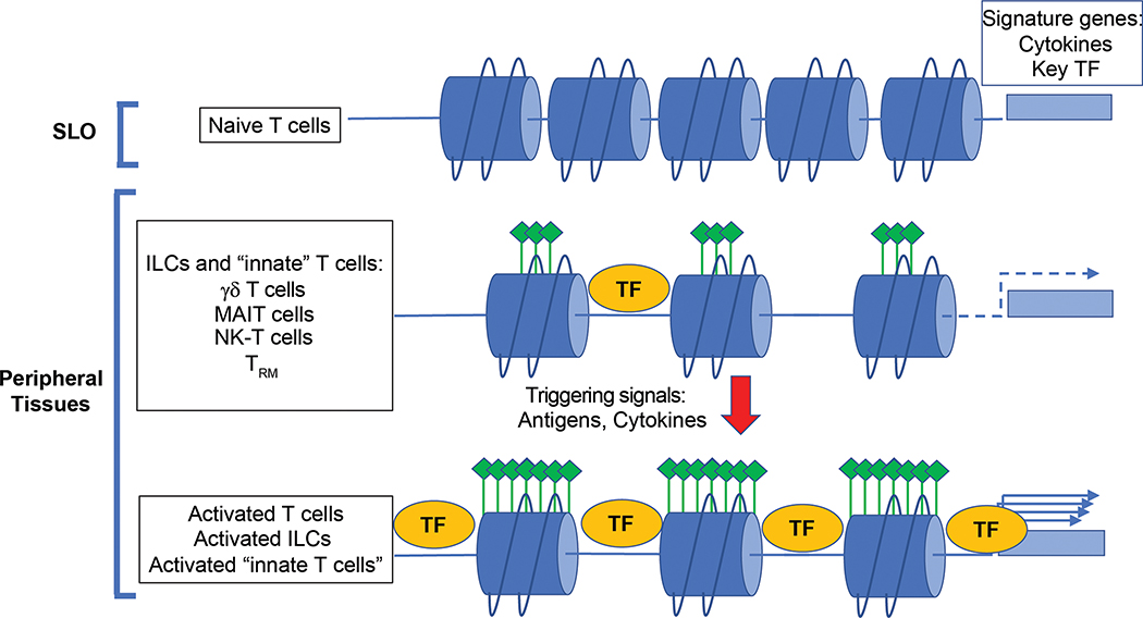
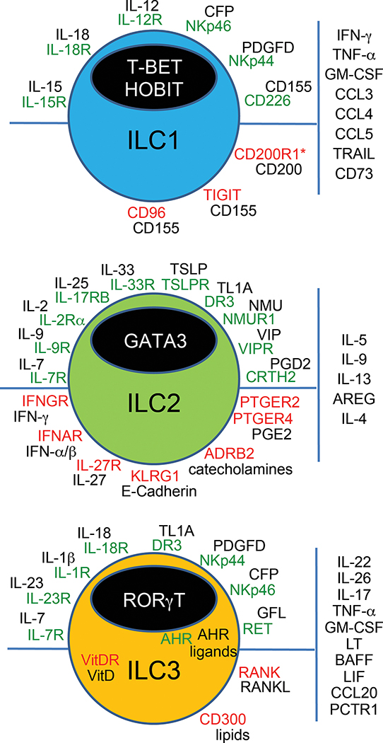
Type 1, 2, and 3 innate lymphoid cells (ILCs) have emerged as tissue-resident innate correlates of T helper 1 (Th1), Th2, and Th17 cells. Recent studies suggest that ILCs are more diverse than originally proposed; this might reflect truly distinct lineages or adaptation of ILCs to disparate tissue microenvironments, known as plasticity. Given that ILCs strikingly resemble T cells, are they redundant? While the regulation, timing, and magnitude of ILC and primary T cell responses differ, tissue-resident memory T cells may render ILCs redundant during secondary responses. The unique impact of ILCs in immunity is probably embodied in the extensive array of surface and intracellular receptors that endow these cells with the ability to distinguish between normal and pathogenic components, interact with other cells, and calibrate their cytokine secretion accordingly. Here I review recent advances in elucidating the diversity of ILCs and discuss their unique and redundant functions.


Figures

Figure 1
Figure 1. Heterogeneity of human and mouse ILC1s
Tissue-resident T-bet+ ILC1s may include: a) bona fide ILC1s derived from ILC progenitors; b) converted ILC2s exposed to IL-12 and IL-1β that downregulate GATA3 and upregulate T-bet; c) converted ILC3s exposed to IL-2, IL-15 and IL-23 that downregulate Rorγt and upregulate T-bet; d) NK cells that downregulate EOMES when exposed to a TGF-β-rich environment. Top panel: in human, tissue resident ILC1s, ILC2s and ILC3s derive from a systemic ILC progenitor (Lim et al., 2018). Bottom panel: in mouse, ILC1s derive from the common helper innate lymphoid progenitor (CHILP) and the downstream innate lymphoid cell progenitor (ILCP), while NK cells derive from an upstream early innate lymphoid progenitor (EILCP) (Constantinides et al., 2014; Klose et al., 2014).
Figure 2
Figure 2. Phenotypic and functional heterogeneity of ILC1s and NK cells
ILC1s and NK cells provide a spectrum of IFN-γ-producing cells with distinct trafficking properties (tissue resident versus circulating, respectively), but partially overlapping markers. Markers used to identify each subset are indicated on the top of each cell. Additional phenotypic features are indicated on the bottom of each cell. Major functions (IFN-γ secretion and release of lytic mediators) are indicated in red. In human, ILC1s include ieILC1s and CD127+ ILC1s. Markers of tissue residency and other markers reflecting TGF-β imprinting are shown below ieILC1s. In mouse, ILC1s show slightly different phenotypic markers depending on the tissue of origin. Liver and small intestine (si) ILC1s express CD127 and CD49a. Salivary gland (sg) ILC1s lack CD127 and express both CD49a and CD49b, as well as CD103. Human NK cells include a CD56bright subset that specializes in IFN-γ secretion, and a CD56dim subset that is capable of cytolysis. Mouse NK subsets with functional features similar to those of CD56bright and CD56dim include thymic NK cells (Vosshenrich et al., 2006) and CD27+CD11b+ NK cells (Hayakawa et al., 2006), respectively. GZM, granzymes.
Figure 3
Figure 3. Differential regulation of gene expression programs in tissue resident ILCs versus T cells
ILCs and Th lineages utilize similar regulatory regions to control expression of signature genes, such as master transcription factors (TF) and effector cytokines. However, in naïve T cells, enhancers are inaccessible to transcription factors until T cells are primed, activated and polarized in secondary lymphoid organs (SLO). In contrast, enhancers are already poised in ILCs, and other “innate” T cells residing in peripheral tissues, as indicated by high density of H3-K27 acetylation or super-enhancers (green symbols). Innate T cells include γδT cells, MAIT cells, and NK-T cells. TRM cells, which are generated after a primary response and reach peripheral tissues, also adopt ILC-like regulatory features. Upon tissue signals caused by pathogenic infections or tissue damage, regulatory regions become even more accessible and TFs downstream of tissue signals promptly bind to enhancers inducing the expression of signature genes.
Figure 4
Figure 4. ILC surface and intracellular receptors
Activating receptors (green, top), inhibitory receptors (red, bottom), and their cognate ligands (black) are indicated for each ILC type. Effector molecules are indicated on the right of each cell type. This scheme is comprehensive of both human and mouse data.

Similar articles

Cited by

References

    1. Allard B, Longhi MS, Robson SC, Stagg J. The ectonucleotidases CD39 and CD73: Novel checkpoint inhibitor targets. Immunol Rev. 2017;276:121–144. - PMC - PubMed
    1. Bando JK, Colonna M. Innate lymphoid cell function in the context of adaptive immunity. Nat Immunol. 2016;17:783–789. - PMC - PubMed
    1. Bando JK, Gilfillan S, Song C, McDonald KG, Huang SCC, Newberry RD, Kobayashi Y, Allan DSJ, Carlyle JR, Cella M, Colonna M. RANKL controls effector cytokine production in group 3 innate lymphoid cells. Immunity. 2018 in press. - PMC - PubMed
    1. Bank U, Deiser K, Finke D, Hammerling GJ, Arnold B, Schuler T. Cutting edge: innate lymphoid cells suppress homeostatic T cell expansion in neonatal mice. J Immunol. 2016;196:3532–3536. - PubMed
    1. Bar-Ephraim YE, Mebius RE. Innate lymphoid cells in secondary lymphoid organs. Immunol Rev. 2016;271:185–199. - PubMed

Publication types