. 2018 Jun 19;23(12):3419-3428.
doi: 10.1016/j.celrep.2018.05.061.

Replication Fork Reversal during DNA Interstrand Crosslink Repair Requires CMG Unloading

Affiliations

Replication Fork Reversal during DNA Interstrand Crosslink Repair Requires CMG Unloading

Ravindra Amunugama et al. Cell Rep. .

Abstract

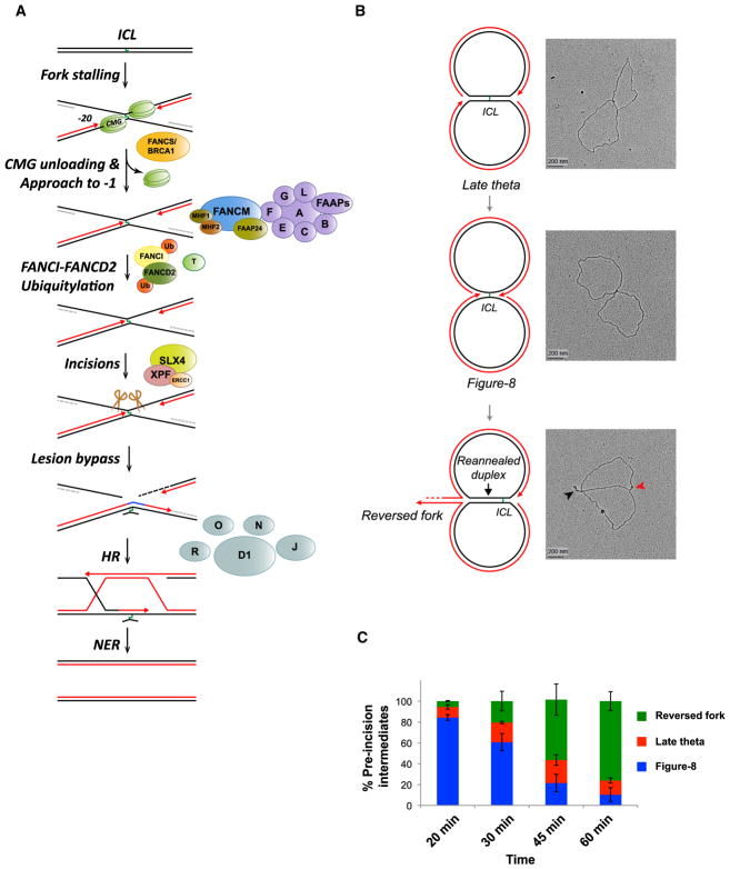
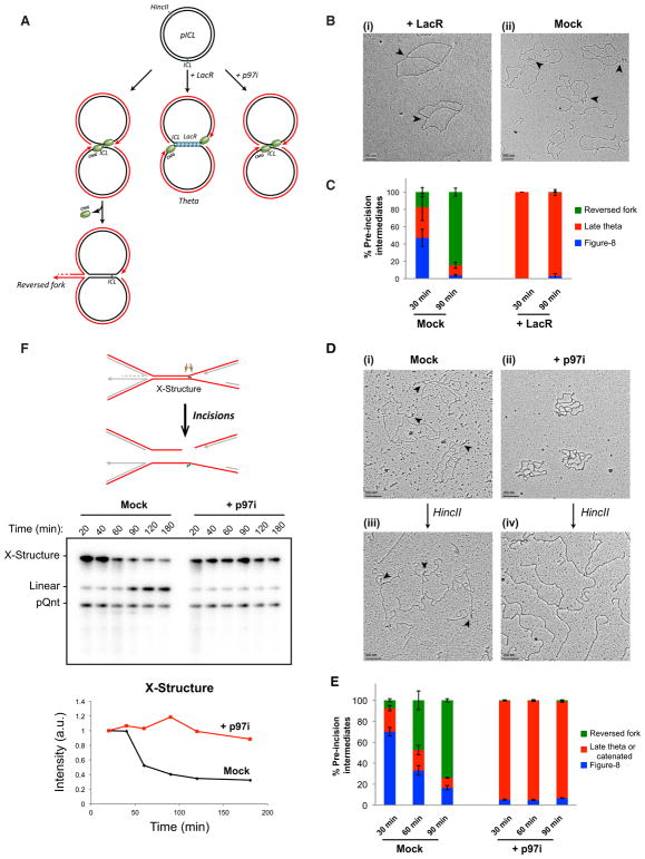
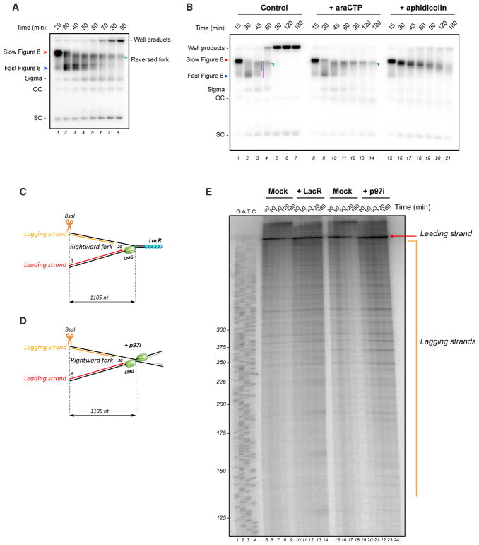
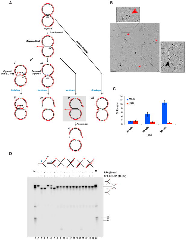
DNA interstrand crosslinks (ICLs) are extremely cytotoxic, but the mechanism of their repair remains incompletely understood. Using Xenopus egg extracts, we previously showed that repair of a cisplatin ICL is triggered when two replication forks converge on the lesion. After CDC45/MCM2-7/GINS (CMG) ubiquitylation and unloading by the p97 segregase, FANCI-FANCD2 promotes DNA incisions by XPF-ERCC1, leading to ICL unhooking. Here, we report that, during this cell-free ICL repair reaction, one of the two converged forks undergoes reversal. Fork reversal fails when CMG unloading is inhibited, but it does not require FANCI-FANCD2. After one fork has undergone reversal, the opposing fork that still abuts the ICL undergoes incisions. Our data show that replication fork reversal at an ICL requires replisome disassembly. We present a revised model of ICL repair that involves a reversed fork intermediate.

Keywords: CMG; DNA interstrand crosslink repair; DNA replication; Fanconi anemia; ICL; XPF; replication fork reversal.


Conflict of interest statement

DECLARATION OF INTERESTS

The authors declare no competing interests.

Figures

Figure 1
Figure 1. Replication Fork Reversal Observed during ICL Repair
(A) Current model of cell-free cisplatin ICL repair. See text for details. (B) pICL was incubated in high-speed supernatant (HSS) of egg cytoplasm to license DNA and then supplemented with nucleoplasmic extract (NPE) to promote replication initiation (Walter et al., 1998; see Supplemental Experimental Procedures). 60 min after NPE addition, DNA was analyzed by EM, and representative images of late theta, figure 8, and reversed-fork intermediates, together with interpretive cartoons, are shown. Black arrowhead, reversed fork; red arrowhead, ssDNA on the lagging strand of the non-reversed fork. (C) Quantification of late theta, figure 8, and reversed-fork intermediates. At the indicated times after NPE addition, samples were analyzed by EM as in (B). At least 100 interpretable molecules were analyzed for the quantification of repair intermediates at each time point. Error bars indicate the range in two independent experiments. A similar time-dependent decrease in figure 8 structures and increase in reversed forks was observed in Figures 2C and 2E.
Figure 2
Figure 2. Fork Convergence and CMG Unloading Are Required for Fork Reversal during ICL Repair
(A) Model depicting replication fork convergence, CMG unloading, and fork reversal in an unperturbed reaction (left), in the presence of lacR (middle), and in the presence of NMS 873 (“p97i”; right). (B) EM image of late theta structures (black arrows) in a LacR-treated reaction (i) or in a mock (buffer)-treated reaction (ii) at 90 min. (C) At the indicated times, late theta, figure 8, and reversed-fork structures from the experiment shown in (B) were quantified and graphed. Error bars indicate the range in two independent experiments. (D) EM images of reversed fork structures or catenated structures in the presence of mock- (i and iii) or p97i-treated (ii and iv) conditions, respectively, before and after HincII digestion. See text for details. (E) Quantification of late theta, catenated molecules, figure 8s, and reversed forks by EM in a mock (DMSO)-treated or p97i-treated reaction. Error bars indicate the range in two independent experiments. (F) pICL incision assay in a mock-treated (buffer) or p97i-treated reaction. pICL and an undamaged, internal control plasmid (pQnt) were nick translated with [α-32P] dATP before addition to extracts to induce replication and repair. Repair intermediates were recovered from extract and digested with HincII, separated by denaturing agarose gel electrophoresis, and visualized by autoradiography. Incisions result in loss of the large parental X-shaped structure (red strands in schematic; quantified in graph) and accumulation of a linear species. A similar result was seen in a second, independent experiment.
Figure 3
Figure 3. Efficient Resection of the Nascent Lagging Strand Is Impaired when CMG Unloading Is Blocked
(A) pICL plasmid was replicated in NPE containing [α-32P] dATP, and repair intermediates were separated on a native agarose gel and visualized by autoradiography. OC, open circular; SC, supercoiled. (B) pICL was replicated in extract with [α-32P]dATP. 15 min after initiation of replication, extract was supplemented with cytosine arabinoside triphosphate (araCTP) (2 mM) or aphidicolin (50 μM). Replication intermediates were separated on a native agarose gel after deproteinization and visualized by autoradiography. Red arrowhead, slow figure 8; Blue arrowhead, fast figure 8; green arrowhead, reversed forks. To determine the absolute fraction of reversed forks, the radioactivity adjacent to the green bar in lane 3 was divided by the total radioactivity in the lane. To determine the fraction of pre-incision intermediates comprising reversed forks, the radioactivity in the green bar was divided by the radioactivity adjacent to the pink bar. The gel is representative of three independent experiments. (C) Model depicting the rightward stalled fork in the presence of LacR, together with the BsaI site and primer used to generate the sequencing ladder. (D) Same as (C) but in the presence of p97i. (E) pICLLacO was replicated with [32P-α]dATP in the presence of LacR or p97i, and nascent strand products were analyzed by denaturing PAGE after digestion with BsaI. Red arrow, stalled leading strand; orange line, lagging strands of the rightward fork. The sequencing ladder was generated with primer R (C) and (D). Similar results were obtained in a second, independent experiment.
Figure 4
Figure 4. The Non-reversed Fork Abutting the ICL Is Subject to DNA Incisions
(A) Model depicting three possible pathways of incision, after one fork has undergone reversal, and one pathway for breakage. The sigma structure expected if the non-reversed fork on the reversed intermediate undergoes incisions is highlighted in gray. See text for other details. (B) EM image of pICL repair intermediates at 90 min in a mock-treated condition. Black arrowheads, sigma structures containing a reversed fork; red arrowheads, linear species. Similar results were seen in two other independent experiments. (C) Quantification of linear structures during ICL repair in a mock-treated or p97i-treated reaction. Error bars indicate the range in two independent experiments. A time-dependent increase in linear species of similar magnitude was observed in two other experiments, but the data were not included in the quantification due to different time points or slightly different conditions. (D) A series of 3′-radiolabeled (red asterisks) splayed arm and X-shaped substrates containing or lacking nascent strands (dotted arrows) were incubated with XPF-ERCC1 in the presence or absence of RPA for 60 min and the DNA analyzed by denaturing PAGE. M, radiolabeled marker oligonucleotides of indicated structures and sizes. Blue arrow, approximate position of incision. See Supplemental Experimental Procedures and Figure S3D for details of model substrate preparation.

Similar articles

Cited by

References

    1. Abdullah UB, McGouran JF, Brolih S, Ptchelkine D, El-Sagheer AH, Brown T, McHugh PJ. RPA activates the XPF-ERCC1 to initiate the processing of DNA interstrand crosslinks. EMBO J. 2017;36:2047–2060. - PMC - PubMed
    1. Budzowska M, Graham TG, Sobeck A, Waga S, Walter JC. Regulation of the Rev1-pol ζ complex during bypass of a DNA interstrand cross-link. EMBO J. 2015;34:1971–1985. - PMC - PubMed
    1. Chrysogelos S, Griffith J. Escherichia coli single-strand binding protein organizes single-stranded DNA in nucleosome-like units. Proc Natl Acad Sci USA. 1982;79:5803–5807. - PMC - PubMed
    1. Clauson C, Schärer OD, Niedernhofer L. Advances in understanding the complex mechanisms of DNA interstrand cross-link repair. Cold Spring Harb Perspect Biol. 2013;5:a012732. - PMC - PubMed
    1. D’Andrea AD. Susceptibility pathways in Fanconi’s anemia and breast cancer. N Engl J Med. 2010;362:1909–1919. - PMC - PubMed

Publication types