. 2018 Jun 19;23(12):3438-3446.e5.
doi: 10.1016/j.celrep.2018.05.059.

Aberrant Striatal Activity in Parkinsonism and Levodopa-Induced Dyskinesia

Affiliations

Aberrant Striatal Activity in Parkinsonism and Levodopa-Induced Dyskinesia

Michael B Ryan et al. Cell Rep. .

Abstract

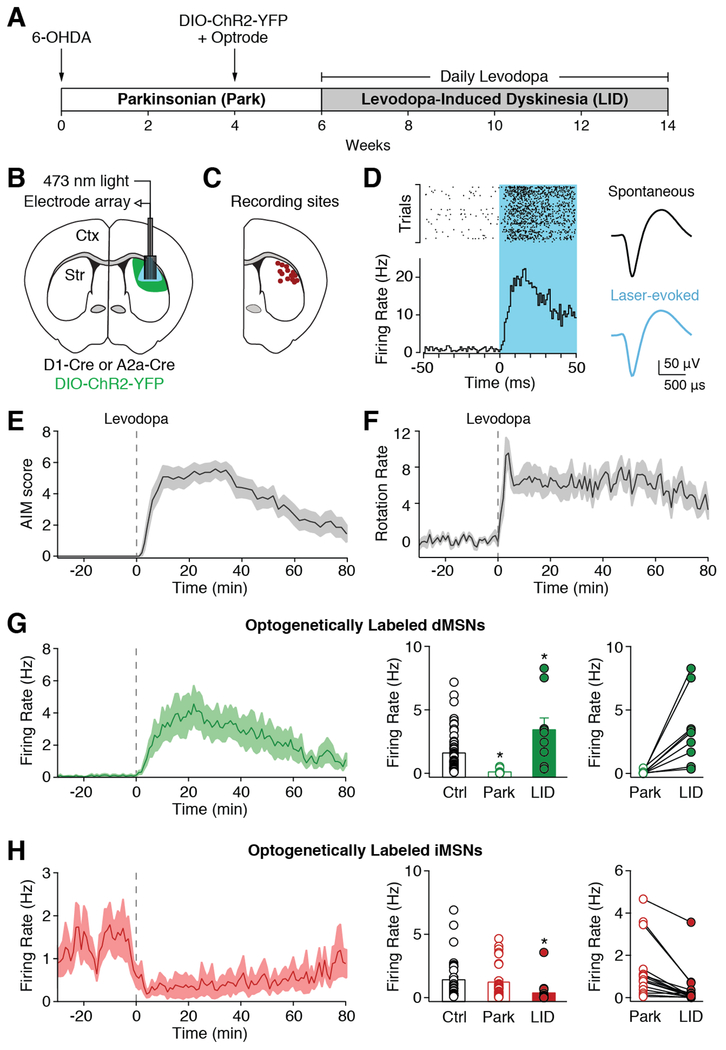
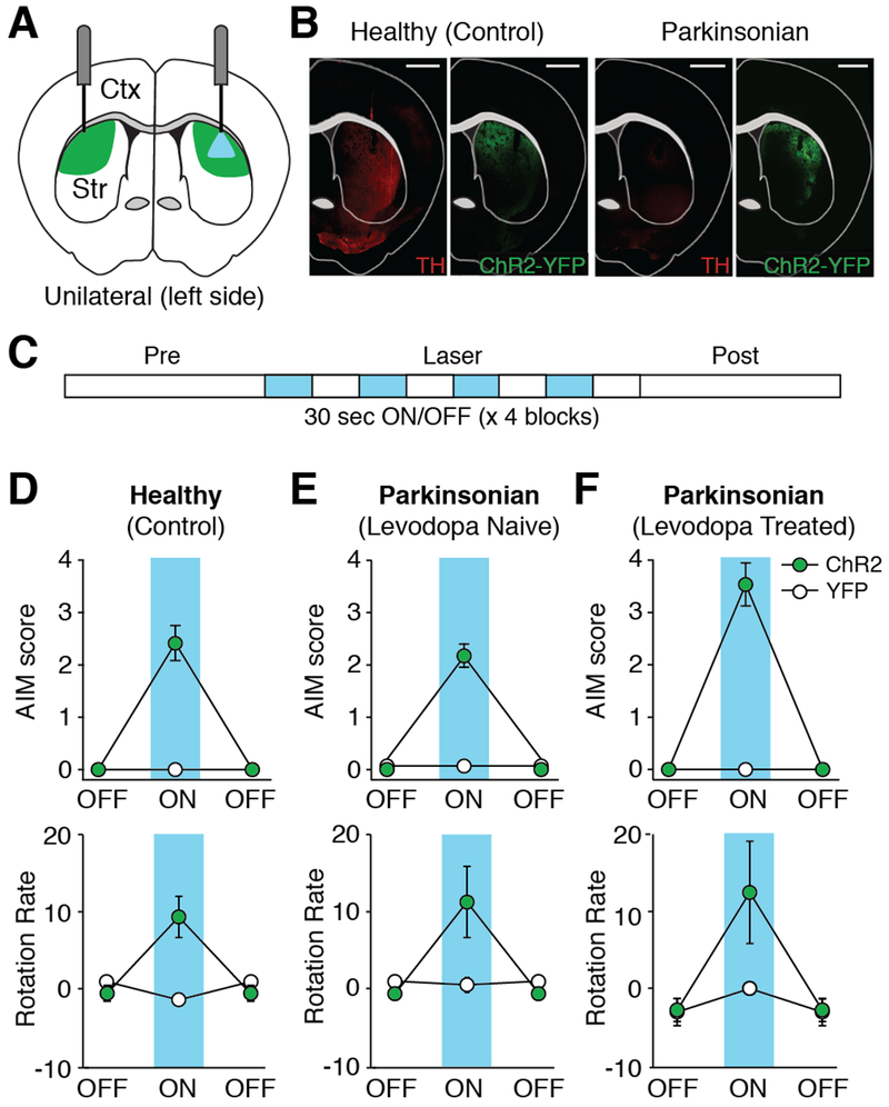
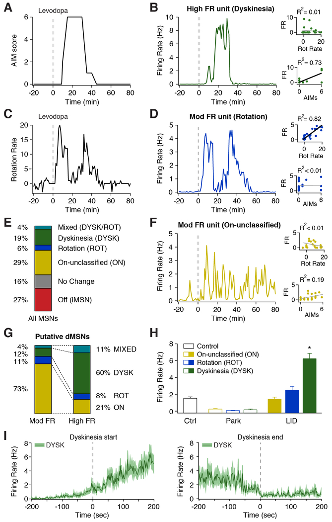
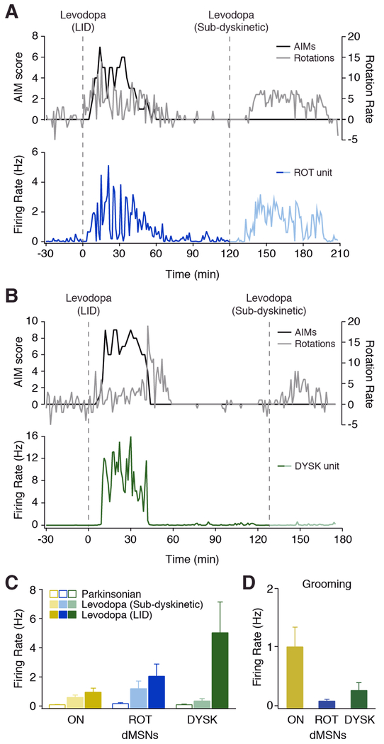
Action selection relies on the coordinated activity of striatal direct and indirect pathway medium spiny neurons (dMSNs and iMSNs, respectively). Loss of dopamine in Parkinson's disease is thought to disrupt this balance. While dopamine replacement with levodopa may restore normal function, the development of involuntary movements (levodopa-induced dyskinesia [LID]) limits therapy. How chronic dopamine loss and replacement with levodopa modulate the firing of identified MSNs in behaving animals is unknown. Using optogenetically labeled striatal single-unit recordings, we assess circuit dysfunction in parkinsonism and LID. Counter to current models, we found that following dopamine depletion, iMSN firing was elevated only during periods of immobility, while dMSN firing was dramatically and persistently reduced. Most notably, we identified a subpopulation of dMSNs with abnormally high levodopa-evoked firing rates, which correlated specifically with dyskinesia. These findings provide key insights into the circuit mechanisms underlying parkinsonism and LID, with implications for developing targeted therapies.

Keywords: Parkinson’s disease; basal ganglia; dopamine; dyskinesia; electrophysiology; levodopa; optogenetics; striatum.


Conflict of interest statement

Declaration of Interests

The authors declare no competing interests.

Figures

Figure 1.
Figure 1.. Alterations in Identified Striatal Neurons Following Dopamine Depletion and Replacement with Levodopa.
(A) Experimental timeline. (B) Schematic of optrode in dorsolateral striatum (DLS). (C) Recording sites verified by electrolytic lesions. (D) Example of optogenetically labeled striatal direct pathway neuron (dMSN). Left: PSTH and peri-event raster aligned to laser onset. Right: average spontaneous and laser-evoked waveforms. (E-H) Levodopa was administered at t=0 (dotted line). (E) Average dyskinesia, as measured by the Abnormal Involuntary Movement (AIM) score (N=12). (F) Average rotations (contralesional-ipsilesional) per minute (N=12). (G) Left: average firing rate of optogenetically labeled dMSNs. Middle: dMSN firing rates in healthy mice (Ctrl, n=64, N=5) and parkinsonian mice before (Park, n=14, N=10) and after (LID, n=9, N=6) levodopa injection. Right: firing rate of individual dMSNs before and after levodopa. (H) Left: average firing rate of optogenetically labeled striatal indirect pathway neurons (iMSNs). Middle: iMSN firing rates in healthy mice (Ctrl, n=34, N=5) and parkinsonian mice before (Park, n=32, N=8) and after (LID, n=16, N=6) levodopa injection. Right: firing rate of individual iMSNs before and after levodopa. n=cells, N=animals. *p<0.05 vs Ctrl. All data presented as mean ± SEM. See also Figures S1 and S2.
Figure 2.
Figure 2.. Optogenetic Activation of dMSNs Produces Dyskinesia in Healthy and Parkinsonian Mice.
(A) Schematic showing optic fiber placement and laser stimulation in the left (ipsilesional) DLS of parkinsonian mice. Green = DIO-ChR2-YFP expression. (B) Representative coronal sections from healthy and parkinsonian D1-Cre mice showing tyrosine hydroxylase (TH) staining and DIO-ChR2-YFP expression. Scale bar = 1mm. (C) Experimental timeline. (D-F) Behavior before (OFF) and during (ON) dMSN stimulation. Top: average dyskinesia (AIM) score. Bottom: average rotation rate. (D) Healthy mice (ChR2: N=12, YFP: N=11). (E-F) Parkinsonian mice (ChR2: N=8, YFP: N=4) (E) before levodopa exposure and (F) after 2 weeks of chronic levodopa. N=animals. All data presented as mean ± SEM. See also Figure S3 and Movie S1.
Figure 3.
Figure 3.. A Subpopulation of dMSNs Show High Firing Rates Correlated to Dyskinesia.
(A-D) Behavior and single unit firing rates from a representative session. (A) Dyskinesia score. (B) Representative putative dMSN classified as High FR whose firing rate was correlated with dyskinesia score (DYSK). (C) Rotation rate. (D) Representative putative dMSN classified as Mod FR whose firing rate was correlated with rotation rate (ROT). (E) Fraction of all putative MSNs (n=255, N=15) classified by behavioral correlation. (F) Representative putative dMSN classified as Norm FR whose firing rate was uncorrelated with rotation rate or AIM score (ON). (B,D,F) Insets: correlation between firing rate and rotation rate (top) and dyskinesia (bottom). (G) Fraction of putative dMSNs classified by rate and their correlation to behavior (n=146, N=15). (H) Average firing rate of putative dMSNs based on correlation to behavior, before (Park) and after (LID) levodopa injection, compared to MSNs from healthy (Ctrl) mice. Ctrl: n=98, N=10; ON: n=74, ROT: n=14, DYSK: n=47, N=15. *p<0.05 vs Ctrl. (I) Average firing rate of DYSK units aligned to dyskinesia start (left, n=43) and end (right, n=37, N=15). n=cells, N=animals. All data presented as mean ± SEM. See also Figure S4 and Movie S2.
Figure 4.
Figure 4.. DYSK Unit Activity is Specific to LID
(A-B) Two doses of levodopa were administered in a single session. Top: dyskinesia score (black) and rotation rate (gray). Bottom: unit firing rate, aligned to levodopa injection (dotted line). Representative (A) ROT unit and (B) DYSK unit. (C) Average firing rate of putative dMSN subtypes before (parkinsonian) and after dyskinetic and sub-dyskinetic doses of levodopa. ON: n=14, N=4; ROT: n=8, N=3; DYSK: n=8, N=3. (D) Average firing rate of putative dMSN subtypes during grooming (ON: n=30, N=4; ROT: n=5, N=2; DYSK: n=44, N=5). n=cells, N=animals. All data presented as mean ± SEM. See also Movie S3 and S4.

Similar articles

Cited by

References

    1. Ahlskog JE, and Muenter MD (2001). Frequency of levodopa-related dyskinesias and motor fluctuations as estimated from the cumulative literature. Mov. Disord. Off. J. Mov. Disord. Soc 16, 448–458. - PubMed
    1. Albin RL, Young AB, and Penney JB (1989). The functional anatomy of basal ganglia disorders. Trends Neurosci 12, 366–375. - PubMed
    1. Alcacer C, Andreoli L, Sebastianutto I, Jakobsson J, Fieblinger T, and Cenci MA (2017). Chemogenetic stimulation of striatal projection neurons modulates responses to Parkinson’s disease therapy. J. Clin. Invest 127, 720–734. - PMC - PubMed
    1. Barbera G, Liang B, Zhang L, Gerfen CR, Culurciello E, Chen R, Li Y, and Lin D-T (2016). Spatially Compact Neural Clusters in the Dorsal Striatum Encode Locomotion Relevant Information. Neuron 92, 202–213. - PMC - PubMed
    1. Bergman H, Wichmann T, Karmon B, and DeLong MR (1994). The primate subthalamic nucleus. II. Neuronal activity in the MPTP model of parkinsonism. J. Neurophysiol 72, 507–520. - PubMed