. 2018 Jun 19;114(12):2811-2819.
doi: 10.1016/j.bpj.2018.05.011.

A Protofilament-Protofilament Interface in the Structure of Mouse α-Synuclein Fibrils

Affiliations

A Protofilament-Protofilament Interface in the Structure of Mouse α-Synuclein Fibrils

Guohua Lv et al. Biophys J. .

Abstract

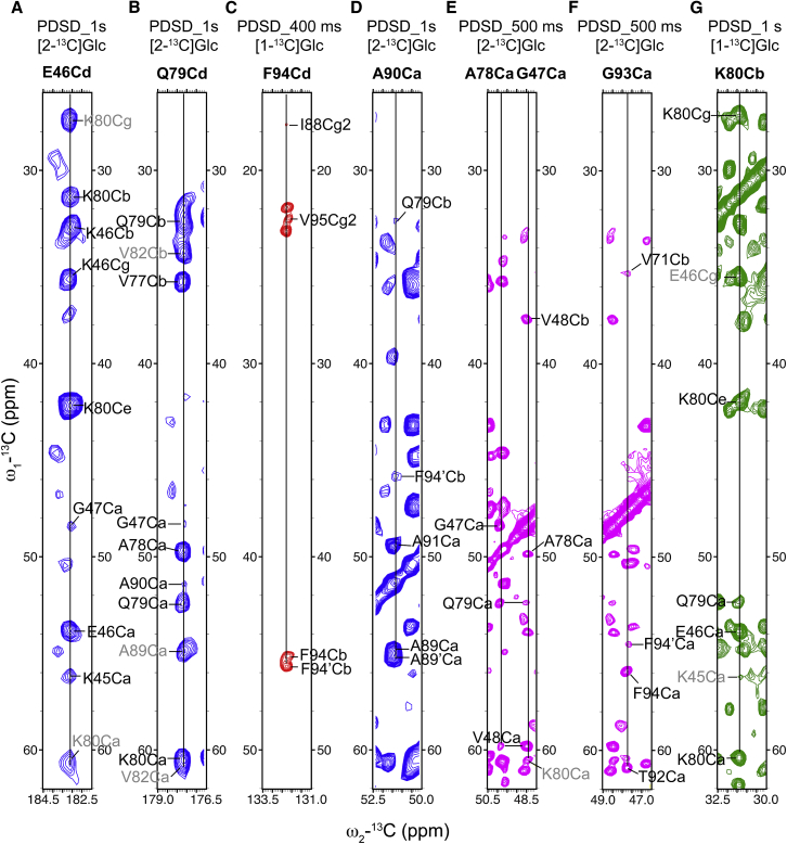
Fibrillar α-synuclein (AS) is the major component of Lewy bodies, the pathological hallmark of Parkinson's disease. Using solid-state nuclear magnetic resonance (ssNMR), we previously reported a structural characterization of mouse AS (mAS) fibrils and found that the secondary structure of the mAS fibrils is highly similar to a form of human AS (hAS) fibrils. Recently, a three-dimensional structure of these same hAS fibrils was determined by ssNMR and scanning transmission electron microscopy. Using medium- and long-range distance restraints obtained from ssNMR spectra, we found that the single protofilament structure of mAS fibrils is also similar to that of the hAS fibrils. However, residue-specific water accessibility of mAS fibrils probed by water polarization transfer ssNMR measurements indicates that residues S42-T44 and G84-V95 are largely protected from water even though they are located at the edge of the protofilament. Some of the corresponding resonances also exhibit peak doubling. These observations suggest that these residues may be involved in, to our knowledge, a novel protofilament-protofilament interface. We propose a structural model of mAS fibrils that incorporates this dimer interface.


Figures

Figure 1
Figure 1
ssNMR structural constraints for mAS fibrils. (A, B, and D) 2D 13C-13C PDSD correlation spectrum is shown with a mixing time of 1 s on [2-13C]Glc mAS fibrils; (C) 2D 13C-13C PDSD correlation spectrum is shown with a mixing time of 400 ms on [1-13C]Glc mAS fibrils; (E and F) 2D 13C-13C PDSD correlation spectrum is shown with a mixing time of 500 ms on [2-13C]Glc mAS fibrils; and (G) 2D 13C-13C PDSD correlation spectrum is shown with a mixing time of 1 s on [1-13C]Glc mAS fibrils. Unambiguous medium-range (1<|i-j|<5) and long-range (|i-j|≥5) restraints are annotated in black, and network unambiguous medium-range and long-range restraints are annotated in gray. The full view of the PDSD spectra on [2-13C]Glc and [1-13C]Glc mAS fibrils is provided in Fig. S3 and Fig. S4, respectively. To see this figure in color, go online.
Figure 2
Figure 2
Structural models of the single protofilament of (A) mAS fibril and (B) hAS fibril central core comprising residues L38–K97. The main fold of the single protofilament of mAS fibrils was that of the hAS fibrils (PDB: 2N0A) (21) and was extracted by using PyMOL 1.3 (http://www.pymol.org). The residues L38–K97 were then added according to the primary sequence of mAS and hAS in illustrator CS6. The two different mutants within the rigid core (residues at positions 53 and 87) are highlighted in italic and bold. The distance constraints obtained from our ssNMR spectra were further plotted on the model of mAS (A), and the distance constraints adapted from Fig. S4a for hAS fibrils (21) were further plotted on the model of hAS (B), confirming that the single protofilament structures of our mAS fibrils and of the hAS fibrils studied by Tuttle et al. are highly similar. Unambiguous restraints and network unambiguous restraints are represented by dark dashed and gray dashed lines, respectively. Residues exhibiting peak doubling for mAS fibrils are colored in gray. To see this figure in color, go online.
Figure 3
Figure 3
(A) Water-edited 2D NCA correlation spectra with a longitudinal 1H-1H mixing time of 2.5 ms (black) and 150 ms (red) of [2-13C]Glc mAS fibrils. (B) Magnetization exchange rates between water and mAS fibrils were calculated from intensity changes in water-edited 2D NCA as a function of 1H-1H mixing time. Only unambiguously assigned peaks with isolated peak center in all water-edited 2D NCA spectra were plotted. The magnetization exchange rates were simply classified into five categories: 0–0.03 (brown), 0.03–0.06 (red), 0.06–0.09 (orange), 0.09–0.12 (green), 0.15–0.18 (blue), and >0.3 (magenta). The prime symbol is used to distinguish a second set of resonances observed for some residues. (C) A proposed two-protofilament structural model of mAS fibrils is shown. Each individual protofilament structure is the same as in Fig. 2A, and the two protofilaments were manually docked to match the water protection data. Residues exhibiting peak doubling are represented by black bold circles. Residues were colored corresponding to the classified magnetization exchange rates in Fig. 3B.
Figure 4
Figure 4
Illustration of peak doublings observed for mAS fibrils using (A) 2D 13C-13C correlation PDSD spectrum with a mixing time of 500 ms and (B) NCA spectrum on [2-13C]Glc mAS fibrils. The second set of sequential resonance assignments observed is indicated with primes, for example, F94′. To see this figure in color, go online.

References

    1. Spillantini M.G., Schmidt M.L., Goedert M. α-synuclein in Lewy bodies. Nature. 1997;388:839–840. - PubMed
    1. Spillantini M.G., Crowther R.A., Goedert M. alpha-Synuclein in filamentous inclusions of Lewy bodies from Parkinson’s disease and dementia with lewy bodies. Proc. Natl. Acad. Sci. U. S. A. 1998;95:6469–6473. - PMC - PubMed
    1. Baba M., Nakajo S., Iwatsubo T. Aggregation of alpha-synuclein in Lewy bodies of sporadic Parkinson’s disease and dementia with Lewy bodies. Am. J. Pathol. 1998;152:879–884. - PMC - PubMed
    1. Der-Sarkissian A., Jao C.C., Langen R. Structural organization of α-synuclein fibrils studied by site-directed spin labeling. J. Biol. Chem. 2003;278:37530–37535. - PubMed
    1. Lemkau L.R., Comellas G., Rienstra C.M. Mutant protein A30P α-synuclein adopts wild-type fibril structure, despite slower fibrillation kinetics. J. Biol. Chem. 2012;287:11526–11532. - PMC - PubMed

Publication types

LinkOut - more resources