. 2018 Jun 19;114(12):2933-2944.
doi: 10.1016/j.bpj.2018.05.002.

Force Spectrum Microscopy Using Mitochondrial Fluctuations of Control and ATP-Depleted Cells

Affiliations

Force Spectrum Microscopy Using Mitochondrial Fluctuations of Control and ATP-Depleted Cells

Wenlong Xu et al. Biophys J. .

Abstract

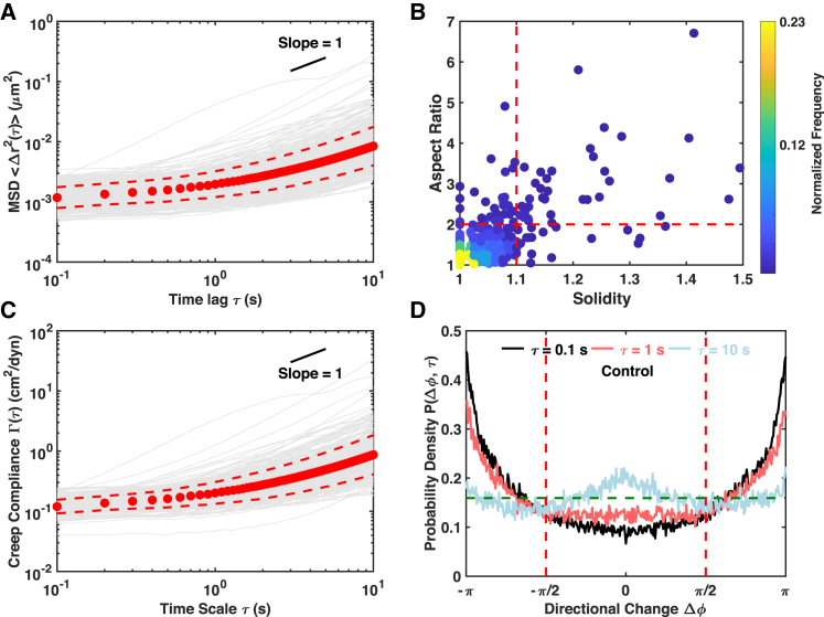
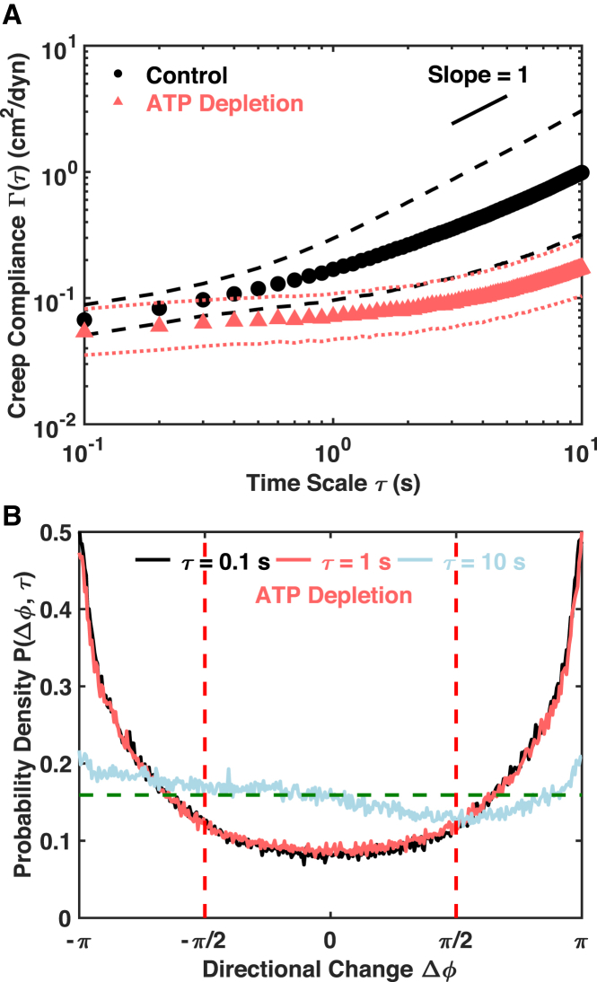
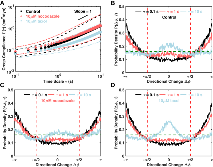
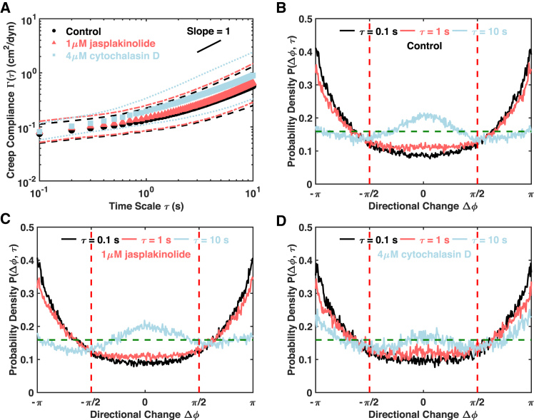
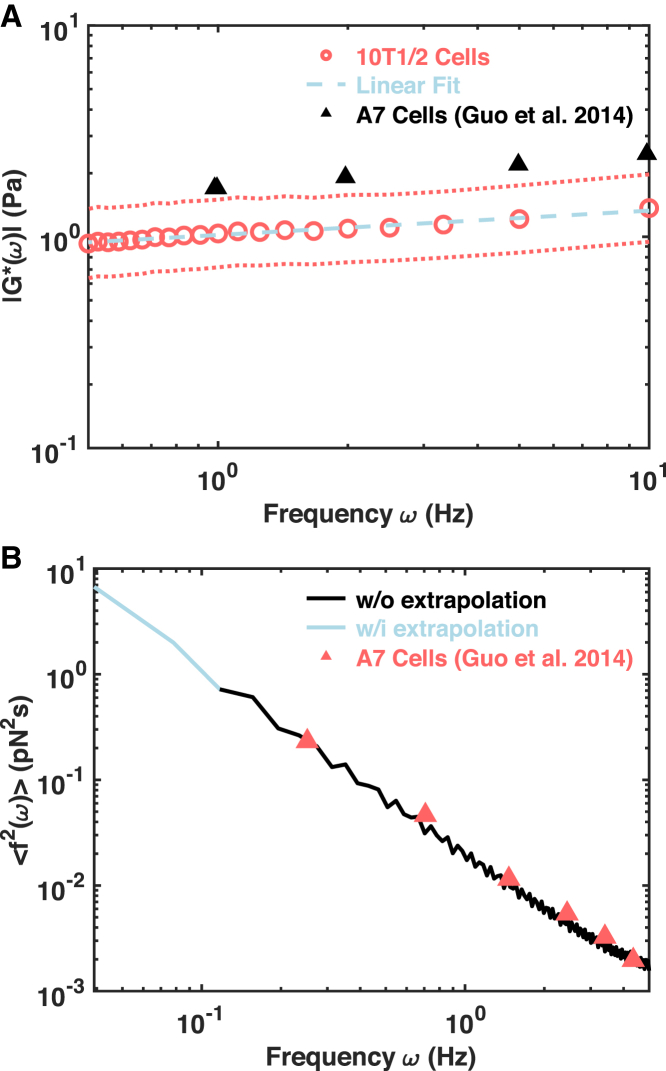
A single-cell assay of active and passive intracellular mechanical properties of mammalian cells could give significant insight into cellular processes. Force spectrum microscopy (FSM) is one such technique, which combines the spontaneous motion of probe particles and the mechanical properties of the cytoskeleton measured by active microrheology using optical tweezers to determine the force spectrum of the cytoskeleton. A simpler and noninvasive method to perform FSM would be very useful, enabling its widespread adoption. Here, we develop an alternative method of FSM using measurement of the fluctuating motion of mitochondria. Mitochondria of the C3H-10T1/2 cell line were labeled and tracked using confocal microscopy. Mitochondrial probes were selected based on morphological characteristics, and their mean-square displacement, creep compliance, and distributions of directional change were measured. We found that the creep compliance of mitochondria resembles that of particles in viscoelastic media. However, comparisons of creep compliance between controls and cells treated with pharmacological agents showed that perturbations to the actomysoin network had surprisingly small effects on mitochondrial fluctuations, whereas microtubule disruption and ATP depletion led to a significantly decreased creep compliance. We used properties of the distribution of directional change to identify a regime of thermally dominated fluctuations in ATP-depleted cells, allowing us to estimate the viscoelastic parameters for a range of timescales. We then determined the force spectrum by combining these viscoelastic properties with measurements of spontaneous fluctuations tracked in control cells. Comparisons with previous measurements made using FSM revealed an excellent match.


Figures

Figure 1
Figure 1
Measurements based on mitochondrial fluctuations. (A and C) Individual and population mean with SDs of (A) MSDs (n = 211) and (C) creep compliance (n = 169) are shown. Gray curves are for individual mitochondria, and filled circles indicate the population mean. Dashed lines are SDs. The short black line is a visual guide of slope one. (B) A distribution of mitochondrial morphologies in a 2D space of solidity and aspect ratio is shown. Filled circles are individual mitochondria color-coded with local normalized frequencies in accordance with the colormap. The two dashed lines represent the morphological filtering criteria of solidity less than 1.1 and aspect ratio less than 2. (D) Distributions of the directional change of control cells are shown at different lag times. The dashed horizontal line indicates a uniform distribution that is characteristic of pure diffusion. To see this figure in color, go online.
Figure 2
Figure 2
Mitochondrial fluctuations are ATP dependent. (A) Population mean creep compliances with SDs of control (n = 169) and ATP-depleted cells (n = 156) are shown. Markers of different shapes and colors indicate population means, and the lines of the same colors are SDs. The short black line is a visual guide of slope one. (B) Distributions of directional change of ATP-depleted cells are shown at different lag times. The dashed horizontal line indicates a uniform distribution that is characteristic of pure diffusion. To see this figure in color, go online.
Figure 3
Figure 3
Microtubule network plays a significant role in mitochondrial fluctuations. (A) Population mean creep compliances with SDs of control cells (n = 87), nocodazole-treated cells (n = 106), and taxol-treated cells (n = 131) are shown. Markers of different shapes and colors indicate population means, and the lines of the same colors are SDs. The short black line is a visual guide of slope one. (BD) Distributions of directional change are shown at different lag times for control, nocodazole-treated, and taxol-treated cells, respectively. The dashed horizontal line indicates a uniform distribution that is characteristic of pure diffusion. To see this figure in color, go online.
Figure 4
Figure 4
Actin network is required for active mitochondrial fluctuations. (A) Population mean creep compliances with SDs of control cells (n = 98), jasplakinolide-treated cells (n = 141), and cytochalasin-D-treated cells (n = 44) are shown. Markers of different shapes and colors indicate population means, and the lines of the same colors are SDs. The short black line is a visual guide of slope one. (BD) Distributions of directional change are shown at different lag times for control, jasplakinolide-treated, and cytochalasin-D-treated cells, respectively. The dashed horizontal line indicates a uniform distribution that is characteristic of pure diffusion. To see this figure in color, go online.
Figure 5
Figure 5
Mitochondrial-fluctuation-based force spectrum calculation. (A) A population mean complex shear modulus (open cycles) with SDs (dotted lines) determined from ATP-depleted cells is shown. The dashed line is a linear fit to the population mean. Black triangles indicate published measurements made on A7 cells using active microrheology (3). (B) The force spectrum is determined by combining the results from control cells and ATP-depleted cells using Eq. 4. Triangles indicate the published measurement of mitochondria using the original FSM (3). To see this figure in color, go online.

Similar articles

Cited by

References

    1. Orr A.W., Helmke B.P., Schwartz M.A. Mechanisms of mechanotransduction. Dev. Cell. 2006;10:11–20. - PubMed
    1. Cross S.E., Jin Y.S., Gimzewski J.K. Nanomechanical analysis of cells from cancer patients. Nat. Nanotechnol. 2007;2:780–783. - PubMed
    1. Guo M., Ehrlicher A.J., Weitz D.A. Probing the stochastic, motor-driven properties of the cytoplasm using force spectrum microscopy. Cell. 2014;158:822–832. - PMC - PubMed
    1. Spill F., Reynolds D.S., Zaman M.H. Impact of the physical microenvironment on tumor progression and metastasis. Curr. Opin. Biotechnol. 2016;40:41–48. - PMC - PubMed
    1. Daniels B.R., Hale C.M., Wirtz D. Differences in the microrheology of human embryonic stem cells and human induced pluripotent stem cells. Biophys. J. 2010;99:3563–3570. - PMC - PubMed

Publication types

LinkOut - more resources