. 2018 Jul;29(7):1900-1916.
doi: 10.1681/ASN.2017050581. Epub 2018 Jun 20.

Caspase-3 Is a Pivotal Regulator of Microvascular Rarefaction and Renal Fibrosis after Ischemia-Reperfusion Injury

Affiliations

Caspase-3 Is a Pivotal Regulator of Microvascular Rarefaction and Renal Fibrosis after Ischemia-Reperfusion Injury

Bing Yang et al. J Am Soc Nephrol. 2018 Jul.

Abstract

Background Ischemia-reperfusion injury (IRI) is a major risk factor for chronic renal failure. Here, we characterize the different modes of programmed cell death in the tubular and microvascular compartments during the various stages of IRI-induced AKI, and their relative importance to renal fibrogenesis.Methods We performed unilateral renal artery clamping for 30 minutes and contralateral nephrectomy in wild-type mice (C57BL/6) or caspase-3-/- mice.Results Compared with their wild-type counterparts, caspase-3-/- mice in the early stage of AKI had high urine cystatin C levels, tubular injury scores, and serum creatinine levels. Electron microscopy revealed evidence of tubular epithelial cell necrosis in caspase-3-/- mice, and immunohistochemistry showed upregulation of the necroptosis marker receptor-interacting serine/threonine-protein kinase 3 (RIPK3) in renal cortical sections. Western blot analysis further demonstrated enhanced levels of phosphorylated RIPK3 in the kidneys of caspase-3-/- mice. In contrast, caspase-3-/- mice had less microvascular congestion and activation in the early and extension phases of AKI. In the long term (3 weeks after IRI), caspase-3-/- mice had reduced microvascular rarefaction and renal fibrosis, as well as decreased expression of α-smooth muscle actin and reduced collagen deposition within peritubular capillaries. Moreover, caspase-3-/- mice exhibited signs of reduced tubular ischemia, including lower tubular expression of hypoxia-inducible factor-1α and improved tubular injury scores.Conclusions These results establish the pivotal importance of caspase-3 in regulating microvascular endothelial cell apoptosis and renal fibrosis after IRI. These findings also demonstrate the predominant role of microvascular over tubular injury as a driver of progressive renal damage and fibrosis after IRI.

Keywords: acute kidney injury; apoptosis; caspase-3; ischemia-reperfusion.


Figures

Figure 1.
Figure 1.
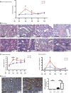
Caspase-3 deficiency aggravates IRI-induced tubular injury. (A) Serum creatinine levels in wild-type (WT) and caspase-3−/− (KO) mice at baseline (pre-op), and 1, 2, 3, and 7 days post-IRI. (B) Representative hematoxylin and eosin (H&E)–stained renal sections from WT and KO mice at 1, 2, 3, and 7 days post-IRI (original magnification ×200). (C) Left panel: mean tubular injury scores of ten randomly chosen high-power fields (original magnification ×200) in mice kidney sections post-IRI. Right panel: urinary levels of cystatin C in WT and KO mice at baseline (pre-op) and 1, 2, 3, or 7 days post-IRI. (D) Left panels: representative Kidney Injury Molecule 1 (KIM-1) immunohistochemistry in renal cortical sections from WT and KO mice at day 1 post-IRI (magnification 200×). Right panel: quantification of KIM-1 immunohistochemistry-stained murine renal cortical sections at baseline (pre-op) and 1 day post-IRI. All scale bars, 50 μm. Values are mean±SEM. *P<0.05, compared between WT and KO at the same time point.
Figure 2.
Figure 2.
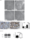
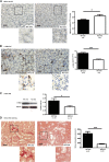
Caspase-3 deficiency increases IRI-induced necroptosis in tubular cells. (A) Representative electron microscopy (EM) showing renal tubules from wild-type (WT) (left) and caspase-3−/− (KO) (right) mice that underwent IRI and were euthanized at 1 day post-IRI (original magnification ×1000). Scale bars, 10 μm. Tubules from KO mice show severe necrotic changes with loss or tubular cell membrane integrity and widespread accumulation of cellular debris within tubules. (B) Left panels: representative RIPK3 immunohistochemistry (IHC) in renal cortical sections from WT and KO mice that underwent IRI and were euthanized at 1 day post-IRI (original magnification ×200). Right panel: quantification of RIPK3 IHC in murine renal cortical sections at baseline (pre-op) and 1 day post-IRI. Scale bars, 50 μm. (C) Left panel: representative Western blot (WB) of phosphorylated RIPK3 (pRIPK3) in renal tissue from WT and KO mice that underwent IRI and were euthanized at 1 day post-IRI. Right panel: quantification of pRIPK3 WB at day 1 post-IRI. Values are mean±SEM. *P<0.05, compared between WT and KO at the same time point.
Figure 3.
Figure 3.
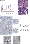
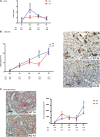
Caspase-3 deficiency attenuates IRI-induced microvascular injury. (A) Left panel: quantification of rouleaux formation in hematoxylin and eosin (H&E)–stained kidney sections at baseline (pre-op) and from mice that underwent IRI and were euthanized at 1, 2, 3, or 7 days post-IRI. Right panel: representative H&E-stained murine renal cortical sections at 3 days post-IRI (original magnification ×400). Scale bars, 20 μm. (B): Left panel: representative images of MECA-32 immunohistochemistry (IHC) in renal cortical sections from wild-type (WT) and caspase-3−/− (KO) mice that underwent IRI and were euthanized at 3 days post-IRI (original magnification ×200 and ×400). Right panel: quantification of MECA-32 in murine renal cortical medullary junction sections at baseline (pre-op) and 1, 2, 3, or 7 days post-IRI. Scale bars, 50 μm. (C) Representative electron microscopy images of renal endothelial cells in WT (left) and KO (right) mice that underwent IRI and were euthanized at 3 days post-IRI. Loss of endothelial fenestration is found in WT mice (arrow), whereas KO show preserved fenestrae (original magnification ×1000). Scale bars, 500 nm. Values are mean±SEM. *P<0.05, compared between WT and KO at the same time point.
Figure 4.
Figure 4.
Caspase-3 deficiency attenuates IRI-induced upregulation of profibrotic markers. (A) Serum levels of CTGF in wild-type (WT) and caspase-3−/− (KO) mice that underwent IRI and were euthanized at 1, 2, 3, or 7 days post-IRI. (B) Left panel: quantification of α-SMA immunohistochemistry (IHC) in renal cortical sections from WT and KO mice at baseline (pre) or at 1, 2, 3, or 7 days post-IRI (original magnification X200). Right panels: representative α-SMA staining in murine renal cortical sections at day 3 after surgery. Scale bars, 50 μm. (C) Left panels: representative images of Sirius Red staining in renal cortical medullary junction sections from WT (top panel) and KO (bottom panel) mice that underwent IRI and were euthanized at 7 days post-IRI (original magnification X200). Scale bars, 50 μm. Right panel: quantification of Sirius Red staining in murine renal cortical medullary junction sections at baseline and 1, 2, 3, or 7 days post-IRI. Values are mean±SEM. *P<0.05, compared between WT and KO at the same time point.
Figure 5.
Figure 5.
Caspase-3 deficiency attenuates IRI-induced microvascular rarefaction and fibrosis. (A) Left panels: representative images of MECA-32 immunohistochemistry (IHC) in cortical medullary junction sections from wild-type (WT) and caspase-3−/− (KO) mice that underwent IRI and were euthanized at 21 days post-IRI (original magnification ×200 and ×400). Right panel: quantification of MECA-32 staining in murine renal cortical sections at 21 days post-IRI. Scale bars, 50 μm. (B) Left panels: representative image of α-SMA IHC of renal cortical sections from WT and KO mice that underwent IRI and were euthanized at 21 days post-IRI (n=10 per group; original magnification X200 and X400). Right panel: quantification of α-SMA staining in PTCs in murine renal cortical sections at 21 days post-IRI. Scale bars, 50 μm. (C) Left panel: representative Western blot (WB) of α-SMA from WT and KO mice that underwent IRI and were euthanized at 21 days post-IRI. Right panel: quantification of α-SMA WB at 21 day post-IRI. (D) Left panels: representative image of Sirius Red staining of renal cortical medullary junction sections from WT and KO mice that underwent IRI and were euthanized at 21 days post-IRI (original magnification ×200 and ×400). Right panel: quantification of Sirius Red staining of murine renal cortical medullary junction sections at 21 days post-IRI. Scale bars, 50 μm. Values are mean±SEM. *P<0.05; ***P<0.001, compared between WT and KO at the same time point.
Figure 6.
Figure 6.
Caspase-3 deficiency prevents IRI-induced long-term endothelial cell death. (A) Representative electronic microscopic images of endothelial cell apoptotic death from wild-type (WT) and caspase-3−/− (KO) mice that underwent IRI and were euthanized at 21 days post-IRI. Red arrows indicate apoptotic bodies and blue arrows indicate exosome-like membrane vesicles (original magnification ×1000). Scale bars, 500 nm. (B) Quantification of apoptotic endothelial cells (ECs) in WT and KO mice that underwent IRI and were euthanized at 21 days post-IRI. Values are mean±SEM. *P<0.05, compared between WT and KO at the same time point.
Figure 7.
Figure 7.
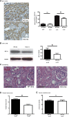
Caspase-3 deficiency prevents IRI-induced long-term tubular injury. (A) Left panels: representative images of HIF-1α staining in murine renal cortical sections 21 days after surgery. Right panel: quantification of HIF-1α immunohistochemistry (IHC) in renal cortical sections from wild-type (WT) and caspase-3−/− (KO) mice at baseline and 21 days post-IRI (original magnification ×200). (B) Left panel: representative Western blot (WB) images of HIF-1α in renal tissue 21 days post-IRI. Right panel: quantification of HIF-1α WB detection at 21 days post-IRI. (C) Representative hematoxylin and eosin (H&E)–stained murine renal cortical sections 21 days post-IRI (original magnification ×200). (D) Mean tubular injury scores of ten randomly chosen high-power fields in wild-type (WT) and caspase-3−/− (KO) mice that underwent IRI and were euthanized at 21 days post-IRI. (E) Serum creatinine levels in WT and KO mice that underwent IRI and were euthanized at 21 days post-IRI. All bar scales=50 μm. Values are mean±SEM. *P<0.05, compared between WT and KO at the same time point.
Figure 8.
Figure 8.
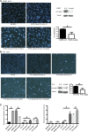
Caspase-3 silencing decreases apoptosis in endothelial cells but increases necrosis in TECs submitted to hypoxia plus serum starvation and reoxygenation. (A) Left panels: representative images of Hoechst 33342 and propidium iodide (Ho-PI) staining in HUVECs exposed to hypoxia (5% O2) for 4 hours, followed by reoxygenation for 1 hour in serum-free medium. Right panels: representative images of western blot (top panel) and quantification (bottom panel) for pro-caspase-3 in HUVECs transfected with siRNA control (si ctl) or caspase-3 (si casp3; n=3 independent experiments). (B) Left panels: representative images of Hoechst 33342 and propidium iodide (Ho-PI) staining of human PT-2 TECs exposed to hypoxia (5% O2) for 4 or 24 hours followed by reoxygenation for 1 hour in serum-free medium. Right panels: representative images of Western blot and quantification for pro-caspase-3 in PT-2 cells transfected with si ctl or caspase-3 (n=3 independent experiments). (C) Left panel: quantification of apoptotic death in HUVECs and PT-2 cells exposed to hypoxia reoxygenation in serum-free medium. Right panel: quantification of necrotic death in HUVECs and PT-2 cells exposed to hypoxia reoxygenation in serum-free medium. Values are mean±SEM. *P<0.05.
Figure 9.
Figure 9.
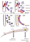
Schematic representation of the effect of caspase-3 deficiency on microvascular rarefaction and renal dysfunction post-IRI. Caspase-3 is a pivotal regulator of PTC rarefaction and renal dysfunction. Early tubular injury and renal dysfunction are increased in caspase-3−/− (KO) mice, whereas microvascular integrity is ameliorated throughout the various phases. In the long term, KO mice show reduced microvascular dropout, decreased tubular injury, and reduced interstitial fibrosis. EC, endothelial cell; WT, wild type.

Similar articles

Cited by

  • Microvascular disease in chronic kidney disease: the base of the iceberg in cardiovascular comorbidity.
    Querfeld U, Mak RH, Pries AR. Querfeld U, et al. Clin Sci (Lond). 2020 Jun 26;134(12):1333-1356. doi: 10.1042/CS20200279. Clin Sci (Lond). 2020. PMID: 32542397 Free PMC article. Review.
  • Ganoderic Acids Prevent Renal Ischemia Reperfusion Injury by Inhibiting Inflammation and Apoptosis.
    Shao G, He J, Meng J, Ma A, Geng X, Zhang S, Qiu Z, Lin D, Li M, Zhou H, Lin S, Yang B. Shao G, et al. Int J Mol Sci. 2021 Sep 23;22(19):10229. doi: 10.3390/ijms221910229. Int J Mol Sci. 2021. PMID: 34638569 Free PMC article.
  • Dynamic cellular changes in acute kidney injury caused by different ischemia time.
    Shan D, Wang YY, Chang Y, Cui H, Tao M, Sheng Y, Kang H, Jia P, Song J. Shan D, et al. iScience. 2023 Apr 11;26(5):106646. doi: 10.1016/j.isci.2023.106646. eCollection 2023 May 19. iScience. 2023. PMID: 37168554 Free PMC article.
  • Effect of fetuin-A on adenine-induced chronic kidney disease model in male rats.
    Ashour WMR, Zamzam MSA, Sayed Ali HEEDE, Ebrahim RH. Ashour WMR, et al. Iran J Basic Med Sci. 2023;26(5):511-516. doi: 10.22038/IJBMS.2023.66346.14584. Iran J Basic Med Sci. 2023. PMID: 37051102 Free PMC article.
  • Apoptotic cell death in disease-Current understanding of the NCCD 2023.
    Vitale I, Pietrocola F, Guilbaud E, Aaronson SA, Abrams JM, Adam D, Agostini M, Agostinis P, Alnemri ES, Altucci L, Amelio I, Andrews DW, Aqeilan RI, Arama E, Baehrecke EH, Balachandran S, Bano D, Barlev NA, Bartek J, Bazan NG, Becker C, Bernassola F, Bertrand MJM, Bianchi ME, Blagosklonny MV, Blander JM, Blandino G, Blomgren K, Borner C, Bortner CD, Bove P, Boya P, Brenner C, Broz P, Brunner T, Damgaard RB, Calin GA, Campanella M, Candi E, Carbone M, Carmona-Gutierrez D, Cecconi F, Chan FK, Chen GQ, Chen Q, Chen YH, Cheng EH, Chipuk JE, Cidlowski JA, Ciechanover A, Ciliberto G, Conrad M, Cubillos-Ruiz JR, Czabotar PE, D'Angiolella V, Daugaard M, Dawson TM, Dawson VL, De Maria R, De Strooper B, Debatin KM, Deberardinis RJ, Degterev A, Del Sal G, Deshmukh M, Di Virgilio F, Diederich M, Dixon SJ, Dynlacht BD, El-Deiry WS, Elrod JW, Engeland K, Fimia GM, Galassi C, Ganini C, Garcia-Saez AJ, Garg AD, Garrido C, Gavathiotis E, Gerlic M, Ghosh S, Green DR, Greene LA, Gronemeyer H, Häcker G, Hajnóczky G, Hardwick JM, Haupt Y, He S, Heery DM, Hengartner MO, Hetz C, Hildeman DA, Ichijo H, Inoue S, Jäättelä M, Janic A, Joseph B, Jost PJ, Kanneganti TD, Karin M, Kashkar H, Kaufmann T, Kelly … See abstract for full author list ➔ Vitale I, et al. Cell Death Differ. 2023 May;30(5):1097-1154. doi: 10.1038/s41418-023-01153-w. Epub 2023 Apr 26. Cell Death Differ. 2023. PMID: 37100955 Free PMC article. Review.

References

    1. Susantitaphong P, Cruz DN, Cerda J, Abulfaraj M, Alqahtani F, Koulouridis I, et al. .: Acute Kidney Injury Advisory Group of the American Society of Nephrology : World incidence of AKI: A meta-analysis. Clin J Am Soc Nephrol 8: 1482–1493, 2013 - PMC - PubMed
    1. Bedford M, Farmer C, Levin A, Ali T, Stevens P: Acute kidney injury and CKD: Chicken or egg? Am J Kidney Dis 59: 485–491, 2012 - PubMed
    1. Ishani A, Xue JL, Himmelfarb J, Eggers PW, Kimmel PL, Molitoris BA, et al. : Acute kidney injury increases risk of ESRD among elderly. J Am Soc Nephrol 20: 223–228, 2009 - PMC - PubMed
    1. Molitoris BA: Therapeutic translation in acute kidney injury: The epithelial/endothelial axis. J Clin Invest 124: 2355–2363, 2014 - PMC - PubMed
    1. Amdur RL, Chawla LS, Amodeo S, Kimmel PL, Palant CE: Outcomes following diagnosis of acute renal failure in U.S. veterans: Focus on acute tubular necrosis. Kidney Int 76: 1089–1097, 2009 - PubMed

Publication types

MeSH terms

Grants and funding

LinkOut - more resources