. 2018 Jul 30;11(7):dmm033563.
doi: 10.1242/dmm.033563.

Octopamine signaling in the metazoan pathogen Schistosoma mansoni: localization, small-molecule screening and opportunities for drug development

Affiliations

Octopamine signaling in the metazoan pathogen Schistosoma mansoni: localization, small-molecule screening and opportunities for drug development

Nelly El-Sakkary et al. Dis Model Mech. .

Abstract

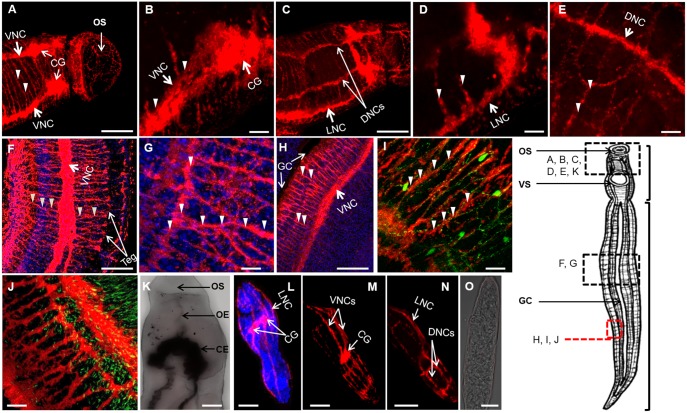
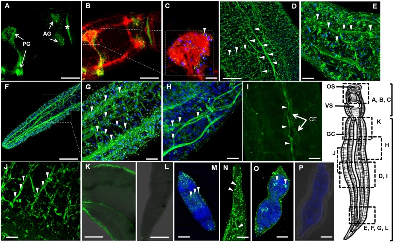
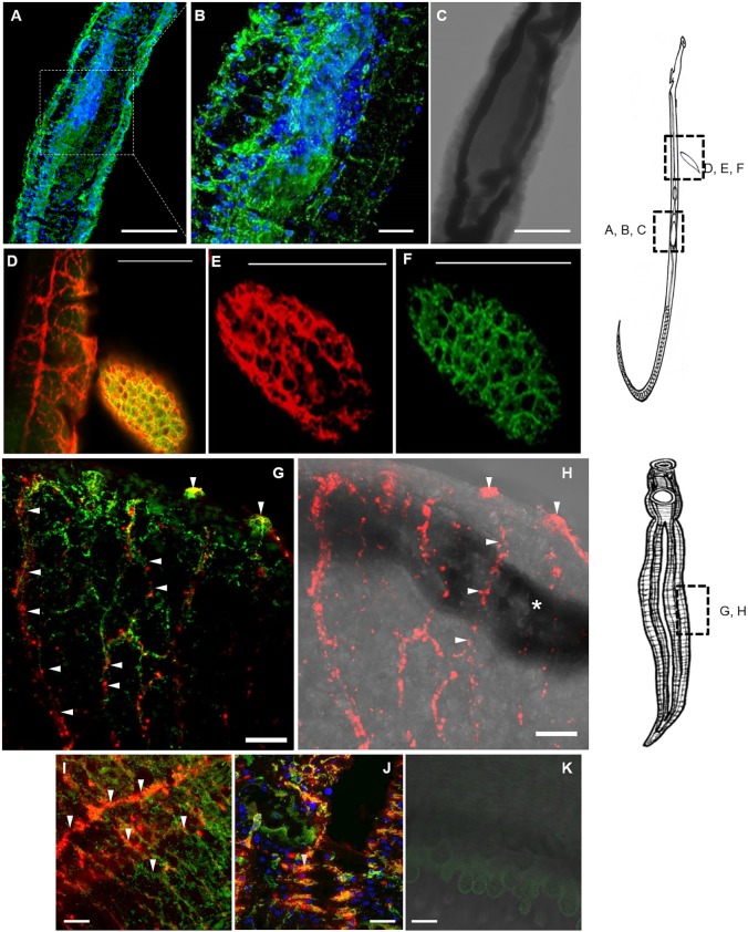
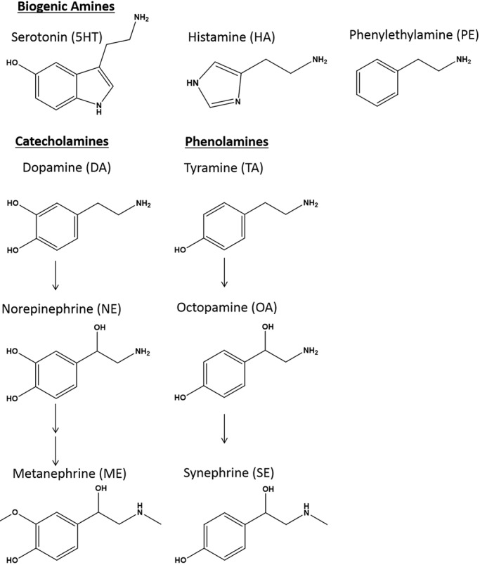
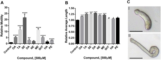
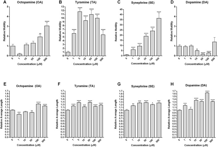
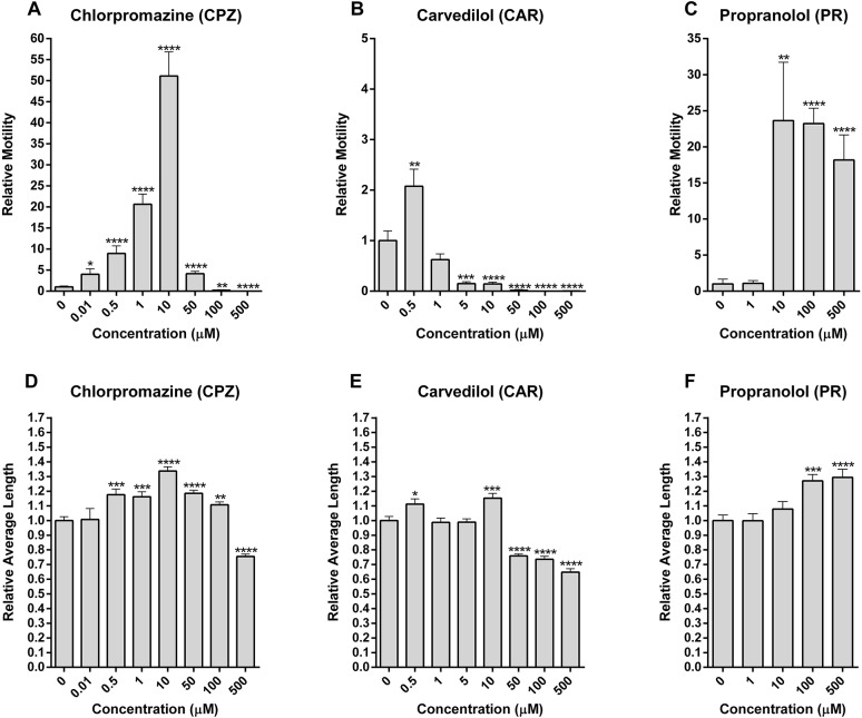
Schistosomiasis is a tropical disease caused by a flatworm trematode parasite that infects over 200 million people worldwide. Treatment and control of the disease rely on just one drug, praziquantel. The possibility of drug resistance coupled with praziquantel's variable efficacy encourages the identification of new drugs and drug targets. Disruption of neuromuscular homeostasis in parasitic worms is a validated strategy for drug development. In schistosomes, however, much remains to be understood about the organization of the nervous system, its component neurotransmitters and potential for drug discovery. Using synapsin as a neuronal marker, we map the central and peripheral nervous systems in the Schistosoma mansoni adult and schistosomulum (post-infective larva). We discover the widespread presence of octopamine (OA), a tyrosine-derived and invertebrate-specific neurotransmitter involved in neuromuscular coordination. OA labeling facilitated the discovery of two pairs of ganglia in the brain of the adult schistosome, rather than the one pair thus far reported for this and other trematodes. In quantitative phenotypic assays, OA and the structurally related tyrosine-derived phenolamine and catecholamine neurotransmitters differentially modulated schistosomulum motility and length. Similarly, from a screen of 28 drug agonists and antagonists of tyrosine-derivative signaling, certain drugs that act on OA and dopamine receptors induced robust and sometimes complex concentration-dependent effects on schistosome motility and length; in some cases, these effects occurred at concentrations achievable in vivo The present data advance our knowledge of the organization of the nervous system in this globally important pathogen and identify a number of drugs that interfere with tyrosine-derivative signaling, one or more of which might provide the basis for a new chemotherapeutic approach to treat schistosomiasis.This article has an associated First Person interview with the first author of the paper.

Keywords: Biogenic amine; Dopamine; Drug discovery; Nervous system; Neuromuscular; Octopamine; Schistosoma mansoni; Synapsin.


Conflict of interest statement

Competing interestsThe authors declare no competing or financial interests.

Figures

Fig. 1.
Fig. 1.
Architecture of the schistosome nervous system. (A) The neuronal marker synapsin was labeled with a monoclonal anti-synapsin antibody (anti-SYNORF1) and a secondary antibody conjugated to HRP followed by tyramide signal amplification, which produces a red color. Arrowheads point to nerve chords, nerve fibers or nerve cell bodies. The position of the worm in each image is approximated relative to the scheme of the worm's body on the right in which the locations of the oral sucker (OS), ventral sucker (VS), gynecophoral canal (GC), the head (upper bracket) and body (lower bracket) are also indicated. (B) The ventral nerve chords (VNCs), joined by transverse commissures, and cerebral ganglia (CG) are visible. (C,D) Lateral nerve chords (LNCs) are visible on either side of the worm. (C,E) The dorsal nerve chords (DNCs) are also observed. (F,G) The fine PNS nerve fibers are visible throughout the body and extend to the surface [tegument (Teg), indicated with arrows in F] and intersect with the main longitudinal nerve chords. (H) Short nerve fibers of the PNS extend from the VNCs to the GC flap, as indicated with arrows. (I) The neurotransmitter 5HT (green), was co-immunolabeled with anti-synapsin and the labeling pattern shows a close juxtaposition of nerve fibers. (J) Actin (green) was co-immunolabeled with synapsin (red) and is apparent along the main nerve chords of the CNS. Incubation of worms with secondary antibody and amplification reagent alone did not yield significant labeling, as demonstrated by the negative control image that was overlaid with the brightfield image (K): the oesophagus (OE) and caecum (CE) are indicated. Scale bars: 100 μm at 20× magnification in panels A, C, F, H and K; 20 μm at 63× magnification in panels B, D, E, G, I, J, L-O. (L,M) In D7 schistosomula, synapsin was labeled in the CG and VNCs that projected longitudinally. (L,N) DNCs and LNCs are also labeled. (O) Incubation with the amplification reagent and secondary antibody alone did not yield significant labeling, as demonstrated with the transmitted image overlay.
Fig. 2.
Fig. 2.
OA is found in the central and peripheral nervous systems. OA was labeled with an OA-specific primary antibody and an Alexa-488 (green)-conjugated secondary antibody; DAPI was used as a counterstain (blue). Arrowheads indicate nerve chords, nerve fibers or cell bodies. (A) OA is localized to anterior ganglia (AG) and posterior ganglia (PG) in the head of the adult male worm, a finding that is supported by synapsin co-labeling (B,C; in red). The AG are located posterior of the oral sucker, whereas the PG are anterior of the ventral sucker. Non-specific surface labeling of OA in (A) is indicated with an asterisk. (D-H) OA is found in the longitudinal nerve chords of the CNS and PNS in both males (D,E) and females (F-H). (E,G,H) At higher magnification, OA is identified in cell bodies. (I) An octopaminergic neuron lines the parasite's caecum (CE), indicated by arrowheads. (J) OA is present in what appears to be the submuscular peripheral nerve net of the PNS. (K) Worms were also probed with anti-OA antibody pre-adsorbed with OA or (L) secondary antibody alone: fluorescence, if noted, was non-specific. OA is also detected in D7 schistosomula, indicated by labeling with Alexa-488-conjugated antibody. Innervation of the PNS (M,N) and possibly of the CG (O) is visible. (P) The secondary antibody alone did not yield non-specific labeling. Scale bars: 100 μm at low magnification (20×) in panels A, B, D, F, K and L; 20 μm at high magnification (63×) in panels C, E, G, H-J, M-P.
Fig. 3.
Fig. 3.
OA and synapsin are found in the ovary of the female, in the developing embryo and innervating the surface of the worm. The region in the adult female worm or adjacent egg is indicated in the diagram of the female worm on the right. OA was labeled with an anti-OA primary antibody and an Alexa-488 (green)-conjugated secondary antibody. (A,B) Octopaminergic neurons run along the length of the ovary, coinciding with a concentration of nuclei (Neves et al., 2005). (C) The ovary is also shown in the brightfield image. Colocalization of OA with (D) anti-synapsin (yellow) and either (E) anti-synapsin (red) or (F) anti-OA (green) alone reveals the organization of the nerve fibers in the developing embryo. For the adult male worm, regions in the panels are indicated in the lower diagram to the right of the image grid. In the male, OA and synapsin are colocalized in the surface tubercles (G) with synapsin alone in the synapsin-transmitted light image (H). The asterisk in H indicates a dark line running from left to right in the brightfield image overlay, which is due to a female worm within the male's gynecophoral canal just below the plane of view. (I) Synapsin appears to be enriched in CNS nerve fibers and OA in PNS nerve fibers. (J) The surface of the gynecophoral canal is innervated by a cluster of short nerve fibers containing both OA and synapsin, as indicated by the labeling in yellow. Scale bars: 100 μm with 20× magnification in panels A, C-F; 20 μm at high magnification (63×) in panels B, G-K.
Fig. 4.
Fig. 4.
Biogenic amine structures. The indicated BAs are derived from histidine [histamine (HA)] or aromatic amino acids such as tyrosine and tryptophan. Structurally related tyrosine derivatives include the catecholamines (DA, NE and ME) and the phenolamines (TA, OA and SE).
Fig. 5.
Fig. 5.
BAs increase motility and body length. D7 schistosomula were incubated with BAs at a concentration of 500 µM for 20 min. The BAs tested were octopamine (OA), tyramine (TA), synephrine (SE), dopamine (DA), noradrenaline (NE), metanephrine (ME), serotonin (5HT), histamine (HA) and phenylethylamine (PE). (A) Relative motility and (B) length were quantified as described in the text and are expressed as the fold change compared with schistosomula in the presence of 0.015% DMSO for SE or H2O for all other compounds. The mean relative motility and length were measured by normalization to each well, at baseline. Means and s.e.m. from a minimum of two experiments with a minimum of two replicates per experiment are shown (n=36). Significance of the mean values, compared with controls, was determined using the unpaired two-tailed Student's t-test: P-values <0.05*, <0.005** and <0.00005**** were considered significant. (C) Representative images of (I) a control and (II) an OA-treated parasite: note the length of the parasite exposed to OA relative to control. Scale bars: 50 µm.
Fig. 6.
Fig. 6.
OA and related amines modulate motility and length of schistosomula. Using D7 schistosomula, concentration-response assays (1-500 μM) were performed over 20 min with four tyrosine derivatives previously determined to cause significant changes in motility at 500 μM (Fig. 5). The mean relative motility (A-D) and length (E-H) were measured by normalization to each well, at baseline. Means and s.e.m. from a minimum of two experiments with a minimum of two replicates per experiment are shown (n=36). The data presented at the 500 µM concentration are distinct from the data presented in Fig. 5. Significance of the mean values, compared with controls, was determined using the unpaired two-tailed Student's t-test: P-values <0.05*, <0.005**, <0.0005*** and <0.00005**** were considered significant.
Fig. 7.
Fig. 7.
Chlorpromazine, carvedilol and propranolol modulate motility and length of schistosomula. Using D7 schistosomula, concentration-response assays (0.01-500 μM) were performed over 20 min for three modulators of tyrosine-derivative signaling that had caused pronounced effects on (A-C) motility and (D-F) length at 65 μM (Table 1). Mean relative motility values were determined by normalization to the corresponding wells at baseline (preceding addition of the drug compound; n≥13). Means and s.e.m. from a minimum of three experiments, each with a minimum of two replicates per experiment, are shown. A minimum of three wells were recorded per treatment. Note that there is no continuity of scale in the y-axis between graphs A-C. Significance of the mean values, compared with controls, was determined using the unpaired two-tailed Student's t-test: P-values <0.05*, <0.005**, 0.0005*** and <0.00005**** were considered significant.

Similar articles

Cited by

References

    1. Abdulla M.-H., Ruelas D. S., Wolff B., Snedecor J., Lim K.-C., Xu F., Renslo A. R., Williams J., McKerrow J. H. and Caffrey C. R. (2009). Drug discovery for schistosomiasis: hit and lead compounds identified in a library of known drugs by medium-throughput phenotypic screening. PLoS Negl. Trop. Dis. 3, e478 10.1371/journal.pntd.0000478 - DOI - PMC - PubMed
    1. Achari R. and Laddu A. (1992). Terazosin: a new alpha adrenoceptor blocking drug. J. Clin. Pharmacol. 32, 520-523. 10.1177/009127009203200605 - DOI - PubMed
    1. Alkema M. J., Hunter-Ensor M., Ringstad N. and Horvitz H. R. (2005). Tyramine functions independently of octopamine in the Caenorhabditis elegans nervous system. Neuron 46, 247-260. 10.1016/j.neuron.2005.02.024 - DOI - PubMed
    1. Andrews P., Thomas H., Pohlke R. and Seubert J. R. (1983). Praziquantel. Med. Res. Rev. 3, 147-200. 10.1002/med.2610030204 - DOI - PubMed
    1. Basch P. F. (1981). Cultivation of Schistosoma mansoni in vitro. I. Establishment of cultures from cercariae and development until pairing. J. Parasitol. 67, 179-185. 10.2307/3280632 - DOI - PubMed

Publication types

MeSH terms

LinkOut - more resources