. 2018 Jul 31;8(8):2723-2733.
doi: 10.1534/g3.118.200486.

Fission Yeast CENP-C (Cnp3) Plays a Role in Restricting the Site of CENP-A Accumulation

Affiliations

Fission Yeast CENP-C (Cnp3) Plays a Role in Restricting the Site of CENP-A Accumulation

Michiko Suma et al. G3 (Bethesda). .

Abstract

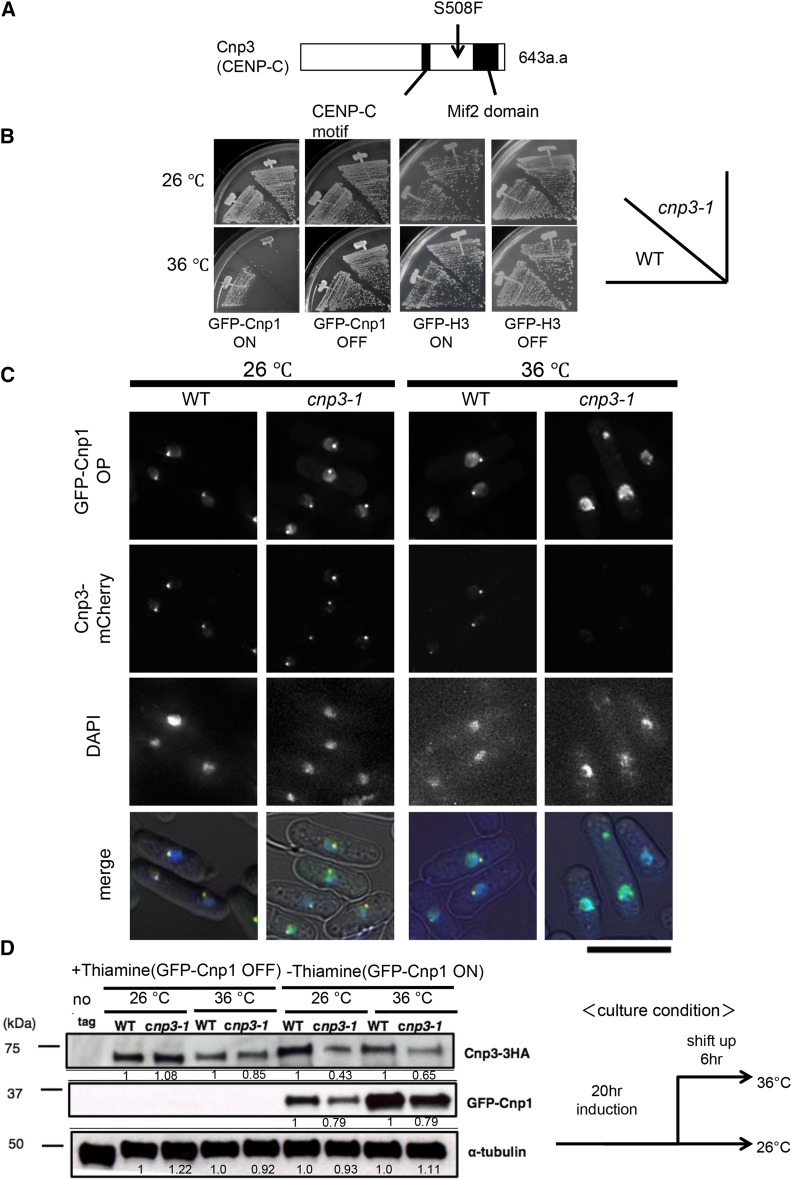
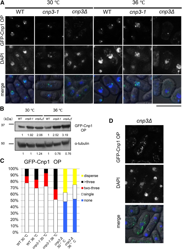
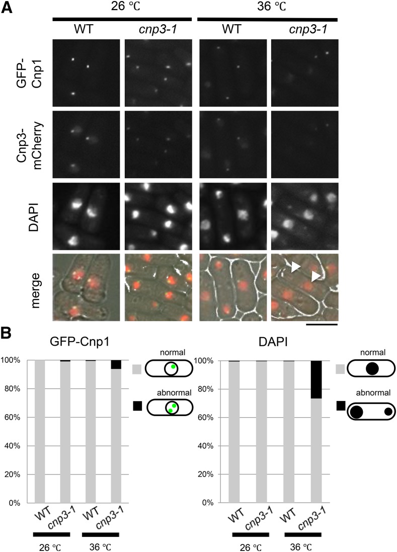
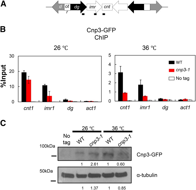
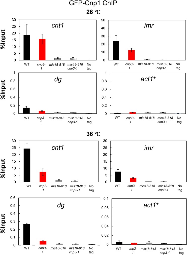
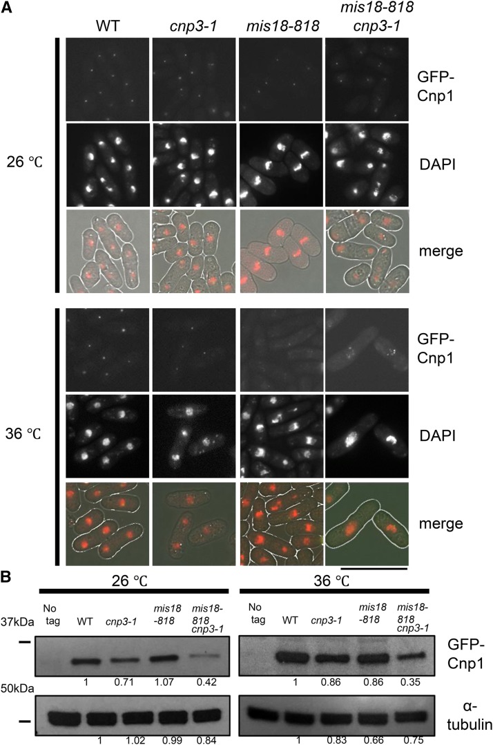
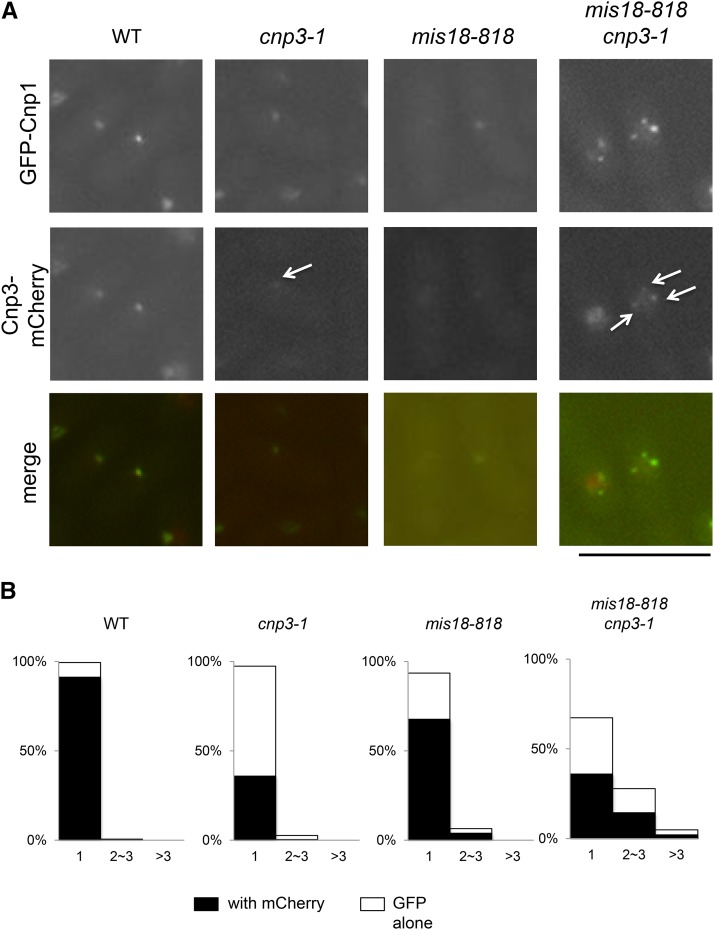
The centromere is a chromosomal locus where a microtubule attachment site, termed kinetochore, is assembled in mitosis. In most eukaryotes, with the exception of holocentric species, each chromosome contains a single distinct centromere. A chromosome with an additional centromere undergoes successive rounds of anaphase bridge formation and breakage, or triggers a cell cycle arrest imposed by DNA damage and replication checkpoints. We report here a study in Schizosaccharomyces pombe to characterize a mutant (cnp3-1) in a gene encoding a homolog of mammalian centromere-specific protein, CENP-C. At the restrictive temperature 36°, the Cnp3-1 mutant protein loses its localization at the centromere. In the cnp3-1 mutant, the level of the Cnp1 (a homolog of a centromere-specific histone CENP-A) also decreases at the centromere. Interestingly, the cnp3-1 mutant is prone to promiscuous accumulation of Cnp1 at non-centromeric regions, when Cnp1 is present in excess. Unlike the wild type protein, Cnp3-1 mutant protein is found at the sites of promiscuous accumulation of Cnp1, suggesting that Cnp3-1 may stabilize or promote accumulation of Cnp1 at non-centromeric regions. From these results, we infer the role of Cnp3 in restricting the site of accumulation of Cnp1 and thus to prevent formation of de novo centromeres.

Keywords: CENP-A; CENP-C; and fission yeast; centromere.


Figures

Figure 1
Figure 1
Distribution of GFP-Cnp1. (A) Schematic illustration of the structure of Cnp3 protein and the position of the cnp3-1 mutation. (B) The wild type strain (WT) and the cnp3-1 mutant (cnp3-1) were transformed with pREP41-GFP- Cnp1 or GFP- H3. The resulting transformants (MS01, MS02, MS03 and MS04) were streaked as indicated and grown at 26°C or 36°C on EMM 2 media with thiamine for repression (GFP-Cnp1 OFF) or without thiamine for derepression (GFP-Cnp1 ON). (C) Localization of GFP-Cnp1 and Cnp3-mCherry were examined in the wild type (MS213) and cnp3-1 cells (MS217). They were grown at 26°C for 20 hr for induction of GFP-Cnp1 in absence of thiamine, and shifted to 36°C for 6 hr in absence of thiamine. The bar indicates 5 μm. (D) The levels of GFP-Cnp1and Cnp3-HA were examined by western blot in the wild type strain (WT: MS525) and the cnp3-1 mutant (cnp3-1: MS527). They were grown as (C) and the samples were taken 20 hr after the induction at 26 °C and 6 hr after the shift to 36 °C. α-tubulin was used as a loading control.
Figure 2
Figure 2
Comparison between cnp3-1 and cnp3 Δ. (A and D) (A) Localization of GFP-Cnp1 were examined in WT (MS216), cnp3-1 (MS262) and cnp3Δ (MS168) cells. They were grown at 30 °C for 20 hr for induction of GFP-Cnp1 in absence of thiamine, and shifted to 36 °C for 6 hr in absence of thiamine. The GFP-Cnp1 appeared as cytoplasmic speckles in the cnp3Δ cells (MS168) was shown in (D). The bar indicates 5 μm. (B) The level of GFP-Cnp1 was examined by western-blot. α-tubulin was used as a loading control. The samples were taken 20 hr after the induction at 30 °C and 6 hr after the shift to 36 °C. (C) The statistic analysis of (A).
Figure 3
Figure 3
Characterization of the cnp3-1 mutant. (A) The strains expressing GFP-tagged Cnp1 and mCherry-tagged Cnp3 (WT, MS548), Cnp3-1 mutant protein (cnp3-1, MS549) from the native promoter were grown at 26°C or 36°C for 24 hr and observed under a fluorescence microscope. The white arrow heads indicate mis-segregated chromatin mass scored as abnormal in (B). The bar indicates 10μm. (B) The statistic analysis of (A).
Figure 4
Figure 4
Loss of Cnp3-1 from the centromere. (A) Schematic illustration of the centromere I (Cen I). Each centromere contains a central domain (cnt), flanked by innermost repeats (imr). This core domain is surrounded by arrays of outer repeats (otr). The black bars indicate the position of the primers used in the ChIP analysis. (B) Localization of Cnp3-GFP in Cen I was examined by ChIP analysis in the wild type stain (WT: MS468) and the cnp3-1 mutant (cnp3-1: MS544). The stains were grown at 26 °C and shifted to 36 °C for 6 hr. (C) The level of Cnp3-GFP was examined by western-blot in the wild type strain (WT: MS468) and the cnp3-1 mutant (cnp3-1: MS544) grown as in (B). α-tubulin was used as a loading control.
Figure 5
Figure 5
Distribution of Cnp1 at the centromere. Distribution of GFP-Cnp1 at Cen I was examined by ChIP analysis in the indicated mutants (WT: MS343, cnp3-1: MS344, mis18-818; MS503 and cnp3-1 mis18-818: MS534) grown as in Fig. 4B.
Figure 6
Figure 6
S508 of Cnp3 is important for centromere-targeting. Localization of a partial fragment of Cnp3 spanning from 414 to 643 amino acids tagged with GFP were examined at 26°C or 36°C in the wild type strain (MS568 and MS569). Sad1 tagged with mCherry was used as a marker for the centromere cluster. These strain were grown as in Fig. 1C. The bar indicates 5 μm.
Figure 7
Figure 7
Multiple foci of GFP-Cnp1 in mis18 cnp3-1. (A) Cnp1 tagged with GFP was expressed from the native promoter. Each strain (WT: MS343, cnp3-1: MS344, mis18-818: MS503 and cnp3-1 mis18-818: MS534) was grown at 26 °C and shifted to 36 °C for 6 hr. The bar indicates 10 μm. (B) The level of Cnp1 in strains used in (A) was examined by immunoblot. α-tubulin was used as a loading control.
Figure 8
Figure 8
Colocalization of Cnp1-foci and Cnp3-1 in mis18 cnp3-1. (A) Both Cnp1 tagged with GFP and Cnp3 (or Cnp3-1) tagged with mCherry were expressed from their native promoters in the four strains (WT: MS548, cnp3-1: MS549, mis18-818: MS546 and cnp3-1 mis18-818: MS545). They were grown as in Fig. 7A. The white arrows indicate the Cnp3 (Cnp3-1)-foci. The bar indicates 10 μm. (B) The statistic analysis of (A).

Similar articles

Cited by

References

    1. Allshire R. C., Karpen G. H., 2008. Epigenetic regulation of centromeric chromatin: old dogs, new tricks? Nat. Rev. Genet. 9: 923–937. 10.1038/nrg2466 - DOI - PMC - PubMed
    1. Allshire R. C., Nimmo E. R., Ekwall K., Javerzat J. P., Cranston G., 1995. Mutations derepressing silent centromeric domains in fission yeast disrupt chromosome segregation. Genes Dev. 9: 218–233. 10.1101/gad.9.2.218 - DOI - PubMed
    1. Arimura Y., Shirayama K., Horikoshi N., Fujita R., Taguchi H., et al. , 2014. Crystal structure and stable property of the cancer-associated heterotypic nucleosome containing CENP-A and H3.3. Sci. Rep. 4: 7115 10.1038/srep07115 - DOI - PMC - PubMed
    1. Black B. E., Cleveland D. W., 2011. Epigenetic centromere propagation and the nature of CENP-a nucleosomes. Cell 144: 471–479. 10.1016/j.cell.2011.02.002 - DOI - PMC - PubMed
    1. Castillo A. G., Pidoux A. L., Catania S., Durand-Dubief M., Choi E. S., et al. , 2013. Telomeric repeats facilitate CENP-A(Cnp1) incorporation via telomere binding proteins. PLoS One 8: e69673 10.1371/journal.pone.0069673 - DOI - PMC - PubMed

Publication types

MeSH terms

Substances