. 2018 Aug 16;11(9):dmm032698.
doi: 10.1242/dmm.032698.

Pgc-1α repression and high-fat diet induce age-related macular degeneration-like phenotypes in mice

Affiliations

Pgc-1α repression and high-fat diet induce age-related macular degeneration-like phenotypes in mice

Meng Zhang et al. Dis Model Mech. .

Abstract

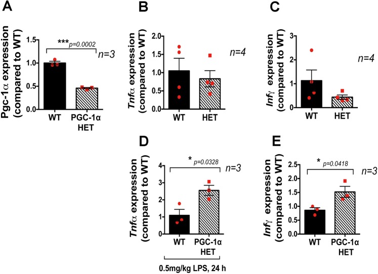
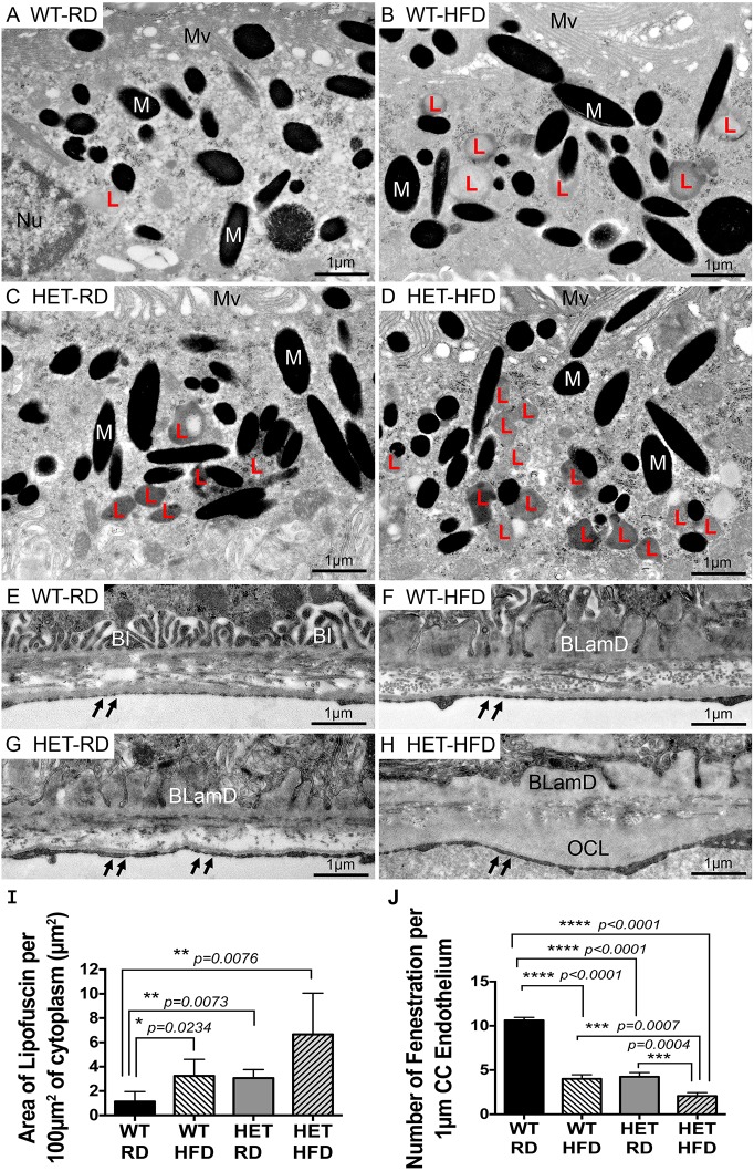
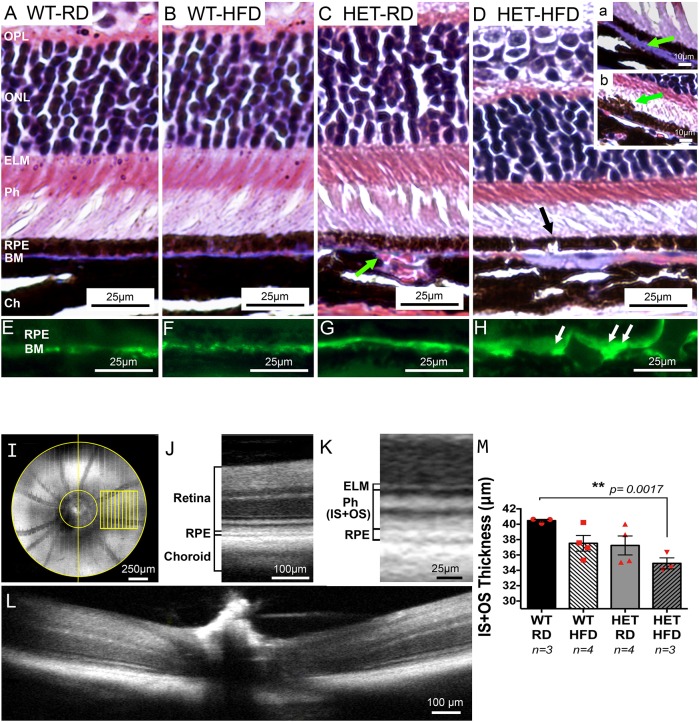
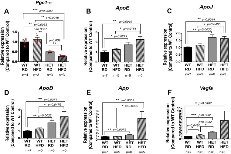
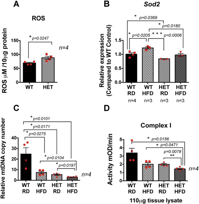
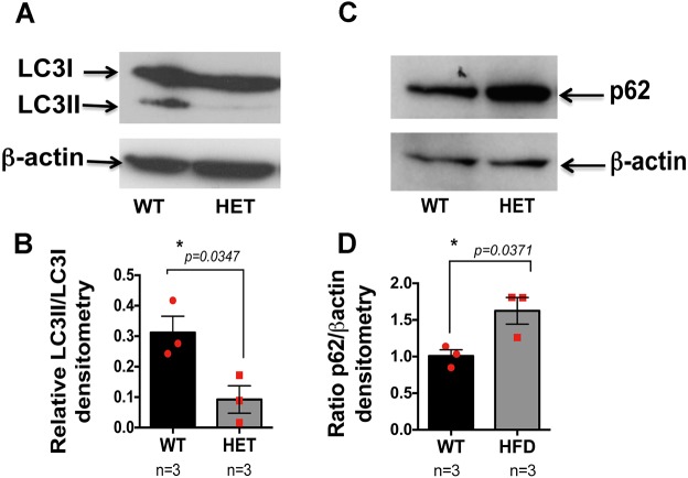
Age-related macular degeneration (AMD) is the major cause of blindness in the elderly in developed countries and its prevalence is increasing with the aging population. AMD initially affects the retinal pigment epithelium (RPE) and gradually leads to secondary photoreceptor degeneration. Recent studies have associated mitochondrial damage with AMD, and we have observed mitochondrial and autophagic dysfunction and repressed peroxisome proliferator-activated receptor-γ coactivator 1α (PGC-1α; also known as Ppargc1a) in native RPE from AMD donor eyes and their respective induced pluripotent stem cell-derived RPE. To further investigate the effect of PGC-1α repression, we have established a mouse model by feeding Pgc-1α+/- mice with a high-fat diet (HFD) and investigated RPE and retinal health. We show that when mice expressing lower levels of Pgc-1α are exposed to HFD, they present AMD-like abnormalities in RPE and retinal morphology and function. These abnormalities include basal laminar deposits, thickening of Bruch's membrane with drusen marker-containing deposits, RPE and photoreceptor degeneration, decreased mitochondrial activity, increased levels of reactive oxygen species, decreased autophagy dynamics/flux, and increased inflammatory response in the RPE and retina. Our study shows that Pgc-1α is important in outer retina biology and that Pgc-1α+/- mice fed with HFD provide a promising model to study AMD, opening doors for novel treatment strategies.

Keywords: AMD; Autophagy; High-fat diet; Mitochondria; PGC-1α; RPE; Retinal degeneration.


Conflict of interest statement

Competing interestsThe authors declare no competing or financial interests.

Figures

Fig. 1.
Fig. 1.
Pgc-1α+/− mice show an increased inflammatory response to LPS injection. (A) RPE/retina of Pgc-1α+/− mice exhibit 50% reduced Pgc-1α mRNA expression as compared with WT. (B,C) Basal levels of Tnfα and Infγ expression in WT and Pgc-1α+/− (HET) mice in the absence of LPS injection. (D,E) Mice were injected with LPS (0.5 mg/kg, i.p.) and euthanized 24 h after injection. Eyes were enucleated and RPE/retinas extracted for RNA isolation followed by qPCR for Tnfα (D) and Infγ (E). A higher inflammatory response was observed in the RPE/retina of Pgc-1α+/− mice compared with WT, as shown by increased expression of Tnfα (D) and Infγ (E) (n=3 WT, n=3 Pgc-1α+/−). One-way ANOVA followed by Student's t-test was performed using GraphPad Prism7.
Fig. 2.
Fig. 2.
Accumulation of lipofuscin, basal deposits, thickening of the outer collagenous layer and loss of fenestrations in CC endothelium in RPE of Pgc-1α+/− mice fed with HFD. (A-D) Representative TEM images of RPE cytoplasm. (A) WT mice fed RD showing a low amount of lipofuscin. (B) WT mice fed HFD showing elevated amounts of lipofuscin. (C) Pgc-1α+/− (HET) mice fed RD showing elevated amounts of lipofuscin. (D) HET mice fed HFD showing high amounts of lipofuscin. (E-H) Representative TEM images of BM. (E) WT mice fed RD showing normal basal infoldings and normal fenestrations of the CC endothelium (double arrows). (F) WT mice fed HFD showing an accumulation of basal laminar deposits and fewer fenestrations of the CC endothelium. (G) HET mice fed RD showing an accumulation of basal laminar deposits and fewer fenestrations of the CC endothelium. (H) HET mice fed HFD showing large accumulations of basal laminar deposits, deposits in the OCL layer and loss of fenestrations of the CC endothelium (n=5 WT/RD, n=5 WT/HFD, n=5 HET/RD, n=5 HET/HFD). BI, basal infoldings; BLamD, basal laminar deposits; L, lipofuscin; M, melanosomes; Mv, microvilli; Nu, nucleus; OCL, outer collagenous layer. (I) Area of lipofuscin per 100 µm2 of cytoplasm, measured from five representative areas from each group and analyzed with Image J analysis software. Pgc-1α+/− mice fed RD and HFD presented higher number of lipofuscin deposits as compared with WT fed RD. HFD increases the number of lipofuscin deposits in WT. (J) Number of fenestrations per 1 µm of CC endothelium, counted from four representative areas from each group. Pgc-1α+/− mice fed HFD present the lowest number of CC endothelium fenestrations. One-way ANOVA followed by Dunnett's multiple comparisons test was performed using GraphPad Prism7.
Fig. 3.
Fig. 3.
Repression of Pgc-1α combined with HFD induces damage to RPE, photoreceptors and BM; enlarged choroidal blood vessels are also observed. Eyes from WT and Pgc-1α+/− mice fed RD and HFD were fixed, paraffin-embedded and stained with Masson's Trichrome. No changes were observed in the RPE or retinal structures in WT fed (A) RD or (B) HFD. (C) Pgc-1α+/− (HET) mice fed RD showed enlarged and congested blood vessels in the interface between the BM and the choroid (green arrow). (D) Pgc-1α+/− mice fed with HFD exhibit enlarged choroidal blood vessels. Changes in the BM, characterized either by atrophy or thickening of the membrane (inset a; green arrow). RPE degeneration was observed with breaks or ‘gaps’ (black arrow) and occasional melanosomes migrating into the outer segment (inset b; green arrow). Photoreceptor degeneration was apparent in the Pgc-1α+/− mice fed HFD (D) presented by reduced thickness of the photoreceptor layer as compared with WT fed RD (A) or HFD (B) and Pgc-1α+/− mice fed with RD (C). (E-H) Immunofluorescence staining with CML, a drusen marker, in WT fed RD (E), WT fed HFD (F), Pgc-1α+/− fed RD (G) and Pgc-1α+/− fed HFD (H), showing accumulation of CML deposits in the BM of Pgc-1α+/− mice fed HFD (H; white arrows). (n=5 WT/RD, n=5 WT/HFD, n=5 HET/RD, n=5 HET/HFD). (I) En face image of a mouse eye obtained by OCT with the 0.5 mm×0.5 mm approximate sampling locations (in yellow) relative to the optic nerve. (J) Representative OCT B-scan with the defined retina and adjacent layers identified. (K) Magnified region of the representative B-scan with the defined IS+OS layers and bounding layers identified. ELM, external limiting membrane; Ph (IS+OS), inner and outer segments of the photoreceptor layer. (L) B-scan corresponding to the vertical yellow line from the en face image (I), demonstrating the 0.5 mm diameter (area between yellow lines) around the optic nerve excluded from sampling to remove bias from layer curvature in this area. (M) Measurement of IS+OS thickness in µm. Pgc-1α repression combined with HFD induces photoreceptor degeneration. One-way ANOVA followed by Dunnett's multiple comparisons test was performed using GraphPad Prism7.
Fig. 4.
Fig. 4.
Increased expression of the drusen-associated genes (ApoE, ApoJ, ApoB and App) and Vegfa in the RPE/retina of Pgc-+/− mice under RD and HFD. (A) The expression of Pgc-1α is reduced in the RPE/retina of Pgc-1α+/− (HET) mice fed HFD as compared with Pgc-1α+/− mice fed RD. (A-E) The expression of drusen-associated genes ApoE (B), ApoJ (C), ApoB (D) and App (E) is increased in the RPE/retina of Pgc-1α+/− fed RD or HFD, as compared with WT mice. (F) Pgc-1α repression induces Vegfa expression, as shown by increased levels of Vegfa in RPE/retina of Pgc-1α+/− mice fed RD as compared with WT. HFD seemed to increase Vegfa expression in WT, but levels were still lower than those in Pgc-1α+/−. One-way ANOVA followed by Dunnett's multiple comparisons test was performed using GraphPad Prism7.
Fig. 5.
Fig. 5.
Reduced antioxidant capacity in RPE/retina of Pgc-1α+/− mice, and decreased mitochondrial activity in RPE/retina of Pgc-1α+/− mice fed HFD. (A) ROS measurement in the RPE/retina extract of WT and Pgc-1α+/− mice showing increased ROS levels in the Pgc-1α+/− mice as compared with WT. (B) Reduced antioxidant capacity in the RPE/retina of Pgc-1α+/− mice, as compared with WT, shown by reduced Sod2 expression and inability to induce Sod2 expression under stress conditions such as HFD. (C) mtDNA copy number measured by qPCR showing reduced mtDNA induced by HFD. Pgc-1α repression combined with HFD further reduced the mtDNA copy number. (D) Pgc-1α repression and HFD significantly reduced mitochondrial complex I activity.
Fig. 6.
Fig. 6.
Autophagy dynamics and flux are reduced in the RPE/retina of the PGC-1α+/− mice. (A,C) Western blot analysis with anti-LC3 (A) and p62 (C) antibodies (compared with β-actin loading control) revealed reduced autophagy dynamics (A) and flux (C) in the RPE/retinal protein extract of Pgc-1α+/− (HET) mice, as compared with WT (a representative image of three independent experiments). (B,D) Densitometry analysis of A and C, respectively, (n=3 WT; n=3 HET) performed using ImageJ. One-way ANOVA followed by Dunnett's multiple comparisons test was performed using GraphPad Prism7.

Similar articles

Cited by

References

    1. Ach T., Tolstik E., Messinger J. D., Zarubina A. V., Heintzmann R. and Curcio C. A. (2015). Lipofuscin redistribution and loss accompanied by cytoskeletal stress in retinal pigment epithelium of eyes with age-related macular degeneration. Invest. Ophthalmol. Vis. Sci. 56, 3242-3252. 10.1167/iovs.14-16274 - DOI - PMC - PubMed
    1. Austin S., Klimcakova E. and St-Pierre J. (2011). Impact of PGC-1alpha on the topology and rate of superoxide production by the mitochondrial electron transport chain. Free Radic. Biol. Med. 51, 2243-2248. 10.1016/j.freeradbiomed.2011.08.036 - DOI - PubMed
    1. Bhutto I. and Lutty G. (2012). Understanding age-related macular degeneration (AMD): relationships between the photoreceptor/retinal pigment epithelium/Bruch's membrane/choriocapillaris complex. Mol. Aspects Med. 33, 295-317. 10.1016/j.mam.2012.04.005 - DOI - PMC - PubMed
    1. Biesemeier A., Taubitz T., Julien S., Yoeruek E. and Schraermeyer U. (2014). Choriocapillaris breakdown precedes retinal degeneration in age-related macular degeneration. Neurobiol. Aging 35, 2562-2573. 10.1016/j.neurobiolaging.2014.05.003 - DOI - PubMed
    1. Bjørkøy G., Lamark T., Brech A., Outzen H., Perander M., Øvervatn A., Stenmark H. and Johansen T. (2005). p62/SQSTM1 forms protein aggregates degraded by autophagy and has a protective effect on huntingtin-induced cell death. J. Cell Biol. 171, 603-614. 10.1083/jcb.200507002 - DOI - PMC - PubMed

Publication types

MeSH terms

Substances