

Three-Tesla MRI does not improve the diagnosis of multiple sclerosis: A multicenter study
- PMID: 29925550
- PMCID: PMC6059032
- DOI: 10.1212/WNL.0000000000005825
Three-Tesla MRI does not improve the diagnosis of multiple sclerosis: A multicenter study
Abstract
Objective: In the work-up of patients presenting with a clinically isolated syndrome (CIS), 3T MRI might offer a higher lesion detection than 1.5T, but it remains unclear whether this affects the fulfilment of the diagnostic criteria for multiple sclerosis (MS).
Methods: We recruited 66 patients with CIS within 6 months from symptom onset and 26 healthy controls in 6 MS centers. All participants underwent 1.5T and 3T brain and spinal cord MRI at baseline according to local optimized protocols and the MAGNIMS guidelines. Patients who had not converted to MS during follow-up received repeat brain MRI at 3-6 months and 12-15 months. The number of lesions per anatomical region was scored by 3 raters in consensus. Criteria for dissemination in space (DIS) and dissemination in time (DIT) were determined according to the 2017 revisions of the McDonald criteria.
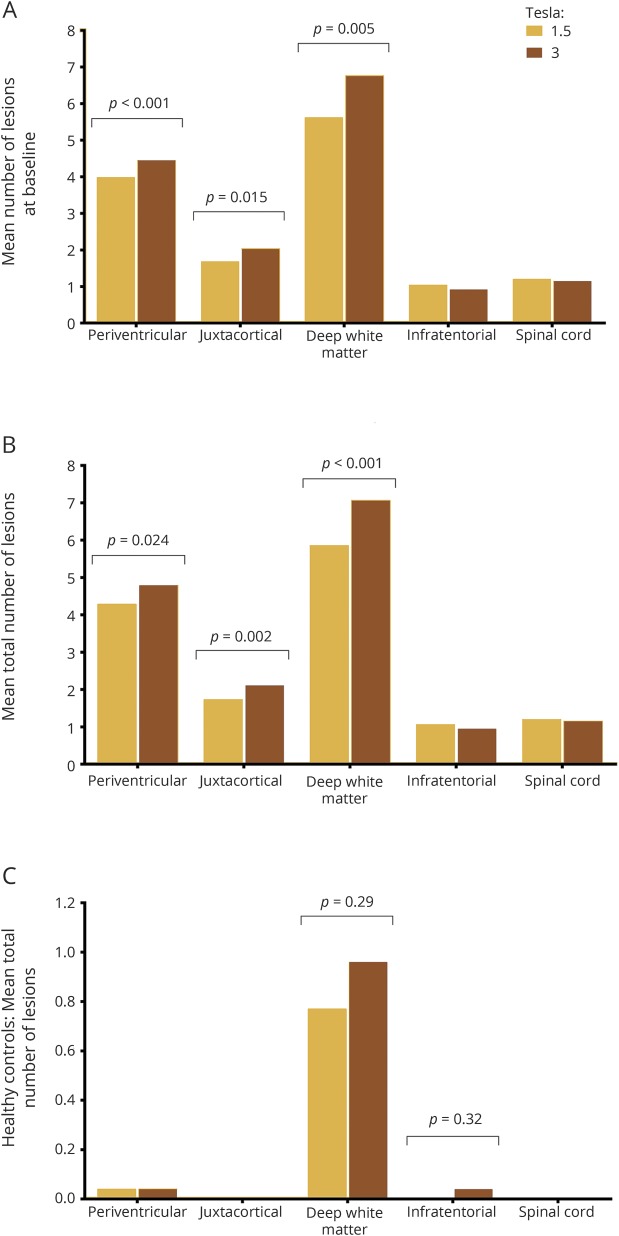
Results: Three-Tesla MRI detected 15% more T2 brain lesions compared to 1.5T (p < 0.001), which was driven by an increase in baseline detection of periventricular (12%, p = 0.015), (juxta)cortical (21%, p = 0.005), and deep white matter lesions (21%, p < 0.001). The detection rate of spinal cord lesions and gadolinium-enhancing lesions did not differ between field strengths. Three-Tesla MRI did not lead to a higher number of patients fulfilling the criteria for DIS or DIT, or subsequent diagnosis of MS, at any of the 3 time points.
Conclusion: Scanning at 3T does not influence the diagnosis of MS according to McDonald diagnostic criteria.
© 2018 The Author(s). Published by Wolters Kluwer Health, Inc. on behalf of the American Academy of Neurology.
Figures
Similar articles
-
Prediction of a multiple sclerosis diagnosis in patients with clinically isolated syndrome using the 2016 MAGNIMS and 2010 McDonald criteria: a retrospective study.Lancet Neurol. 2018 Feb;17(2):133-142. doi: 10.1016/S1474-4422(17)30469-6. Epub 2017 Dec 21. Lancet Neurol. 2018. PMID: 29275979
-
Added value of optic nerve lesions for multiple sclerosis diagnostic criteria.J Neurol. 2025 Apr 24;272(5):358. doi: 10.1007/s00415-025-13036-w. J Neurol. 2025. PMID: 40272570
-
Conversion of clinically isolated syndrome to multiple sclerosis: a prospective study.Mult Scler Relat Disord. 2020 Sep;44:102262. doi: 10.1016/j.msard.2020.102262. Epub 2020 Jun 4. Mult Scler Relat Disord. 2020. PMID: 32570179
-
The role of MRI in the diagnosis of multiple sclerosis.Adv Neurol. 2006;98:125-46. Adv Neurol. 2006. PMID: 16400831 Review.
-
Revised diagnostic criteria of multiple sclerosis.Autoimmun Rev. 2014 Apr-May;13(4-5):518-24. doi: 10.1016/j.autrev.2014.01.012. Epub 2014 Jan 12. Autoimmun Rev. 2014. PMID: 24424194 Review.
Cited by
-
Relevance of brain lesion location for cognition in vascular mild cognitive impairment.Neuroimage Clin. 2019;22:101789. doi: 10.1016/j.nicl.2019.101789. Epub 2019 Mar 23. Neuroimage Clin. 2019. PMID: 30927600 Free PMC article.
-
Exploring Spinal Cord Changes in Multiple Sclerosis Patients Using MRI.NeuroSci. 2024 Mar 12;5(1):87-97. doi: 10.3390/neurosci5010006. eCollection 2024 Mar. NeuroSci. 2024. PMID: 39483810 Free PMC article.
-
Scanner-specific optimisation of automated lesion segmentation in MS.Neuroimage Clin. 2024;44:103680. doi: 10.1016/j.nicl.2024.103680. Epub 2024 Oct 2. Neuroimage Clin. 2024. PMID: 39378750 Free PMC article.
-
Spontaneous spinal cord infarction in Austria: a two-center comparative study.Ther Adv Neurol Disord. 2022 Mar 11;15:17562864221076321. doi: 10.1177/17562864221076321. eCollection 2022. Ther Adv Neurol Disord. 2022. PMID: 35299778 Free PMC article.
-
Sensitivity of portable low-field magnetic resonance imaging for multiple sclerosis lesions.Neuroimage Clin. 2022;35:103101. doi: 10.1016/j.nicl.2022.103101. Epub 2022 Jun 27. Neuroimage Clin. 2022. PMID: 35792417 Free PMC article.
References
-
- Paty DW, Oger JJ, Kastrukoff LF, et al. . MRI in the diagnosis of MS: a prospective study with comparison of clinical evaluation, evoked potentials, oligoclonal banding, and CT. Neurology 1988;38:180–185. - PubMed
-
- Dekker I, Wattjes MP. Brain and spinal cord MR imaging features in multiple sclerosis and variants. Neuroimaging Clin N Am 2017;27:205–227. - PubMed
-
- Fazekas F, Barkhof F, Filippi M, et al. . The contribution of magnetic resonance imaging to the diagnosis of multiple sclerosis. Neurology 1999;53:448–456. - PubMed
-
- McDonald WI, Compston A, Edan G, et al. . Recommended diagnostic criteria for multiple sclerosis: guidelines from the International Panel on the Diagnosis of Multiple Sclerosis. Ann Neurol 2001;50:121–127. - PubMed
Publication types
MeSH terms
LinkOut - more resources
Full Text Sources
Other Literature Sources
Medical