. 2018 Aug 10;293(32):12415-12428.
doi: 10.1074/jbc.RA118.001922. Epub 2018 Jun 20.

Consequences of cathepsin C inactivation for membrane exposure of proteinase 3, the target antigen in autoimmune vasculitis

Affiliations

Consequences of cathepsin C inactivation for membrane exposure of proteinase 3, the target antigen in autoimmune vasculitis

Seda Seren et al. J Biol Chem. .

Abstract

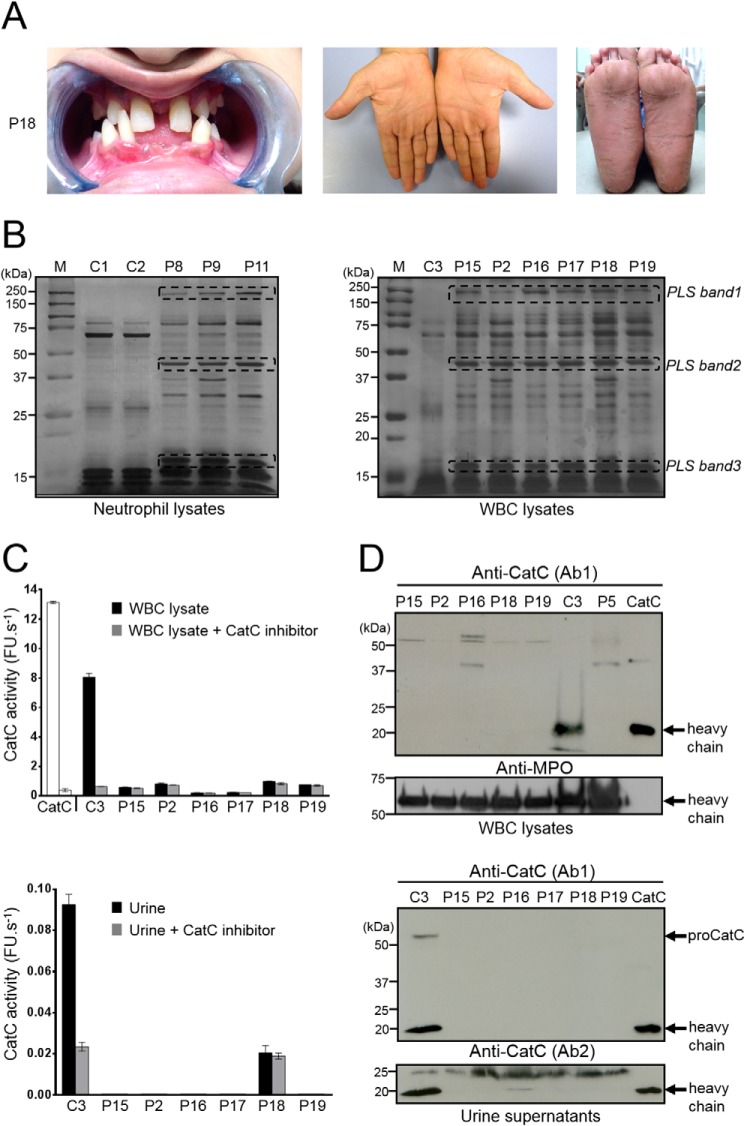
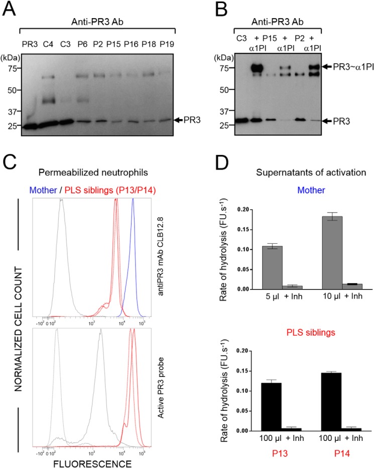
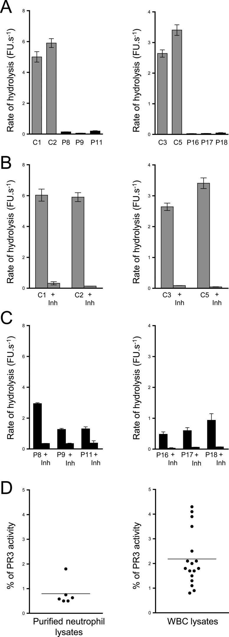
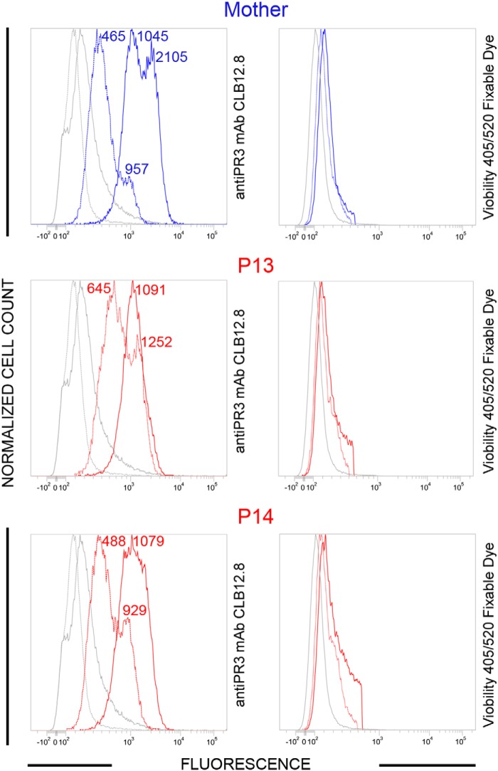
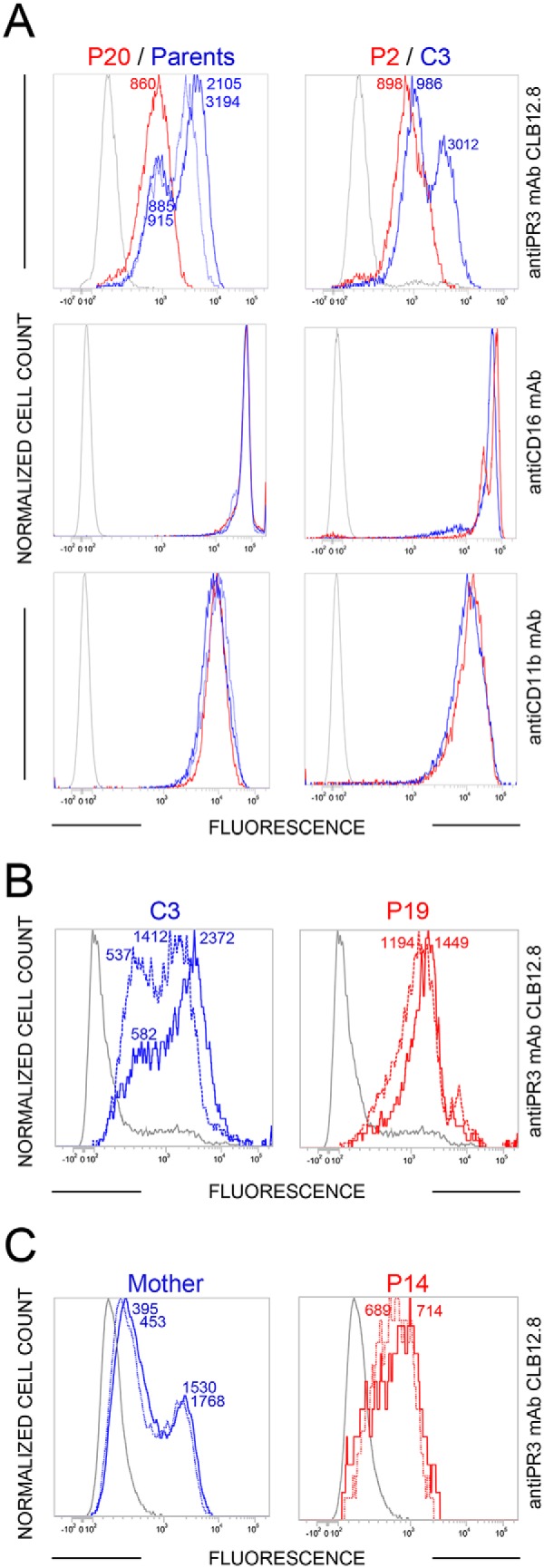
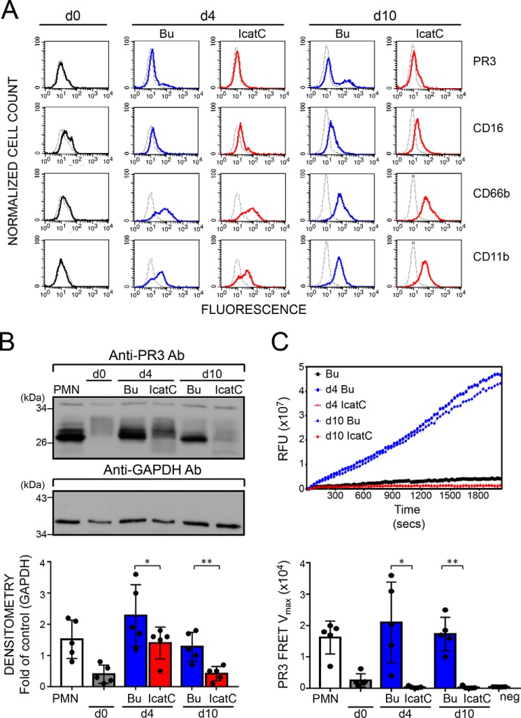
Membrane-bound proteinase 3 (PR3m) is the main target antigen of anti-neutrophil cytoplasmic autoantibodies (ANCA) in granulomatosis with polyangiitis, a systemic small-vessel vasculitis. Binding of ANCA to PR3m triggers neutrophil activation with the secretion of enzymatically active PR3 and related neutrophil serine proteases, thereby contributing to vascular damage. PR3 and related proteases are activated from pro-forms by the lysosomal cysteine protease cathepsin C (CatC) during neutrophil maturation. We hypothesized that pharmacological inhibition of CatC provides an effective measure to reduce PR3m and therefore has implications as a novel therapeutic approach in granulomatosis with polyangiitis. We first studied neutrophilic PR3 from 24 patients with Papillon-Lefèvre syndrome (PLS), a genetic form of CatC deficiency. PLS neutrophil lysates showed a largely reduced but still detectable (0.5-4%) PR3 activity when compared with healthy control cells. Despite extremely low levels of cellular PR3, the amount of constitutive PR3m expressed on the surface of quiescent neutrophils and the typical bimodal membrane distribution pattern were similar to what was observed in healthy neutrophils. However, following cell activation, there was no significant increase in the total amount of PR3m on PLS neutrophils, whereas the total amount of PR3m on healthy neutrophils was significantly increased. We then explored the effect of pharmacological CatC inhibition on PR3 stability in normal neutrophils using a potent cell-permeable CatC inhibitor and a CD34+ hematopoietic stem cell model. Human CD34+ hematopoietic stem cells were treated with the inhibitor during neutrophil differentiation over 10 days. We observed strong reductions in PR3m, cellular PR3 protein, and proteolytic PR3 activity, whereas neutrophil differentiation was not compromised.

Keywords: Papillon-Lefèvre syndrome; aminopeptidase; antigen; autoimmune disease; cathepsin C; genetic disease; granulomatosis with polyangiitis; neutrophil; protease; protease inhibitor; proteinase 3.


Conflict of interest statement

The authors declare that they have no conflicts of interest with the contents of this article. Responsibility for the information and views set out in this article lies entirely with the authors

Figures

Figure 1.
Figure 1.
CatC in biological samples of PLS patients. A, characteristic dental and palmoplantar features of PLS (patient 18, P18). Photos show early loss of teeth and hyperkeratosis of the palms and soles. B, neutrophil and WBC lysates from PLS patients and from healthy controls, lysed in 50 mm HEPES buffer, 750 mm NaCl, 0.05% Nonidet P-40, pH 7.4, during 5 min at room temperature and analyzed by SDS-PAGE (12%)/silver staining under reducing conditions (10 μg/lane) strongly differ by their protein profile. Lane M shows protein molecular weight size markers. PLS bands 1, 2, and 3 correspond to proteins that are not cleaved by NSPs in PLS WBC lysates but are hydrolyzed in WBC lysates from healthy controls. Similar results were found in three independent experiments. C, measurement of CatC activity in WBC lysates (10 μg of protein) (top) and concentrated urines (bottom) in the presence or not of a selective CatC inhibitor. The residual proteolytic activity was not inhibited by the CatC inhibitor, which demonstrates the absence of CatC activity in PLS samples. Bars, mean ± S.D. (error bars) of experiments performed in triplicates. D, Western blot analysis of WBC lysates (10 μg of protein) and concentrated urines of PLS samples and controls using anti-CatC antibodies shows the absence of the CatC heavy chain in all PLS samples. The urines were collected and analyzed as in Hamon et al. (34). Similar results were found in three independent experiments. C, control; P, PLS patient; FU, fluorescence units.
Figure 2.
Figure 2.
Active PR3 in PLS blood samples. A, Western blotting of WBC lysates (10 μg of protein) from PLS patients and healthy controls using anti-PR3 antibodies: low amounts of PR3 are present in PLS samples. PR3 antigen was 3–10% of the healthy controls as determined by quantifying protein bands from Western blotting films. B, Western blotting of WBC lysates (10 μg of protein) from PLS patients and a healthy control incubated with α1PI (5 μm) in 50 mm HEPES buffer, 750 mm NaCl, 0.05% Nonidet P-40, pH 7.4, for 3 h at 37 °C. The de novo formation of irreversible α1PI–PR3 complexes of about 75 kDa reveals that PR3 is proteolytically active despite the absence of active CatC. C, top, flow cytometry analysis of the expression of PR3 in permeabilized PLS neutrophils. Using anti-PR3 antibodies, a lesser fluorescence is observed in permeabilized neutrophils from two PLS siblings (P13 and P14) (red) as compared with their mother used here as a control (blue). The gray peak corresponds to the isotype control. Bottom, the use of PR3 activity probe Biotin-PEG66-PYDAP(O-C6H4-4-Cl)2 as in Guarino et al. (50) shows that the residual PLS PR3 is enzymatically active. The gray peak indicates the fluorescence of permeabilized neutrophils incubated with streptavidin-Alexa Fluor® 488. The dotted gray peak corresponds to the (auto)fluorescence of permeabilized neutrophils. D, PR3 activity in supernatants of calcium ionophore A23187 (Sigma-Aldrich, St. Quentin Fallavier, France)–activated WBC in the presence or absence of the specific PR3 inhibitor Ac-PYDAP(O-C6H4-4-Cl)2. PR3 activity in PLS cell supernatants is about one-twentieth of that in control cells and is almost totally inhibited in the presence of the selective PR3 inhibitor. Bars, mean ± S.D. (error bars) of experiments performed in duplicates. Similar results were found in three independent experiments. C, control; Inh, inhibitor; P, PLS patient; FU, fluorescence units.
Figure 3.
Figure 3.
PR3 activity in PLS blood samples. PR3 activities in purified neutrophil (A, left) or in WBC lysates (A, right). PR3 activities were measured with the selective substrate ABZ-VAD(nor)VADYQ-EDDnp. Samples were also incubated with the selective PR3 inhibitor Ac-PYDAP(O-C6H4-4-Cl)2 to distinguish PR3 activity and nonspecific signal (B and C). Low levels of active PR3 were found in PLS cell lysates (5 μg) compared with control cell lysates (0.25 μg). D, percentage of PR3 activity in neutrophils and whole-blood samples. We calculated the percentage of PR3 activity in purified neutrophils and in WBC of PLS patients (n = 23) compared with healthy controls cells (n = 11). We estimated that PLS cells contained 0.5–4% of PR3 activities compared with healthy cells. Bars, mean ± S.D. (error bars) of experiments performed in triplicates. Similar results were found in three independent experiments. C, control; Inh, inhibitor; P, PLS patient; FU, fluorescence units.
Figure 4.
Figure 4.
Flow cytometry analysis of the membrane exposure of PR3 on quiescent and chemically activated PLS neutrophils. WBC from PLS siblings (local patients P13 and P14) and their mother were activated using A23187, 30 min after blood collection. Both viable quiescent PLS cells (red dotted line) and control cells (blue dotted line) express PR3 on their surface. After chemical activation (continuous lines), membrane PR3 was largely increased on control cells, whereas PR3 on PLS neutrophils did not increase significantly but obeyed a monomodal distribution after activation. We used Viobility 405/520 fixable dye to discriminate between live and apoptotic/dead cells. Flow cytometry revealed 81 ± 5% viable neutrophils in all samples. No statistically significant difference between quiescent and activated neutrophils was observed (t test). P, PLS patient. Numbers indicate mean fluorescence intensity values.
Figure 5.
Figure 5.
Flow cytometry analysis of the membrane exposure of PR3 on transported PLS (P) and control (C) neutrophils before and after activation. A and B, the unimodal distribution of PR3 at the surface of PLS cells (continuous red line, n = 14 PLS patients) (A) and the absence of further significant PR3 exposure following A23187 treatment (continuous line) (B) indicate that the cells were spontaneously activated or at least primed before they were analyzed (n = 4 PLS patients). C, the same result was obtained using a local PLS sample (P14) stored for 48 h at room temperature before (dotted line) and after (continuous line) ionophore treatment. Cell surface markers CD11b and CD16 were used as quantitative controls. It is noteworthy that the flow cytometry analyses of samples collected from the same individuals 9 months apart are almost fully superimposable (Fig. 5 (B and C) versus Fig. 4 and Fig. S5).
Figure 6.
Figure 6.
Effect of pharmacological CatC inhibition on PR3 expression and proteolytic activity in neutrophil-differentiated CD34+ HSC. CD34+ HSC were differentiated over 10 days into neutrophils in the presence of DMSO buffer control (Bu, blue) or a 1 μm concentration of the CatC inhibitor (IcatC, red). A, flow cytometry indicates that differentiating cells acquired the typical neutrophil surface markers CD16, CD66b, and CD11b, together with a bimodal membrane PR3 phenotype. Color lines, staining with the specific antibodies; dotted lines, the corresponding isotype control. A representative of five independent differentiation experiments is shown. B, PR3 protein was assessed in cell lysates (7.0 μg/lane) by immunoblotting at the indicated time points using a specific anti-PR3 antibody. PR3 protein was strongly induced during CD34+ HSC differentiation, and this effect was significantly reduced with IcatC. A representative Western blot and the densitometric analysis from five independent differentiation experiments are shown. Bars, mean ± S.D. (error bars) of each condition; asterisks, p value of t test (*, p < 0.05; **, p < 0.01). C, proteolytic PR3 activity was assessed in cell lysates (2.5 μg of protein) at the indicated time points, using the PR3-specific FRET substrate ABZ-VAD(nor)VADYQ-EDDnp. Representative PR3 substrate conversion curves from one of five independent differentiation experiments are depicted together with the corresponding statistics for the mean Vmax values ± S.D. (n = 5 independent differentiation experiments). The data show a complete loss of proteolytic PR3 activity with CatC inhibition. Isolated normal blood neutrophils (PMN) served as a positive control, and an endothelial cell line served as a negative control (neg ctrl). Asterisks, p value of t test (*, p < 0.05; **, p < 0.01). GAPDH, glyceraldehyde-3-phosphate dehydrogenase.

References

    1. Millet A., Pederzoli-Ribeil M., Guillevin L., Witko-Sarsat V., and Mouthon L. (2013) Antineutrophil cytoplasmic antibody-associated vasculitides: is it time to split up the group? Ann. Rheum. Dis. 72, 1273–1279 10.1136/annrheumdis-2013-203255 - DOI - PubMed
    1. Pagnoux C. (2016) Updates in ANCA-associated vasculitis. Eur. J. Rheumatol. 3, 122–133 10.5152/eurjrheum.2015.0043 - DOI - PMC - PubMed
    1. Jenne D. E., Tschopp J., Lüdemann J., Utecht B., and Gross W. L. (1990) Wegener's autoantigen decoded. Nature 346, 520 10.1038/346520a0 - DOI - PubMed
    1. Thieblemont N., Wright H. L., Edwards S. W., and Witko-Sarsat V. (2016) Human neutrophils in auto-immunity. Semin. Immunol. 28, 159–173 10.1016/j.smim.2016.03.004 - DOI - PubMed
    1. Kettritz R. (2016) Neutral serine proteases of neutrophils. Immunol. Rev. 273, 232–248 10.1111/imr.12441 - DOI - PubMed

Publication types

MeSH terms