. 2018 Jul 31;92(16):e00415-18.
doi: 10.1128/JVI.00415-18. Print 2018 Aug 15.

Pigs Lacking the Scavenger Receptor Cysteine-Rich Domain 5 of CD163 Are Resistant to Porcine Reproductive and Respiratory Syndrome Virus 1 Infection

Affiliations

Pigs Lacking the Scavenger Receptor Cysteine-Rich Domain 5 of CD163 Are Resistant to Porcine Reproductive and Respiratory Syndrome Virus 1 Infection

Christine Burkard et al. J Virol. .

Erratum in

Abstract

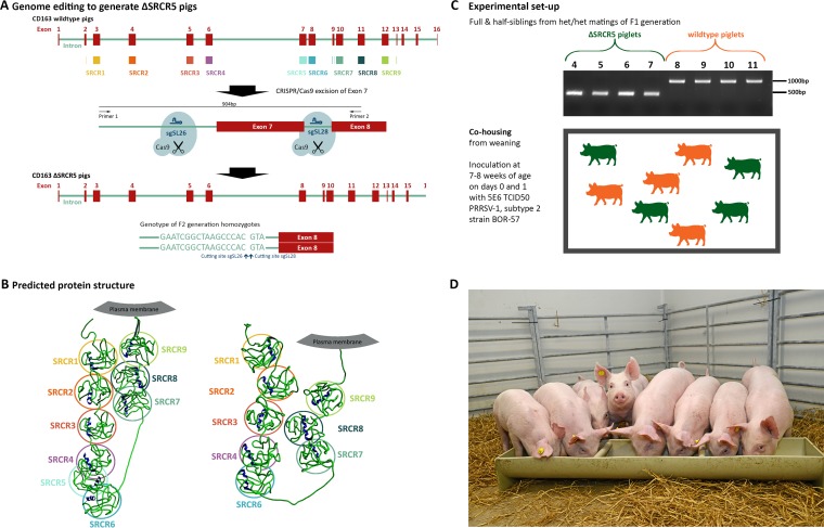
Porcine reproductive and respiratory syndrome virus (PRRSV) has a narrow host cell tropism, limited to cells of the monocyte/macrophage lineage. CD163 protein is expressed at high levels on the surface of specific macrophage types, and a soluble form is circulating in blood. CD163 has been described as a fusion receptor for PRRSV, with the scavenger receptor cysteine-rich domain 5 (SRCR5) region having been shown to be the interaction site for the virus. As reported previously, we have generated pigs in which exon 7 of the CD163 gene has been deleted using CRISPR/Cas9 editing in pig zygotes. These pigs express CD163 protein lacking SRCR5 (ΔSRCR5 CD163) and show no adverse effects when maintained under standard husbandry conditions. Not only was ΔSRCR5 CD163 detected on the surface of macrophage subsets, but the secreted, soluble protein can also be detected in the serum of the edited pigs, as shown here by a porcine soluble CD163-specific enzyme-linked immunosorbent assay (ELISA). Previous results showed that primary macrophage cells from ΔSRCR5 CD163 animals are resistant to PRRSV-1 subtype 1, 2, and 3 as well as PRRSV-2 infection in vitro Here, ΔSRCR5 pigs were challenged with a highly virulent PRRSV-1 subtype 2 strain. In contrast to the wild-type control group, ΔSRCR5 pigs showed no signs of infection and no viremia or antibody response indicative of a productive infection. Histopathological analysis of lung and lymph node tissue showed no presence of virus-replicating cells in either tissue. This shows that ΔSRCR5 pigs are fully resistant to infection by the virus.IMPORTANCE Porcine reproductive and respiratory syndrome (PRRS) virus (PRRSV) is the etiological agent of PRRS, causing late-term abortions, stillbirths, and respiratory disease in pigs, incurring major economic losses to the worldwide pig industry. The virus is highly mutagenic and can be divided into two species, PRRSV-1 and PRRSV-2, each containing several subtypes. Current control strategies mainly involve biosecurity measures, depopulation, and vaccination. Vaccines are at best only partially protective against infection with heterologous subtypes and sublineages, and modified live vaccines have frequently been reported to revert to virulence. Here, we demonstrate that a genetic-control approach results in complete resistance to PRRSV infection in vivo CD163 is edited so as to remove the viral interaction domain while maintaining protein expression and biological function, averting any potential adverse effect associated with protein knockout. This research demonstrates a genetic-control approach with potential benefits in animal welfare as well as to the pork industry.

Keywords: CD163; CRISPR/Cas9; PRRSV; arterivirus; exon deletion; genome editing; nidovirus; porcine reproductive and respiratory syndrome virus; resistance.


Figures

FIG 1
FIG 1
Generation of ΔSRCR5 pigs and experimental setup. (A) Genome editing to generate ΔSRCR5 pigs. Genome-edited founder animals were generated by zygote injection of CRISPR/Cas9 editing reagents using Cas9 mRNA and two guide RNAs, sgSL26 and sgSL28, in combination to generate a deletion of exon 7 in CD163. Animals were bred to generate F1 and F2 generations, focusing on one genotype showing clean religation at the cutting sites of both guide RNAs. Homozygous F2 animals carry this genotype in both alleles (bottom). (B) Structure prediction and expression of ΔSRCR5 in pulmonary alveolar macrophages of F2 animals. Protein structure prediction using RaptorX points toward an intact protein product upon the deletion of SRCR5. (C) Experimental design of the challenge study. Four homozygous (green) and 4 wild-type (orange) siblings from heterozygous/heterozygous mating of the F1 generation animals were cohoused from weaning. Genotypes were confirmed by PCR amplification across exon 7 (see panel A) and by Sanger sequencing. Piglets were cohoused after weaning and after acclimation to the specific-pathogen-free unit for 1 week and throughout the 14-day challenge experiment that was initiated by inoculating each pig intranasally with 5E6 TCID50 of PRRSV-1 subtype 2 strain BOR-57 at day 0 and day 1 of the challenge. The piglets were 7 to 8 weeks of age at the start of the acclimation period. (D) Piglets 1 day before the start of the challenge.
FIG 2
FIG 2
Serum levels of soluble C163. Serum samples collected 2 weeks prior to and on day 0 of the challenge were assessed for the level of sCD163 using a commercial ELISA (n = 4 pigs per genotype, serum collected at 2 different time points, assessed in duplicate in 3 replicates). Minima/maxima and 90th percentiles are displayed. Statistical analysis using an unpaired t test showed no significant difference.
FIG 3
FIG 3
ΔSRCR5 pigs show no clinical signs or pathology of PRRSV-1 infection. (A) Rectal temperatures of ΔSRCR5 (green) and wild-type (orange) piglets during challenge with BOR-57. Rectal temperatures were measured daily during feeding. Error bars represent standard errors of the means (SEM) (n = 4). (B) Average daily weight gain based on weight measurements at day 0, 7, and 14 of the challenge. For panels A and B, statistical analysis was performed using two-way ANOVA and Sidak's multiple-comparison test. ns, not significant. (C) Viremia during challenge with BOR-57. Serum samples were collected at days 0, 3, 7, 10, and 14 from the jugular vein using vacuum tubes, and viral RNA was isolated and quantified using RT-qPCR with primers specific to ORF5 of BOR-57. (D) Antibody response to PRRSV-1 during the challenge. Serum samples were analyzed for the presence of PRRSV antibodies using the Idexx PRRSV X3 ELISA, where a value of <0.40 is negative and a value of ≧0.4 is positive. Each data point/line represents data for a single animal, with 4 animals per genotype group. (E) Lung and lymph node pathology, histopathology, and immunohistochemistry (IHC) scores. Lung pathology was assessed in a blind fashion, and a subjective score for the severity of gross lung lesions using an established scoring system was applied (scale, 0 to 100). Lung histopathology sections were scored for the presence and severity of interstitial pneumonia, ranging from 0 to 6 (0, normal; 1, mild multifocal; 2, mild diffuse; 3, moderate multifocal; 4, moderate diffuse; 5, severe multifocal; 6, severe diffuse). Immunohistochemistry staining against PRRSV-N of lung and lymph node sections was scored, ranging from 0 to 3 (0, no signal; 1, low numbers of positive cells; 2, moderate numbers of positive cells; 3, abundant). Numbers represent averages (n = 4) ± SEM. (F) Lung histology and immunohistochemistry. (Top) Formalin-fixed, paraffin-embedded, hematoxylin-and-eosin-stained lung sections from necropsy on day 14 postchallenge. (Left) ΔSRCR5 piglets; (right) wild-type piglets. Bar, 100 μm. (Bottom) Formalin-fixed, paraffin-embedded immunohistochemical staining against PRRSV antigen (brown) and hematoxylin counterstain. (Left) ΔSRCR5 piglets; (right) wild-type piglets. The scale bar represents 50 μm. (G) Lung pathology. Shown are lungs from pigs at necropsy at 14 days postchallenge. (Left) Lungs from two ΔSRCR5 pigs; (right) lungs from two wild-type pigs.
FIG 4
FIG 4
Cytokine response to BOR-57 PRRSV infection. Cytokine levels in serum samples collected prior to challenge on day 0 and on challenge days 3, 7, 10, and 14 were measured using cytokine antibody arrays. (A) Alpha interferon (IFN-α); (B) interleukin-17A (IL-17A); (C) interleukin-1 receptor antagonist (IL-1ra); (D) IL-4; (E) IL-6; (F) IL-8; (G) monokine induced by gamma interferon (MIG/CXCL9); (H) macrophage inflammatory protein 1β (MIP-1β/CCL4); (I) chemokine ligand 3-like 1 (CCL3L1); (J) granulocyte-macrophage colony-stimulating factor (GM-CSF); (K) tumor necrosis factor alpha (TNF-α); (L) IL-12; (M) IL-1β; (N) IL-10; (O) transforming growth factor β1 (TGFβ1); (P) IFN-γ; (Q) IL-18; (R) platelet endothelial cell adhesion molecule 1 (PECAM-1/CD31); (S) IL-1α; (T) IL-13. Error bars represent SEM (duplicates of 4 replicates). Statistical analysis was performed using two-way ANOVA and Sidak's multiple-comparison test. *, P ≤ 0.5; **, P ≤ 0.01; ***, P ≤ 0.001; ****, P ≤ 0.0001.

References

    1. Karniychuk UU, Saha D, Geldhof M, Vanhee M, Cornillie P, Van den Broeck W, Nauwynck HJ. 2011. Porcine reproductive and respiratory syndrome virus (PRRSV) causes apoptosis during its replication in fetal implantation sites. Microb Pathog 51:194–202. doi:10.1016/j.micpath.2011.04.001. - DOI - PubMed
    1. Pejsak Z, Stadejek T, Markowska-Daniel I. 1997. Clinical signs and economic losses caused by porcine reproductive and respiratory syndrome virus in a large breeding farm. Vet Microbiol 55:317–322. doi:10.1016/S0378-1135(96)01326-0. - DOI - PubMed
    1. Christianson WT, Joo HS. 1994. Porcine reproductive and respiratory syndrome: a review. J Swine Health Prod 2:10–28.
    1. Wagstrom EA, Chang CC, Yoon KJ, Zimmerman JJ. 2001. Shedding of porcine reproductive and respiratory syndrome virus in mammary gland secretions of sows. Am J Vet Res 62:1876–1880. doi:10.2460/ajvr.2001.62.1876. - DOI - PubMed
    1. Holtkamp DJ, Kliebenstein JB, Neumann EJ, Zimmerman JJ, Rotto HF, Yoder TK, Wang C, Yeske PE, Mowrer CL, Hayley CA. 2013. Assessment of the economic impact of porcine reproductive and respiratory syndrome virus on United States pork producers. J Swine Health Prod 21:72–84.

Publication types

MeSH terms