. 2018 Aug 16;92(17):e00971-18.
doi: 10.1128/JVI.00971-18. Print 2018 Sep 1.

Recurrent Loss of APOBEC3H Activity during Primate Evolution

Affiliations

Recurrent Loss of APOBEC3H Activity during Primate Evolution

Erin I Garcia et al. J Virol. .

Abstract

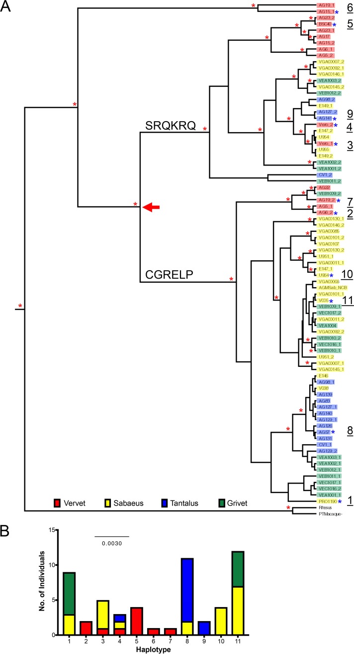
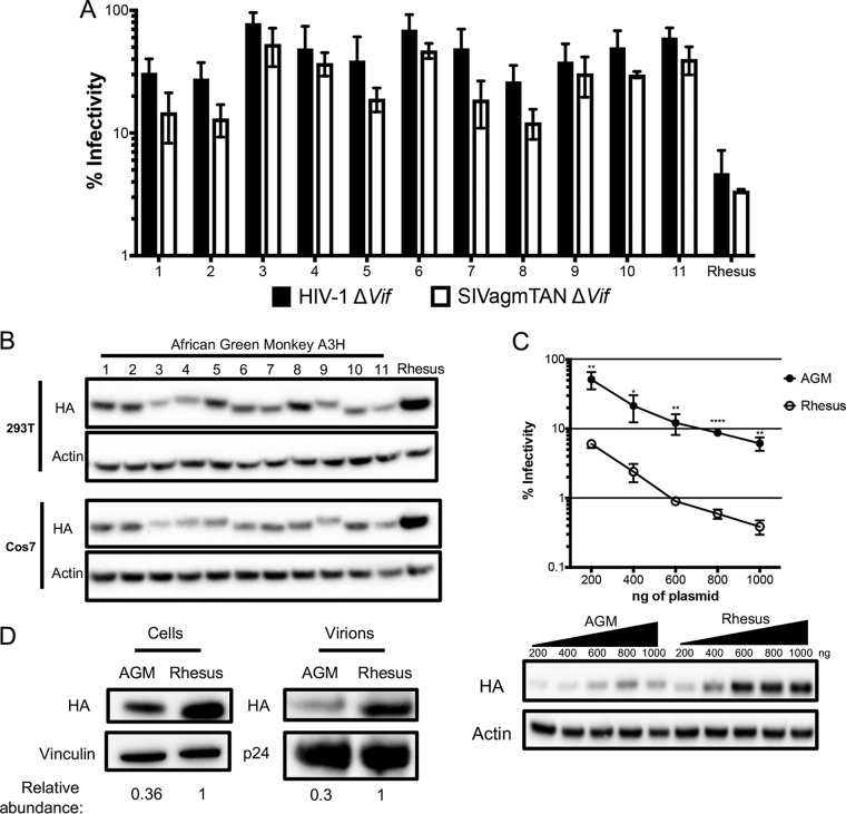
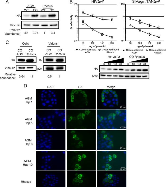
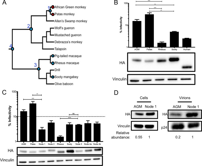
Genes in the APOBEC3 family encode cytidine deaminases that provide a barrier against viral infection and retrotransposition. Of all the APOBEC3 genes in humans, APOBEC3H (A3H) is the most polymorphic: some genes encode stable and active A3H proteins, while others are unstable and poorly antiviral. Such variation in human A3H affects interactions with the lentiviral antagonist Vif, which counteracts A3H via proteasomal degradation. In order to broaden our understanding of A3H-Vif interactions, as well as its evolution in Old World monkeys, we characterized A3H variation within four African green monkey (AGM) subspecies. We found that A3H is highly polymorphic in AGMs and has lost antiviral activity in multiple Old World monkeys. This loss of function was partially related to protein expression levels but was also influenced by amino acid mutations in the N terminus. Moreover, we demonstrate that the evolution of A3H in the primate lineages leading to AGMs was not driven by Vif. Our work suggests that the activity of A3H is evolutionarily dynamic and may have a negative effect on host fitness, resulting in its recurrent loss in primates.IMPORTANCE Adaptation of viruses to their hosts is critical for viral transmission between different species. Previous studies had identified changes in a protein from the APOBEC3 family that influenced the species specificity of simian immunodeficiency viruses (SIVs) in African green monkeys. We studied the evolution of a related protein in the same system, APOBEC3H, which has experienced a loss of function in humans. This evolutionary approach revealed that recurrent loss of APOBEC3H activity has taken place during primate evolution, suggesting that APOBEC3H places a fitness cost on hosts. The variability of APOBEC3H activity between different primates highlights the differential selective pressures on the APOBEC3 gene family.

Keywords: APOBEC3H; African green monkey; evolution; human immunodeficiency virus; innate immunity; lentiviruses.


Figures

FIG 1
FIG 1
Sequence and phylogenetic analyses of A3H in African green monkeys. (A) The evolutionary relationship among 80 full-length AGM A3H genes was inferred by Bayesian MCMC phylogenetic reconstruction. The red asterisks mark nodes that have a posterior probability of >0.5. The colored boxes indicate the subspecies of origin (red, vervet; yellow, sabaeus; blue, tantalus; and green, grivet). The blue asterisks mark cloned haplotypes, numbered 1 to 11 on the right. The arrow marks divergence of the phylogenetic tree between two clades. (B) Numbers of AGM individuals carrying genes encoding haplotypes 1 to 11, color coded by subspecies, similar to A3H phylogeny.
FIG 2
FIG 2
Antiviral activity of A3H is lower in AGMs than in another Old World monkey. (A) Single-cycle infectivity assays were performed in the presence or absence of A3 proteins against HIVΔvif and SIVagmΔvif. Rhesus macaque was included as a positive control. Relative infection was normalized to viral infectivity in the absence of A3 proteins. Averages of three replicates, each with triplicate infections (±SEM), are shown. All the samples were statistically significantly different than rhesus macaque A3H, except AGM haplotype 7 against SIVagm.TAN (P = 0.0502), as measured by unpaired t tests. Furthermore, no significant differences in restriction between HIV and SIVagm.TAN within individual haplotypes were found. (B) Western blot analysis of HA-tagged AGM A3H protein expression in human (HEK293T) and AGM (Cos7) cell lines. The different-size bands for different AGM A3H haplotypes were reproducible. β-Actin is shown as a loading control. (C) (Top) Single-cycle infectivity assay of HIVΔvif in the presence of increasing amounts of A3H-expressing plasmids. AGM A3H haplotype 1 (black circles) and rhesus macaque A3H (open circles) were compared. Relative infection was normalized to viral infectivity in the absence of A3 proteins. Averages of three replicates, each with triplicate infections (±SEM), are shown. Statistical differences were determined by unpaired t tests: *, P 0.05; **, P 0.01; ****, P 0.0001. (Bottom) Western blot analysis of protein expression levels with the same amounts of plasmid as in the graph above. β-Actin is shown as a loading control. (D) Packaging of A3H proteins into virions analyzed by Western blotting. Relative abundances in cellular expression (left) and virion incorporation (right) were determined compared to rhesus macaque A3H.
FIG 3
FIG 3
Codon optimization increases protein expression and antiviral activity. (A) Western blot analysis for the expression of AGM A3H haplotype 1, codon-optimized haplotype 1 A3H, rhesus macaque A3H, and codon-optimized rhesus macaque A3H. Vinculin was used as a protein-loading control. Quantification was done relative to rhesus macaque A3H (normalized to 1). (B) (Top) Single-cycle infectivity assay of HIVΔvif and SIVagm.TANΔvif in the presence of increasing amounts of A3H plasmid comparing codon-optimized AGM haplotype 1 A3H (black) and codon-optimized rhesus macaque A3H (open). Relative infection was normalized to viral infectivity in the absence of A3 proteins. Averages of three replicates, each with triplicate infections (±SEM), are shown. Statistical differences were determined by unpaired t tests: **, P 0.01; ***, P 0.001; ****, P 0.0001. (Bottom) Western blot analysis of protein expression levels with amounts of plasmid added as for the graph above. β-Actin is shown as a loading control. (C) Packaging of A3H proteins into virions analyzed by Western blotting. Relative abundances in cellular expression (left) and virion incorporation (right) were determined compared to codon-optimized rhesus macaque A3H. (D) Subcellular localization of wild-type rhesus macaque and wild-type AGM A3H haplotypes (Hap) 1, 5, 6, and 10 in HeLa cells. A3H proteins were detected with an anti-HA antibody (green), and DAPI staining was used to detect the nucleus (blue). The images are representative of 135 total images over 3 replicates.
FIG 4
FIG 4
AGM ancestors produce potent antiviral proteins. (A) Phylogeny, depicted as a cladogram, based on the accepted species tree of all sequenced Old World primates included in the study (28). The blue circles denote active antiviral proteins; the red circles denote less active antiviral proteins. Ancestral nodes are labeled with numbers (1 to 4). (B) (Top) Single-cycle infectivity assay for HIVΔvif against extant primate A3H proteins. Relative infection was normalized to viral infectivity in the absence of A3 proteins. Averages of three replicates, each with triplicate infections (+SEM), are shown. Statistical differences were determined by unpaired t tests. (Bottom) Western blot analysis of the protein expression level. Vinculin was used as a loading control. Human A3H is missing 28 C-terminal amino acids due to a natural premature stop codon (15) and thus ran lower on the blot. (C) (Top) Single-cycle infectivity assay for HIVΔvif against ancestral A3H proteins and their extant descendants. Relative infection was normalized to viral infectivity in the absence of A3 proteins. Averages of three replicates, each with triplicate infections (+SEM), are shown. The dashed line at 10% is an arbitrary reference point. (Bottom) Western blot analysis of the protein expression level. Vinculin was used as a loading control. (D) Packaging of A3H proteins into virions analyzed by Western blotting. Relative abundance in cellular expression (left) and virion incorporation (right) were determined compared to the node 1 A3H ancestor. **, P 0.01; ***, P 0.001; ****, P 0.0001; ns, not significant.
FIG 5
FIG 5
Multiple amino acid mutations required for an increase in antiviral activity. (A) Schematic of the A3H protein. The black bars outline the A3H catalytic site. Numbered amino acid residues that are different between AGM A3H, patas A3H, and the node 1 ancestor are in red in the protein sequence alignment, and ancestral residues are in blue. Amino acids that are different in other primates are similarly colored. (B) (Top) Single-cycle infectivity assay for HIVΔvif against extant mutants. Relative infection was normalized to viral infectivity in the absence of A3 proteins. Averages of three replicates, each with triplicate infections (+SEM), are shown. Statistical differences were determined by unpaired t tests: *, P 0.05; **, P 0.01; ***, P 0.001; ns, not significant. Statistically significant differences from the node 1 ancestor are depicted. (Bottom) Western blot analysis of protein expression levels of HA-tagged extant mutants made in the AGM and patas A3H backgrounds. Vinculin was used as a loading control. (C) (Top) Single-cycle infectivity assay for HIVΔvif against AGM haplotype 1, node 1 ancestor, and node 1 mutant A3H. Relative infection was normalized to viral infectivity in the absence of A3 proteins. Averages of three replicates, each with triplicate infections (+SEM), are shown. Statistical differences were determined by unpaired t tests: *, P 0.05; **, P 0.01; ns, not significant. (Bottom) Western blot analysis of protein expression levels of HA-tagged proteins made in the AGM and patas A3H backgrounds. Vinculin was used as a loading control.
FIG 6
FIG 6
Evolution of A3H is not driven by Vif. (A) Results of positive-selection analyses of primate A3H. The far-right column lists sites under positive selection with dN/dS values of >1 with a posterior probability of >0.98 under M8 Bayes empirical Bayes (BEB) implemented in PAML model 8. The sites are relative to African green monkey A3H. Sites that had a posterior probability of >0.98 in both codon models (F3x4 and F61) are in boldface. (B) A3H from individual PR01190 was modeled onto the pig-tailed macaque A3H structure (Protein Data Bank [PDB] accession no. 5W3V). Locations of positively selected sites are indicated by red arrowheads. The blue arrowheads indicate sites also under positive selection in Old World primates. Site 208 is part of a region missing from the crystallized protein and is therefore not resolved in the model. (C) Single-cycle infectivity assay done with HIVΔvif and HIV-1 expressing SIVagm Vif in the presence of codon-optimized AGM A3H haplotype 1 (CO AGM), the node 1 ancestor, and the node 2 ancestor. Relative infection was normalized to viral infectivity in the absence of A3 proteins. Averages of three replicates, each with triplicate infections (+SEM), are shown. Statistical differences were determined by unpaired t tests: **, P 0.01; ***, P 0.001; ****, P 0.0001. Statistically significant differences from the restriction of HIVΔvif are depicted. (D) Western blot analyses demonstrating virion incorporation of A3H proteins in the absence and presence of Vif. p24 was used as a loading control.

Similar articles

Cited by

References

    1. Harris RS, Dudley JP. 2015. APOBECs and virus restriction. Virology 479-480: 131–145. doi:10.1016/j.virol.2015.03.012. - DOI - PMC - PubMed
    1. Harris RS, Liddament MT. 2004. Retroviral restriction by APOBEC proteins. Nat Rev Immunol 4:868–877. doi:10.1038/nri1489. - DOI - PubMed
    1. Stavrou S, Ross SR. 2015. APOBEC3 proteins in viral immunity. J Immunol 195:4565–4570. doi:10.4049/jimmunol.1501504. - DOI - PMC - PubMed
    1. Hultquist JF, Lengyel JA, Refsland EW, LaRue RS, Lackey L, Brown WL, Harris RS. 2011. Human and rhesus APOBEC3D, APOBEC3F, APOBEC3G, and APOBEC3H demonstrate a conserved capacity to restrict Vif-deficient HIV-1. J Virol 85:11220–11234. doi:10.1128/JVI.05238-11. - DOI - PMC - PubMed
    1. Belanger K, Savoie M, Rosales Gerpe MC, Couture JF, Langlois MA. 2013. Binding of RNA by APOBEC3G controls deamination-independent restriction of retroviruses. Nucleic Acids Res 41:7438–7452. doi:10.1093/nar/gkt527. - DOI - PMC - PubMed

Publication types

LinkOut - more resources