. 2018 Jun 20;3(3):e00105-18.
doi: 10.1128/mSphere.00105-18. Print 2018 Jun 27.

Bacterial RecA Protein Promotes Adenoviral Recombination during In Vitro Infection

Affiliations

Bacterial RecA Protein Promotes Adenoviral Recombination during In Vitro Infection

Jeong Yoon Lee et al. mSphere. .

Abstract

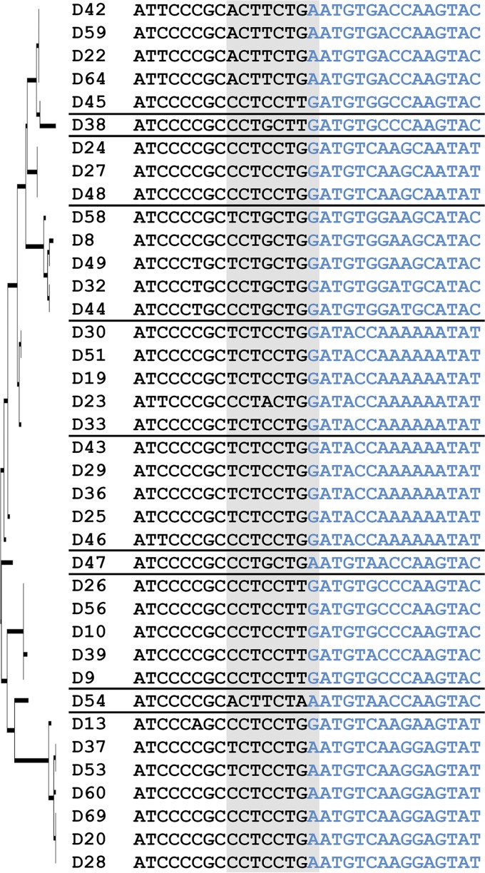
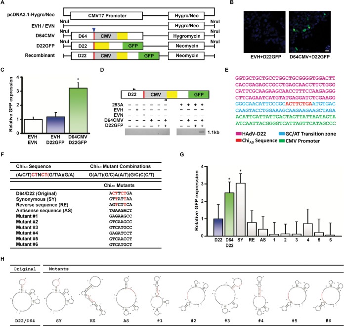
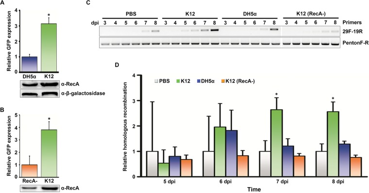
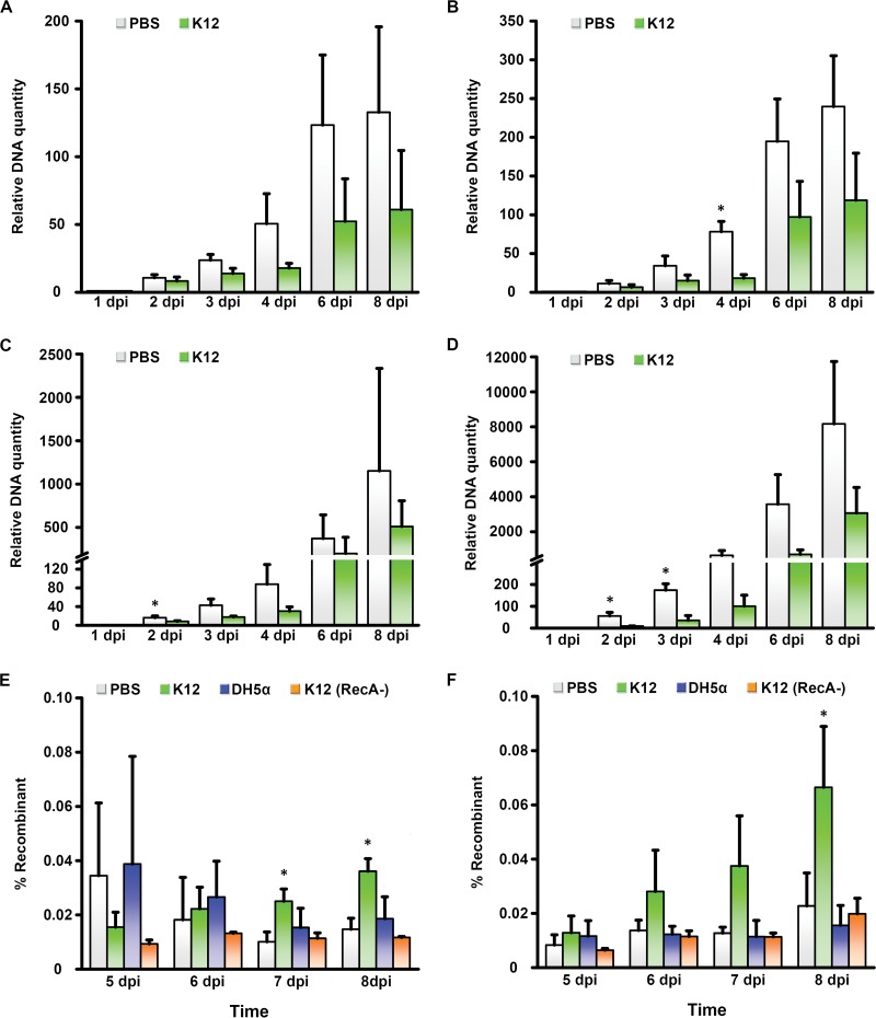
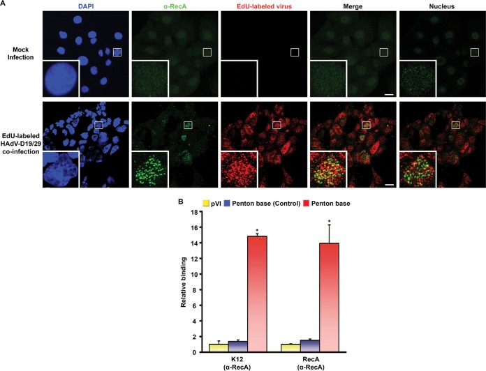
Adenovirus infections in humans are common and sometimes lethal. Adenovirus-derived vectors are also commonly chosen for gene therapy in human clinical trials. We have shown in previous work that homologous recombination between adenoviral genomes of human adenovirus species D (HAdV-D), the largest and fastest growing HAdV species, is responsible for the rapid evolution of this species. Because adenovirus infection initiates in mucosal epithelia, particularly at the gastrointestinal, respiratory, genitourinary, and ocular surfaces, we sought to determine a possible role for mucosal microbiota in adenovirus genome diversity. By analysis of known recombination hot spots across 38 human adenovirus genomes in species D (HAdV-D), we identified nucleotide sequence motifs similar to bacterial Chi sequences, which facilitate homologous recombination in the presence of bacterial Rec enzymes. These motifs, referred to here as ChiAD, were identified immediately 5' to the sequence encoding penton base hypervariable loop 2, which expresses the arginine-glycine-aspartate moiety critical to adenoviral cellular entry. Coinfection with two HAdV-Ds in the presence of an Escherichia coli lysate increased recombination; this was blocked in a RecA mutant strain, E. coli DH5α, or upon RecA depletion. Recombination increased in the presence of E. coli lysate despite a general reduction in viral replication. RecA colocalized with viral DNA in HAdV-D-infected cell nuclei and was shown to bind specifically to ChiAD sequences. These results indicate that adenoviruses may repurpose bacterial recombination machinery, a sharing of evolutionary mechanisms across a diverse microbiota, and unique example of viral commensalism.IMPORTANCE Adenoviruses are common human mucosal pathogens of the gastrointestinal, respiratory, and genitourinary tracts and ocular surface. Here, we report finding Chi-like sequences in adenovirus recombination hot spots. Adenovirus coinfection in the presence of bacterial RecA protein facilitated homologous recombination between viruses. Genetic recombination led to evolution of an important external feature on the adenoviral capsid, namely, the penton base protein hypervariable loop 2, which contains the arginine-glycine-aspartic acid motif critical to viral internalization. We speculate that free Rec proteins present in gastrointestinal secretions upon bacterial cell death facilitate the evolution of human adenoviruses through homologous recombination, an example of viral commensalism and the complexity of virus-host interactions, including regional microbiota.

Keywords: adenoviruses; commensal; homologous recombination.


Figures

FIG 1
FIG 1
Chi (ChiAD) nucleotide sequences in human adenovirus species D (HAdV-D). Thirty-eight HAdV-D genomes were aligned by maximum likelihood analysis, a tree (left) was built based on the amino acid sequences of penton base hypervariable loop 2 (HVL2), and the HAdV-D genomes were divided into proteotypes as previously described (14), shown by a horizontal line separating the virus type names. The 30 nucleotides shown include the junction between conserved nucleotide sequence (black) and HVL2 nucleotide sequence (blue), with 15 nucleotides on either side of the junction. The ChiAD motifs (shown on gray background) fall predominantly within the conserved sequence but include one nucleotide within the hypervariable sequence.
FIG 2
FIG 2
Modeling homologous recombination through ChiAD. (A) Constructs for in vitro recombination were generated on the pcDNA3.1-hygromycin/neomycin by insertion of either the penton base gene of HAdV-D64 while retaining the hygromycin resistance gene or of HAdV-D22 with coding sequence for green fluorescent protein (GFP) without ATG site, no CMVT7 promoter, and retaining the neomycin resistance gene. HVL2 (yellow boxes), GC/AT transition zone (red boxes with blue arrowhead above), CMVT7 promoter (gray boxes), and GFP open reading frames (green boxes) are indicated. EVH and EVN are hygromycin and neomycin empty vectors minus their CMVT7 promoters, respectively. Modified vectors were linearized with NruI prior to transfection. (B) Fluorescence microscopy for GFP expression in transfected 293A cells show green signal indicating recombination only when both HAdV-D sequence constructs are cotransfected (right micrograph). Original magnification, ×40. Bar, 25 µm. (C) Fluorescent signal graphically represented relative to GFP with cotransfection of an empty vector, demonstrating approximately threefold increase in signal upon cotransfection of ChiAD-containing constructs. The value that is significantly different (P < 0.05) by ANOVA is indicated by an asterisk. (D) Conventional PCR performed on transfected 293A cells with primer pairs designed to amplify only the hypothesized recombinant (forward primer from HAdV-D22 and a reverse primer for the CMVT7 promoter) shows a band only when both HAdV-D sequence constructs are cotransfected and at the predicted 1.1-kb size. (E) Sanger sequencing of PCR product (from panel D) demonstrating the specific nucleotide sequence predicted for the recombinant construct. (F) A consensus ChiAD sequence was generated in silico from 38 HAdV-Ds, and sequences not seen in known viruses were then generated, including D64/22 synonymous (SY), reverse (RE), and antisense (AS) mutants, and six randomly chosen mutants. The consensus nucleotides remaining in the chosen mutants are shown in red. (G) Fluorescent signal relative to GFP expression upon cotransfection of empty vector and D22GFP show recombination with the constructs containing native ChiAD and also with a mutant (from panel F) containing synonymous changes to ChiAD (SY) (*, P < 0.05 by ANOVA). Each experiment was performed in triplicate and repeated three times. Error bars represent standard deviations of the means. (H) Secondary ssDNA sequences as predicted by mFold, showing that the original ChiAD and SY mutants, which each recombined in panel G, have highly similar predicted secondary structures.
FIG 3
FIG 3
Promotion of homologous recombination by bacterial lysate in intestinal epithelial cells. (A) Quantification of GFP expression by ChiAD-containing constructs in the presence of a lysate of E. coli K-12 strain or its RecA mutant DH5α demonstrate reduced recombination in 293A cells pretreated with RecA mutant (*, P < 0.05 by Student’s t test). The Western blot below the graph shows expression of RecA in both E. coli lysates and β-galactosidase loading control. α-RecA, anti-RecA antibody. (B) Expression of GFP in cotransfected 293A cells pretreated with either E. coli K-12 lysate depleted of RecA (RecA-) by immunomagnetic beads, or unmodified K-12 lysate, showing reduction of recombination when RecA is depleted (*, P < 0.05 by Student’s t test). Western blot of depleted and native lysates is shown. (C) Conventional PCR performed on C2BBe1 cells pretreated with either PBS, E. coli K-12 lysate, DH5α lysate, or RecA-depleted K-12 lysate, and coinfected with HAdV-D19 and HAdV-D29, from 3 to 8 days postinfection (dpi). The PCR band generated by primers specific to the predicted recombinant (29F-19R, forward primer for HAdV-D29 HVL1 and reverse primer for HAdV-D19 HVL2) was greater in K-12 lysate-pretreated cells. Control PCR with primers that do not distinguish recombinants from parent viruses is shown below (PentonF-R). (D) Quantitative PCR under the same treatment conditions in panel C shows relative homologous recombination levels, as normalized to PBS-treated cells (*, P < 0.05 by ANOVA). Each experiment was performed in triplicate and repeated three times. Error bars represent standard deviations of the means.
FIG 4
FIG 4
Relative viral replication and recombination in the presence of bacterial protein. Using primers specific to hexon sequence in HAdV-D19 (A and C) and HAdV-D29 (B and D), quantitative PCR was used to quantify total viral DNA from 1 to 8 days postinfection in PBS-treated versus E. coli K-12 lysate-treated C2BBe1 cells (A and B) and A549 cells (C and D). DNA quantity is graphed relative to the levels at 1 day postinfection. (E and F) C2BBe1 (E) and A549 (F) cells pretreated with PBS, K-12 lysate, DH5α lysate, or K-12 lysate depleted of RecA were coinfected with HAdV-D19 and HAdV-D29 and subjected to quantitative PCR at 5 to 8 days postinfection, with primers chosen to amplify only the HVL2 recombinant. Values that are significantly different (P < 0.05) by ANOVA are indicated by an asterisk. Each experiment was performed in triplicate and repeated three times. Error bars represent standard deviations of the means.
FIG 5
FIG 5
Binding of RecA to ChiAD in intestinal epithelial cells. (A) Confocal microscopy in C2BBe1 cells pretreated with E. coli K-12 lysate for 24 h and then either mock infected or coinfected with EdU-labeled (red) HAdV-D19 and HAdV-D29. Samples were fixed at 12 h postinfection and stained with DAPI (blue) and anti-RecA (green). Stacked images without blue color are shown in the Merge panels (bars, 25 µm). To reduce any artifact of perinuclear localization, a single image centered on the nucleus in the inset with one image on either side is also shown in the Nucleus panels. Colocalization of viral DNA and RecA is suggested by the yellow color. The small white boxes in the micrographs show the locations of the insets. Original magnification, ×63. (B) ChIP analysis performed on C2BBe1 cells pretreated with either E. coli K-12 lysate or recombinant RecA prior to coinfection with HAdV-D19 and HAdV-D29. Binding of RecA to ChiAD was compared to IgG (not shown) and randomly chosen regions in protein VI (pVI) and penton base (Control). Binding affinities were normalized to pVI binding of RecA protein. Each experiment was repeated three times. Error bars represent standard deviations of the means. For each experiment, *, P < 0.05 by ANOVA.

References

    1. Kapusinszky B, Minor P, Delwart E. 2012. Nearly constant shedding of diverse enteric viruses by two healthy infants. J Clin Microbiol 50:3427–3434. doi:10.1128/JCM.01589-12. - DOI - PMC - PubMed
    1. Nishio O, Sakae K, Ishihara Y, Isomura S, Inouye S. 1992. Adenovirus infection and specific secretory IgA responses in the intestine of infants. Microbiol Immunol 36:623–631. doi:10.1111/j.1348-0421.1992.tb02062.x. - DOI - PubMed
    1. Hilleman MR, Werner JH. 1954. Recovery of new agent from patients with acute respiratory illness. Proc Soc Exp Biol Med 85:183–188. doi:10.3181/00379727-85-20825. - DOI - PubMed
    1. Rowe WP, Huebner RJ, Gilmore LK, Parrott RH, Ward TG. 1953. Isolation of a cytopathogenic agent from human adenoids undergoing spontaneous degeneration in tissue culture. Proc Soc Exp Biol Med 84:570–573. doi:10.3181/00379727-84-20714. - DOI - PubMed
    1. Horwitz MS, Valderrama G, Hatcher V, Korn R, deJong P, Spigland I. 1984. Characterization of adenovirus isolates from AIDS patients. Ann N Y Acad Sci 437:161–174. doi:10.1111/j.1749-6632.1984.tb37132.x. - DOI - PubMed

Publication types

LinkOut - more resources