. 2018 Jun 21;3(12):e120474.
doi: 10.1172/jci.insight.120474.

Double-stranded RNA innate immune response activation from long-term adeno-associated virus vector transduction

Affiliations

Double-stranded RNA innate immune response activation from long-term adeno-associated virus vector transduction

Wenwei Shao et al. JCI Insight. .

Abstract

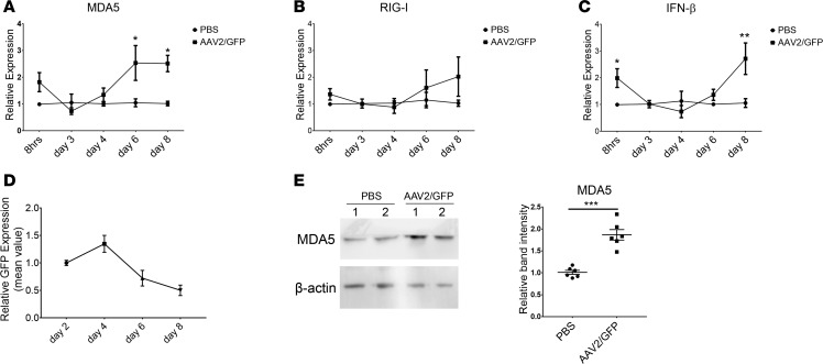
Data from clinical trials for hemophilia B using adeno-associated virus (AAV) vectors have demonstrated decreased transgenic coagulation factor IX (hFIX) expression 6-10 weeks after administration of a high vector dose. While it is likely that capsid-specific cytotoxic T lymphocytes eliminate vector-transduced hepatocytes, thereby resulting in decreased hFIX, this observation is not intuitively consistent with restored hFIX levels following prednisone application. Although the innate immune response is immediately activated following AAV vector infection via TLR pathways, no studies exist regarding the role of the innate immune response at later time points after AAV vector transduction. Herein, activation of the innate immune response in cell lines, primary human hepatocytes, and hepatocytes in a human chimeric mouse model was observed at later time points following AAV vector transduction. Mechanistic analysis demonstrated that the double-stranded RNA (dsRNA) sensor MDA5 was necessary for innate immune response activation and that transient knockdown of MDA5, or MAVS, decreased IFN-β expression while increasing transgene production in AAV-transduced cells. These results both highlight the role of the dsRNA-triggered innate immune response in therapeutic transgene expression at later time points following AAV transduction and facilitate the execution of effective strategies to block the dsRNA innate immune response in future clinical trials.

Keywords: Gene therapy; Immunology; Innate immunity; Therapeutics.


Conflict of interest statement

Conflict of interest: RJS is the founder of and a shareholder at Asklepios BioPharmaceutical and Bamboo Therapeutics Inc. He holds a patent (9475845) that has been licensed by the University of North Carolina at Chapel Hill to Asklepios BioPharmaceutical, for which he receives royalties. He has consulted for Baxter and has received payment for speaking. MLH has also received royalties from Asklepios BioPharmaceutical related to a patent (9447433).

Figures

Figure 1
Figure 1. IFN-β inhibited AAV transgene expression in the HeLa cell line.
HeLa cells were transduced with 5 × 10³ particles of AAV2/luciferase per cell. (A) After 24 hours, recombinant human IFN-β was added to the medium at a different dose. Transgene expression was detected by luciferase assay at days 1, 2, 4, and 6 after supplementation of IFN-β. (B) Recombinant human IFN-β was added to the medium every day at 0.5 ng/ml. Transgene expression was detected by luciferase assay at days 1, 2, 4, and 6. ***P < 0.001, when compared with no IFN-β treatment, as measured by 2-way ANOVA. Results are representative of 3 independent experiments.
Figure 2
Figure 2. Poly (I:C) inhibited AAV transgene expression in cell lines.
HeLa or Huh7 cells were transduced with 5 × 10³ particles of AAV2/luciferase per cell. 2 μg/ml poly (I:C) was added at different time points: 18 hours before AAV transduction, day 0, or day 3. Luciferase expression was detected 3 days after poly (I:C) transfection. *P < 0.05, **P < 0.01, ***P < 0.001, when compared with control group, as measured by 2-way ANOVA. Results are representative of 4 independent experiments.
Figure 3
Figure 3. Minus strand transcript generation in AAV-transduced cells.
(A) Overview of the gene-specific reverse transcription to detect either plus or minus strand transcripts. HeLa cells were harvested at day 8 after AAV2/luciferase transduction. The RNA was extracted and treated with DNase. Specific primers for plus strand or minus strand luciferase were used to synthesize different orientations of the cDNA. PCR was performed to detect the transcripts in different orientations of cDNA using primer pair 1 (F1 and R1) and primer pair 2 (F2 and R2). (B) PCR products are shown. PBS was used as a negative control with no AAV virus. The pTR/luciferase plasmid served as positive control for the PCR. RNA was used as a template to eliminate the possibility of AAV genome DNA contamination in extracted RNA. To measure the yield of transcripts, cDNA in different orientations was diluted to 20-, 200-, or 2,000-fold as PCR templates.
Figure 4
Figure 4. dsRNA immune response is activated at a later time point after AAV transduction.
HeLa cells were transduced with 5 × 10³ particles of scAAV2/GFP per cell. The expression of MDA5 (A), RIG-I (B), and IFN-β (C) in HeLa cells was detected by Q-PCR at different time points after transduction. *P < 0.05, **P < 0.01, when compared with the PBS group, as measured by 2-way ANOVA. The data represent the mean ± SEM from 3 independent experiments. For each experiment, the PBS- or AAV-infected group contained 2 or 3 wells of cells. For Q-PCR data analysis, one sample from the PBS group was normalized to one in each time point of each experiment. (D) GFP expression level in HeLa cells was detected by flow cytometry at different time points after AAV2/GFP transduction. (E) MDA5 expression in HeLa cells in each group was detected by Western blot 8 days after scAAV2/GFP transduction. The relative level of MDA5 expression was calculated based on the intensity of β-actin protein from 3 independent experiments. ***P < 0.001, when compared with the PBS group, as measured by 2-tailed Student’s t test.
Figure 5
Figure 5. dsRNA response profile in different cell lines and different transgenes.
(A) Huh7, HEK293, and HepG2 cells were transduced with 5 × 10³ particles of AAV2/GFP per cell. The expression of MDA5, RIG-I, and IFN-β was detected by Q-PCR at day 7. *P < 0.05, **P < 0.01, when compared with the PBS group, as measured by 2-way ANOVA. The data present the mean ± SEM from 3 independent experiments. (B) HeLa cells were transduced with AAV2 containing different transgenes at 5 × 10³ particles per cell. The expression of MDA5, RIG-I, and IFN-β was detected by Q-PCR at day 7 after AAV transduction. For Q-PCR data analysis, samples from the PBS group were normalized to 1 in each experiment. *P < 0.05, **P < 0.01, ***P < 0.001, when compared with the PBS group, as measured by 2-tailed Student’s t test. (C) HeLa cells were transduced with different doses of scAAV/GFP (1,000, 5,000, or 250,000 particles of AAV2/GFP per cell). The expression of MDA5, RIG-I, and IFN-β was detected by Q-PCR at day 7. *P < 0.05, **P < 0.01, ***P < 0.001, when compared with the PBS group, as measured by 2-way ANOVA. The data present the mean ± SEM from 3 independent experiments, and each group contained 2 or 3 wells of cells for each experiments.
Figure 6
Figure 6. dsRNA innate immune response in human primary hepatocytes after scAAV2/GFP transduction.
Fresh human primary hepatocytes from 12 individuals were transduced by scAAV2/GFP with 5 × 10³ particles per cell. The expression of MDA5, RIG-I, and IFN-β was detected by Q-PCR at different time points after AAV transduction. For relative gene expression calculation, the gene expression of the PBS group at each time point was normalized to 1, which is not shown in the graph. Biological duplicates were used in each sample. (A) MDA5 expression was ≥2-fold upregulated following AAV administration at day 5 or later. (B) MDA5 expression was similar to PBS following AAV administration at day 5 or later.
Figure 7
Figure 7. dsRNA innate immune response in human primary hepatocytes after scAAV2/hFIX-opt transduction.
Fresh human primary hepatocytes from 10 individuals were transduced by scAAV2/hFIX-opt with 5 × 10³ particles per cell. The expression of MDA5, RIG-I, and IFN-β was detected by Q-PCR at different time points after AAV transduction. For relative gene expression calculation, the gene expression of the PBS group at each time point was normalized to 1, which is not shown in the graph. Biological duplicates were used in each sample. (A) MDA5 expression was ≥2-fold upregulated following AAV administration at day 5 or later. (B) MDA5 expression was similar to PBS following AAV administration at day 5 or later.
Figure 8
Figure 8. dsRNA response in human hepatocytes from xenografted mice after scAAV8/hFIX-opt transduction.
(A) Two xenografted mice with human hepatocytes were injected with 3 × 10¹¹ particles of scAAV8/hFIX-opt. The expression of MDA5, RIG-I, and IFN-β in human hepatocytes in mice was detected by Q-PCR at 8 weeks after AAV transduction. Three to four pieces of xenografted liver were chosen randomly for RNA extraction. MDA5 protein in the liver was detected by Western blot after 8 weeks. The band intensity was measured to show the relative MDA5 expression based on β-actin; the data were from 4 separate experiments. *P < 0.05, **P < 0.01, when compared with the control group; data for multiple comparisons were compared using 2-way ANOVA, and data for single comparisons were evaluated by the 2-tailed Student’s t test. (B) Two xenografted mice with human hepatocytes from another donor were injected with a dose of scAAV8/hFIX-opt. The expression of MDA5, RIG-I, and IFN-β in human hepatocytes in mice was detected by Q-PCR at 4 and 8 weeks after AAV transduction. *P < 0.05, **P < 0.01, ***P < 0.001, when compared with the control group, as measured by 2-way ANOVA. (C) MDA5 protein in livers from the second experiment was detected by Western blot, and the relative expression level of MDA5 was calculated based on β-actin intensity; the data were from 4 separate experiments. *P < 0.05, when compared with the control group, as measured by 2-way ANOVA.
Figure 9
Figure 9. Knock out of dsRNA activation pathway increased AAV transgene expression.
(A) HeLa cells were transfected with siControl, siMDA5, or siMAVS. The knockdown efficiency was detected by Western blot and Q-PCR. (B) At day 0, HeLa cells were transduced with 5 × 10³ particles of AAV2/luciferase per cell. siRNA was transfected into HeLa cells at day 4, and luciferase expression was detected 48 or 72 hours later. As a control, 2 μg/ml poly (I:C) was added at day 3 and siRNA was transfected to HeLa cells at day 4. *P < 0.05, **P < 0.01, ***P < 0.001, when compared with the PBS group, as measured by 2-way ANOVA. The data present the mean ± SEM from 4 independent experiments. (C) After 4 days of AAV transduction, siRNA was transfected to HeLa cells, and IFN-β expression was detected by Q-PCR at 48 hours after siRNA transfection. ***P < 0.001, when compared with the PBS group, as measured by 2-way ANOVA. The data present the mean ± SEM from 3 independent experiments. (D) After 4 days of AAV transduction, siRNA and IFN-β promoter reporter plasmids were cotransfected to HeLa cells and then luciferase activity was measured after 72 hours. **P < 0.01, when compared with the PBS group, as measured by 2-way ANOVA. (E) After 4 days of AAV transduction, siRNA was transfected to HeLa cells, and MDA5 expression was detected by Q-PCR at 48 hours after siRNA transfection. *P < 0.05, when compared with the PBS group, as measured by 2-way ANOVA. The data present the mean ± SEM from 4 independent experiments.

Similar articles

Cited by

References

    1. Manno CS, et al. Successful transduction of liver in hemophilia by AAV-factor IX and limitations imposed by the host immune response. Nat Med. 2006;12(3):342–347. doi: 10.1038/nm1358. - DOI - PubMed
    1. Nathwani AC, et al. Adenovirus-associated virus vector-mediated gene transfer in hemophilia B. N Engl J Med. 2011;365(25):2357–2365. doi: 10.1056/NEJMoa1108046. - DOI - PMC - PubMed
    1. Nathwani AC, et al. Long-term safety and efficacy of factor IX gene therapy in hemophilia B. N Engl J Med. 2014;371(21):1994–2004. doi: 10.1056/NEJMoa1407309. - DOI - PMC - PubMed
    1. Nathwani AC, Davidoff AM, Tuddenham EGD. Advances in gene therapy for hemophilia. Hum Gene Ther. 2017;28(11):1004–1012. doi: 10.1089/hum.2017.167. - DOI - PubMed
    1. Wu Z, et al. Optimization of self-complementary AAV vectors for liver-directed expression results in sustained correction of hemophilia B at low vector dose. Mol Ther. 2008;16(2):280–289. doi: 10.1038/sj.mt.6300355. - DOI - PubMed

Publication types

MeSH terms