. 2018 Jun 20;9(7):724.
doi: 10.1038/s41419-018-0748-x.

SERPINB2 is a novel indicator of stem cell toxicity

Affiliations

SERPINB2 is a novel indicator of stem cell toxicity

Na-Hee Lee et al. Cell Death Dis. .

Abstract

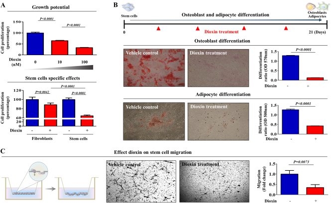
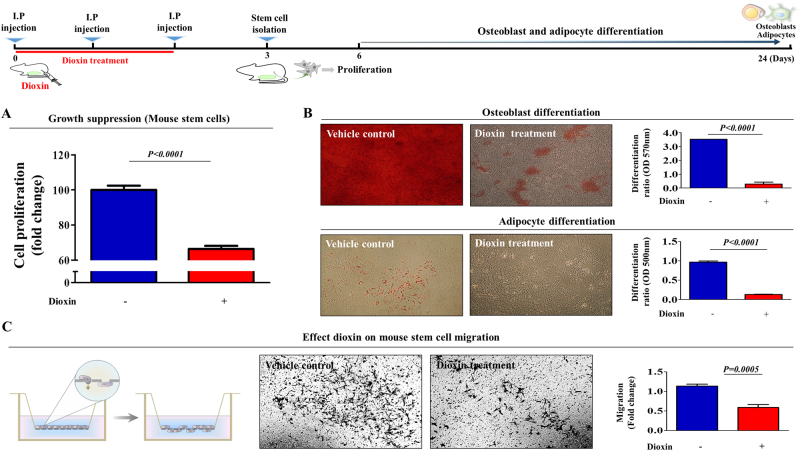
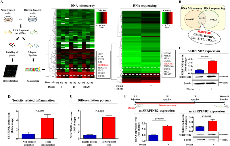
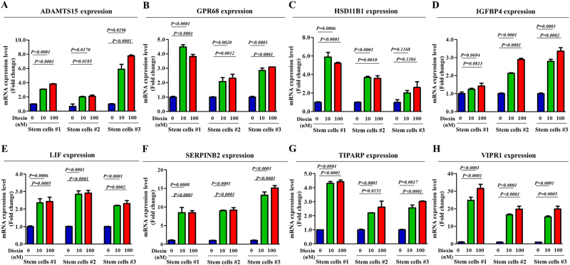
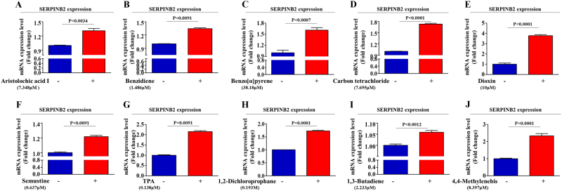
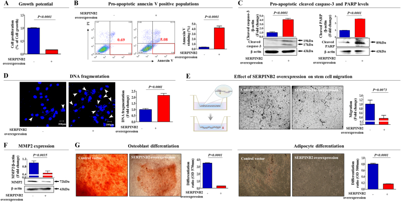
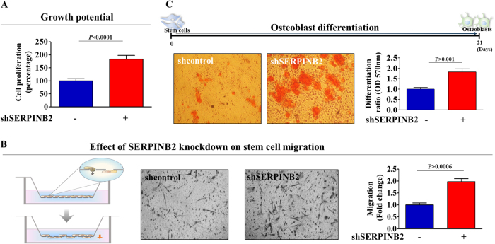
The toxicological evaluation of potential drug candidates is very important in the preclinical phase of drug development. Toxic materials may cause serious decline in stem cell function and loss of stemness. Indeed, we found that toxic exposure more profoundly suppressed the growth of stem cells than terminally differentiated fibroblasts. Importantly, toxic exposure suppressed stem cell migration and multi-lineage differentiation potential in vitro and in vivo. Moreover, early-response genes involved in stem cell properties such as self-renewal and differentiation capabilities can be used as specific markers to predict toxicity. In the present study, we also identified a labile toxic response gene, SERPINB2, which is significantly increased in response to various toxic agents in human stem cells in vitro and in vivo. Consistently, self-renewal, migration, and multi-lineage differentiation potential were markedly decreased following SERPINB2 overexpression. To the best of our knowledge, this is the first study to focus on the functions of SERPINB2 on the regenerative potential of stem cells in response to various existing chemicals, and the findings will facilitate the development of promising toxicity test platforms for newly developed chemicals.


Conflict of interest statement

The authors declare that they have no conflict of interest.

Figures

Fig. 1
Fig. 1. The effects of dioxin on the various functions of stem cells in vitro.
The inhibition of cell viability by dioxin treatment for 72 h was determined via an MTT assay in both stem cells and fibroblasts. The cell viability (%) was calculated as the percent of the vehicle control (a). Schematic representation described the experimental protocol for differentiation and treatment. Confluent stem cells were cultured for 21 days in osteogenic or adipogenic medium with or without dioxin (10 nM). The effect of dioxin on osteoblast or adipocyte differentiation was determined by alizarin red or oil red O staining, respectively. Relative quantification of calcium mineral content or lipid droplet formation was determined by absorbance measurements at 570 nm or 500 nm, respectively (b). The effect of dioxin on the MSC migration ability was evaluated using a transwell migration assay. Dioxin treatment significantly decreased MSC migration across the membrane compared with the negative controls (c). The results are presented as the mean ± SD from at least three independent experiments
Fig. 2
Fig. 2. Toxic exposure affects the in vivo growth and differentiation potential of stem cells.
Schematic representation described the experimental protocol for dioxin treatment in vivo. Mice were treated with dioxin (15 µg/kg, intraperitoneally, three times daily, for 3 days) or vehicle (DMSO), and then after 3 days stem cells from mouse adipose tissue were isolated. The effect of dioxin on cell viability was determined via an MTT assay (a). Stem cells derived from mouse adipose tissues were cultured in osteogenic or adipogenic differentiation medium for 21 days without additional dioxin treatment. The effect of dioxin on osteoblast or adipocyte differentiation in vivo was determined by alizarin red or oil red O staining, respectively (b). The effect of dioxin on the migration ability of mouse adipose tissue-derived MSCs in vivo was evaluated using a transwell migration assay (c). The results are presented as the mean ± SD from three independent experiments
Fig. 3
Fig. 3. Dioxin stimulates SERPINB2 expression in stem cells in vitro and in vivo.
The data from both large-scale DNA microarray and RNA sequencing are presented as a heatmap of differentially expressed genes in non-treated stem cells compared to those treated with dioxin; decreased (green) or increased (red) expression compared to the mean mRNA expression are indicated (a). Among the genes that were analyzed, we observed a positive correlation between dioxin exposure and enhanced SERPINB2 expression in stem cells (b). Western blotting was used to verify enhanced SERPINB2 expression in DNA microarray and RNA sequencing (c). Clinical big data were analyzed using the Seiber dataset (GSE43996 and GSE9452) from ‘R2: Genomics Analysis and Visualization Platform (http://r2.amc.ml)’. The gene datasets were filtered by SERPINB2 expression profiles and the differentiation potency of stem cells (d) or toxicity-related diseases (e). Mice were treated with dioxin (15 µg/kg, intraperitoneally) or vehicle (DMSO), and then stem cells from the adipose tissue of the mice were isolated and expanded as described in the materials and methods section. Real-time PCR and western blotting were performed to confirm the increased SERPINB2 expression in vivo by dioxin exposure (f). β-actin was used as an internal control. The results represent the means ± SD from three independent experiments
Fig. 4
Fig. 4. Effects of dioxin on the expression of various potential response genes.
The stimulatory effect of dioxin on the expression levels of potential response genes, such as ADAMTS15, GPR68M, HSD11B1, IGFBP4, LIF, SERPINB2, TIPARP, and VIPR1, were assessed in stem cells isolated from three different individuals using real-time PCR (ah). The results represent the means ± SD from three independent experiments
Fig. 5
Fig. 5. Effects of multiple test substances on SERPINB2 expression levels in stem cells.
The stimulatory effect of multiple test substances on SERPINB2 expression levels in stem cells was assessed by real-time PCR (aj). The results represent the means ± SD from three independent experiments
Fig. 6
Fig. 6. The effects of overexpression of SERPINB2 on the various functions of stem cells.
Transfection of MSCs with a SERPINB2 vector led to a significant decrease in the number of cells compared with transfection using a control vector (a). SERPINB2 overexpression-mediated cytotoxicity was evaluated by flow cytometry using PE-labeled Annexin-V (b). Elevated levels of caspase-3 fragment following SERPINB2 overexpression were assessed by western blotting (c). SERPINB2 overexpression-mediated apoptotic DNA fragmentation and condensation were visualized using DAPI staining (d). The effects of SERPINB2 overexpression on the cell migration ability were evaluated using a transwell migration assay (e) and by western blotting using an MMP2 antibody (f). The effect of SERPINB2 overexpression on osteoblast or adipocyte differentiation was determined by alizarin red or oil red O staining, respectively. Relative quantification of the calcium mineral content or lipid droplet formation was determined by absorbance measurements at 570 nm or 500 nm, respectively (g). β-actin was used as an internal control. The results represent the means ± SD from three independent experiments
Fig. 7
Fig. 7. The effects of knockdown of SERPINB2 on the various functions of stem cells.
Transfection of MSCs with a shSERPINB2 led to an increase in the number of cells compared with transfection using a control shRNA (a). The effects of SERPINB2 knockdown on the cell migration ability were evaluated using a transwell migration assay (b). The effect of SERPINB2 knockdown on osteoblast differentiation was determined by alizarin red staining. Relative quantification of the calcium mineral content or lipid droplet formation was determined by absorbance measurements at 570 nm (c). The results represent the means ± SD from three independent experiments

References

    1. Sakolish CM, Esch MB, Hickman JJ, Shuler ML, Mahler GJ. Modeling barrier tissues in vitro: methods, achievements, and challenges. EBioMedicine. 2016;5:30–39. doi: 10.1016/j.ebiom.2016.02.023. - DOI - PMC - PubMed
    1. Kumar N, Sharma U, Singh C, Singh B. Thalidomide: chemistry, therapeutic potential and oxidative stress induced teratogenicity. Curr. Top. Med. Chem. 2012;12:1436–1455. doi: 10.2174/156802612801784407. - DOI - PubMed
    1. Papapetrou EP. Patient-derived induced pluripotent stem cells in cancer research and precision oncology. Nat. Med. 2016;22:1392–1401. doi: 10.1038/nm.4238. - DOI - PMC - PubMed
    1. Morrison SJ, Spradling AC. Stem cells and niches: mechanisms that promote stem cell maintenance throughout life. Cell. 2008;132:598–611. doi: 10.1016/j.cell.2008.01.038. - DOI - PMC - PubMed
    1. Kang KS, Trosko JE. Stem cells in toxicology: fundamental biology and practical considerations. Toxicol. Sci. 2011;120(Suppl. 1):S269–S289. doi: 10.1093/toxsci/kfq370. - DOI - PubMed

Publication types

LinkOut - more resources