. 2018 Jun 20;8(1):9401.
doi: 10.1038/s41598-018-27760-z.

Bacterial nanocellulose stimulates mesenchymal stem cell expansion and formation of stable collagen-I networks as a novel biomaterial in tissue engineering

Affiliations

Bacterial nanocellulose stimulates mesenchymal stem cell expansion and formation of stable collagen-I networks as a novel biomaterial in tissue engineering

Martin Vielreicher et al. Sci Rep. .

Abstract

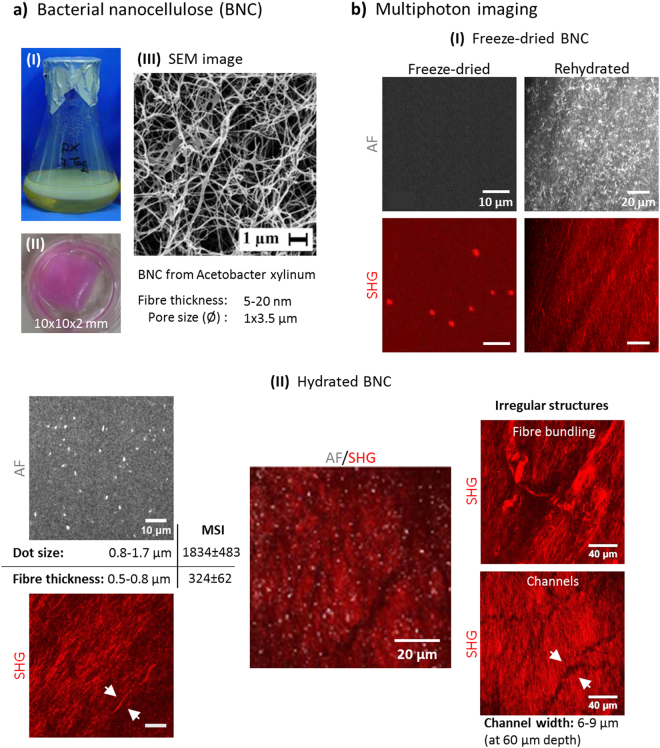
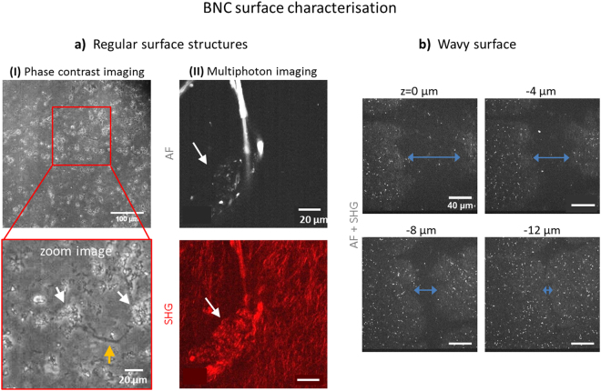
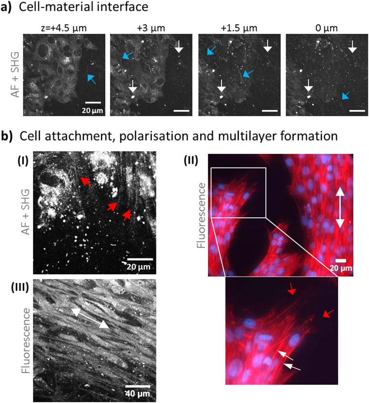
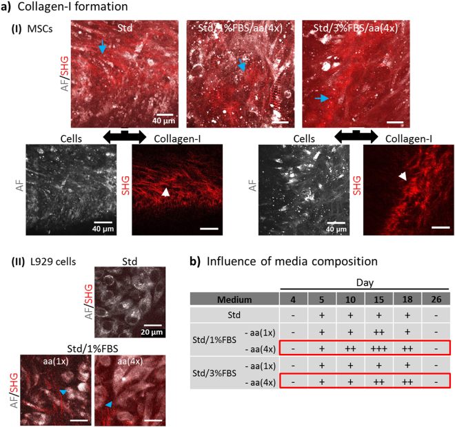
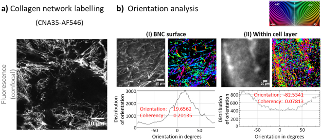
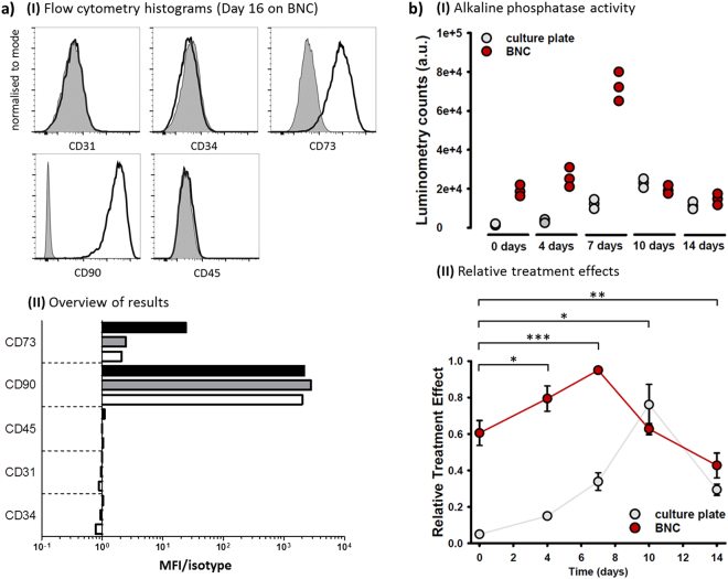
Biomimetic scaffolds are of great interest to tissue engineering (TE) and tissue repair as they support important cell functions. Scaffold coating with soluble collagen-I has been used to achieve better tissue integration in orthopaedy, however, as collagen persistence was only temporary such efforts were limited. Adequate coverage with cell-derived ECM collagen-I would promise great success, in particular for TE of mechanically challenged tissues. Here, we have used label-free, non-invasive multiphoton microscopy (MPM) to characterise bacterial nanocellulose (BNC) - a promising biomaterial for bone TE - and their potency to stimulate collagen-I formation by mesenchymal stem cells (MSCs). BNC fleeces were investigated by Second Harmonic Generation (SHG) imaging and by their characteristic autofluorescence (AF) pattern, here described for the first time. Seeded MSCs adhered fast, tight and very stable, grew to multilayers and formed characteristic, wide-spread and long-lasting collagen-I. MSCs used micron-sized lacunae and cracks on the BNC surface as cell niches. Detailed analysis using a collagen-I specific binding protein revealed a highly ordered collagen network structure at the cell-material interface. In addition, we have evidence that BNC is able to stimulate MSCs towards osteogenic differentiation. These findings offer new options for the development of engineered tissue constructs based on BNC.


Conflict of interest statement

The authors declare no competing interests.

Figures

Figure 1
Figure 1
BNC characterisation. (a) BNC biomaterial is formed as fleeces during static culture at the air-liquid interface (I). BNC fleece patch used for cell seeding (II). (III) SEM image (magnification M = 3,000x) providing insight into the nanocellulose network ultra-structure. Images I and III were provided by D. Kralisch. (b) Multiphoton pattern of dried BNC. The pattern of rolled BNC rehydrated with PBS dramatically changes within minutes. (c) Simultaneous AF/SHG images from within the BNC exhibit both dot- to elliptic-shaped AF signals and fibre-shaped SHG signals with different signal strengths (MSI, mean signal intensity). Representative overlay image shown in the middle. Irregular structures within the cellulose network were observed as well.
Figure 2
Figure 2
Micro-structure of the BNC surface. In (a), regular surface structures are presented. (I) Phase contrast images (M = 10x) reveal structures reminding of cracks (orange arrow) and cavities (~10–20 µm in diameter, white arrows) all over the surface. In (II), a typical structural islet with an agglomerate multiphoton pattern is shown (white arrows in AF and SHG images). In (b), a trench is displayed in various depths which is large enough to act as cell seeding core (diameter of > 60 µm at surface, unseparated multiphoton image, M = 40x).
Figure 3
Figure 3
Cell-material interface: attachment, anchoring and orientation on BNC. In (a), multiphoton images (unseparated, AF + SHG) highlight the MSC attachment site and a few µm above. Signals derived from BNC are visible as dots (white arrows) and fibrous structures (blue arrows). The cells become visible by their AF signal. In (b), cell anchoring structures (filopodia) are shown in both an MPM (I, red arrows) and a fluorescence image (II, phalloidin-PF546 and DAPI). The zoom image in (II) highlights anchoring structures (red arrows) and multilayer cell arrangements (white arrows directed to DAPI-stained nuclei). Cell orientation patterns (white arrow) were observed in (II) and in (III) in between cell layers (staining with Calcein AM).
Figure 4
Figure 4
Collagen-I formation by cells seeded on BNC. (a) Two-colour multiphoton live cell images with AF (grey) showing the cells and SHG (red) representing cell-derived collagen-I. MSCs were cultured in various media of which standard growth (Std) and serum-reduced media (1% and 3% FBS) with elevated ascorbic acid (aa, 4x) resulted in collagen-I fibre formation (I, blue arrows). Images below (separated AF and SHG channel) reveal the collagen fibre structure and location relative to the cells. (II) Detection of collagen formed by L929 cells grown in Std and serum-reduced media. All images are representative for five individual experiments. (b) The table presents the time-frame of collagen-I formation by MSCs with various stimulation media. SHG signals were imaged using identical laser excitation and detection conditions. Analysis was performed semi-quantitatively by defining three categories of intensity (+, ++, +++).
Figure 5
Figure 5
Collagen-I labelling and network analysis. (a) Collagen-I networks were independently imaged by confocal fluorescence microscopy (CFM) after specific staining with CNA35-AlexaFluor546 collagen-I binding protein. Much more details of fibre network structure compared to MPM images are visible (M = 20x). (b) Comparison of collagen fibre networks at the cell-BNC interface (I) and in between cells in a multilayer (II) using a fluorescence image and the corresponding S-colour survey image (colour code expressed by colour wheel at top right). Histograms mapping the incidence of fibre orientations from −90° to +90° including values for Orientation and Coherency are shown below.
Figure 6
Figure 6
Differentiation status of MSCs. (a) MSCs were cultured on BNC or culture plate for 16 days and analysed for expression of CD31, CD34, CD45, CD73, and CD90 by flow cytometry. (I) Overlays show expression of indicated molecules (black) and corresponding isotype controls (grey, filled) at day 16 on BNC. (II) Differentiation of MSCs was analysed at day 0 (2 days after starting cell culture (white bars) and after 16 days on plastic plates (grey bars) or BNC (black bars). Graph represents mean fluorescence intensity (MFI) of surface markers relative to their isotype controls. (b) (I) AP activity (luminometry) data from MSCs seeded on BNC compared to standard cell culture plates (control) are displayed versus days after seeding. In (II), relative treatment effects, calculated based on the non-parametric model, reveal a significantly enhanced AP activity with major differences up to day 7, at which point activity already decreases for cells growing on BNC, while a similar effect takes up to day 10 for culture plate growing cells. ‘*’ Signs indicate time accumulating group differences between cells on BNC and culture plate and denote significances as follows: *p < 0.05; **p < 0.005; ***p < 0.0005.

Similar articles

Cited by

References

    1. Leo AJ, Grande DA. Mesenchymal stem cells in tissue engineering. Cells. Tissues. Organs. 2006;183:112–22. doi: 10.1159/000095985. - DOI - PubMed
    1. Pittenger MF, et al. Multilineage Potential of Adult Human Mesenchymal Stem Cells. Science (80-.). 1999;284:143–147. doi: 10.1126/science.284.5411.143. - DOI - PubMed
    1. Berthiaume F, Maguire TJ, Yarmush ML. Tissue Engineering and Regenerative Medicine: History, Progress, and Challenges. Annu. Rev. Chem. Biomol. Eng. 2011;2:403–430. doi: 10.1146/annurev-chembioeng-061010-114257. - DOI - PubMed
    1. Salgado AJ, Coutinho OP, Reis RL. Bone tissue engineering: state of the art and future trends. Macromol. Biosci. 2004;4:743–65. doi: 10.1002/mabi.200400026. - DOI - PubMed
    1. Kemp KC, Hows J, Donaldson C. Bone marrow-derived mesenchymal stem cells. Leuk. Lymphoma. 2005;46:1531–1544. doi: 10.1080/10428190500215076. - DOI - PubMed

Publication types

MeSH terms

LinkOut - more resources