

Corticobasal degeneration with TDP-43 pathology presenting with progressive supranuclear palsy syndrome: a distinct clinicopathologic subtype
- PMID: 29926172
- PMCID: PMC6309287
- DOI: 10.1007/s00401-018-1878-z
Corticobasal degeneration with TDP-43 pathology presenting with progressive supranuclear palsy syndrome: a distinct clinicopathologic subtype
Abstract
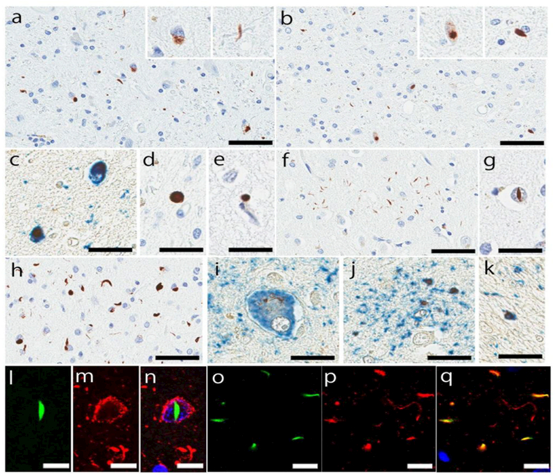
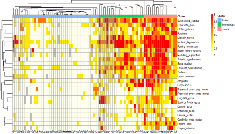
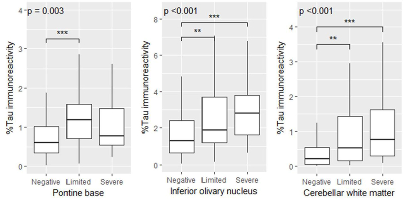
Corticobasal degeneration (CBD) is a clinically heterogeneous tauopathy, which has overlapping clinicopathologic and genetic characteristics with progressive supranuclear palsy (PSP). This study aimed to elucidate whether transactive response DNA-binding protein of 43 kDa (TDP-43) pathology contributes to clinicopathologic heterogeneity of CBD. Paraffin-embedded sections of the midbrain, pons, subthalamic nucleus, and basal forebrain from 187 autopsy-confirmed CBD cases were screened with immunohistochemistry for phospho-TDP-43. In cases with TDP-43 pathology, additional brain regions (i.e., precentral, cingulate, and superior frontal gyri, hippocampus, medulla, and cerebellum) were immunostained. Hierarchical clustering analysis was performed based on the topographical distribution and severity of TDP-43 pathology, and clinicopathologic and genetic features were compared between the clusters. TDP-43 pathology was observed in 45% of CBD cases, most frequently in midbrain tegmentum (80% of TDP-43-positive cases), followed by subthalamic nucleus (69%). TDP-43-positive CBD was divided into TDP-limited (52%) and TDP-severe (48%) by hierarchical clustering analysis. TDP-severe patients were more likely to have been diagnosed clinically as PSP compared to TDP-limited and TDP-negative patients (80 vs 32 vs 30%, P < 0.001). The presence of downward gaze palsy was the strongest factor for the antemortem diagnosis of PSP, and severe TDP-43 pathology in the midbrain tectum was strongly associated with downward gaze palsy. In addition, tau burden in the olivopontocerebellar system was significantly greater in TDP-positive than TDP-negative CBD. Genetic analyses revealed that MAPT H1/H1 genotype frequency was significantly lower in TDP-severe than in TDP-negative and TDP-limited CBD (65 vs 89 vs 91%, P < 0.001). The homozygous minor allele frequencies in GRN rs5848 and TMEM106B rs3173615 were not significantly different between the three groups. In conclusion, the present study indicates that CBD with severe TDP-43 pathology is a distinct clinicopathologic subtype of CBD, characterized by PSP-like clinical presentations, severe tau pathology in the olivopontocerebellar system, and low frequency of MAPT H1 haplotype.
Keywords: Argyrophilic grain disease; Corticobasal degeneration; Corticobasal syndrome; MAPT; Progressive supranuclear palsy; TDP-43.
Figures
Similar articles
-
Machine learning-based decision tree classifier for the diagnosis of progressive supranuclear palsy and corticobasal degeneration.Neuropathol Appl Neurobiol. 2021 Dec;47(7):931-941. doi: 10.1111/nan.12710. Epub 2021 Apr 7. Neuropathol Appl Neurobiol. 2021. PMID: 33763863 Free PMC article.
-
Corticobasal degeneration with olivopontocerebellar atrophy and TDP-43 pathology: an unusual clinicopathologic variant of CBD.Acta Neuropathol. 2013 May;125(5):741-52. doi: 10.1007/s00401-013-1087-8. Epub 2013 Jan 31. Acta Neuropathol. 2013. PMID: 23371366 Free PMC article.
-
Case report of a patient with unclassified tauopathy with molecular and neuropathological features of both progressive supranuclear palsy and corticobasal degeneration.Acta Neuropathol Commun. 2023 Jun 1;11(1):88. doi: 10.1186/s40478-023-01584-z. Acta Neuropathol Commun. 2023. PMID: 37264457 Free PMC article.
-
Chameleons and mimics: Progressive supranuclear palsy and corticobasal degeneration.Neuropathology. 2020 Feb;40(1):57-67. doi: 10.1111/neup.12590. Epub 2019 Sep 12. Neuropathology. 2020. PMID: 31515852 Review.
-
Neuropathology of variants of progressive supranuclear palsy.Curr Opin Neurol. 2010 Aug;23(4):394-400. doi: 10.1097/WCO.0b013e32833be924. Curr Opin Neurol. 2010. PMID: 20610990 Review.
Cited by
-
TDP-43 Is Efficiently Transferred Between Neuron-Like Cells in a Manner Enhanced by Preservation of Its N-Terminus but Independent of Extracellular Vesicles.Front Neurosci. 2020 Jun 11;14:540. doi: 10.3389/fnins.2020.00540. eCollection 2020. Front Neurosci. 2020. PMID: 32595443 Free PMC article.
-
Cerebrovascular pathology presenting as corticobasal syndrome: An autopsy case series of "vascular CBS".Parkinsonism Relat Disord. 2019 Nov;68:79-84. doi: 10.1016/j.parkreldis.2019.09.001. Epub 2019 Sep 2. Parkinsonism Relat Disord. 2019. PMID: 31621626 Free PMC article.
-
TDP-43 Proteinopathy and Tauopathy: Do They Have Pathomechanistic Links?Int J Mol Sci. 2022 Dec 12;23(24):15755. doi: 10.3390/ijms232415755. Int J Mol Sci. 2022. PMID: 36555399 Free PMC article. Review.
-
APOE2 Exacerbates TDP-43 Related Toxicity in the Absence of Alzheimer Pathology.Ann Neurol. 2023 Apr;93(4):830-843. doi: 10.1002/ana.26580. Epub 2023 Jan 10. Ann Neurol. 2023. PMID: 36546684 Free PMC article.
-
Pathomechanisms of cognitive and behavioral impairment in corticobasal degeneration.J Neural Transm (Vienna). 2023 Dec;130(12):1509-1522. doi: 10.1007/s00702-023-02691-w. Epub 2023 Sep 2. J Neural Transm (Vienna). 2023. PMID: 37659990 Review.
References
-
- Baker M, Litvan I, Houlden H, Adamson J, Dickson D, Perez-Tur J, Hardy J, Lynch T, Bigio E, Hutton M (1999) Association of an extended haplotype in the tau gene with progressive supranuclear palsy. Hum Mol Genet 8:711–715. doi:ddc083 - PubMed
-
- Boeve BF, Lang AE, Litvan I (2003) Corticobasal degeneration and its relationship to progressive supranuclear palsy and frontotemporal dementia. Ann Neurol 54 Suppl 5:S15–19. doi:10.1002/ana.10570 - PubMed
Publication types
MeSH terms
Substances
Grants and funding
LinkOut - more resources
Full Text Sources
Other Literature Sources
Medical
Miscellaneous
