Review
. 2019 Mar 4;58(10):2958-2978.
doi: 10.1002/anie.201804067. Epub 2018 Sep 26.

Cubosomes: The Next Generation of Smart Lipid Nanoparticles?

Affiliations
Review

Cubosomes: The Next Generation of Smart Lipid Nanoparticles?

Hanna M G Barriga et al. Angew Chem Int Ed Engl. .

Abstract

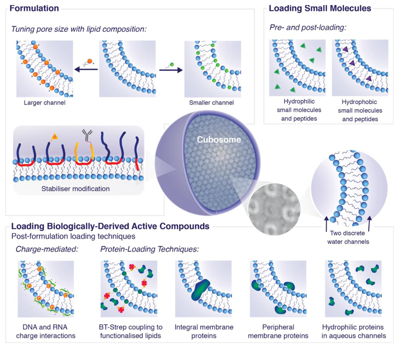
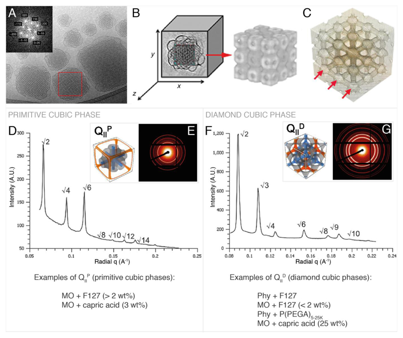
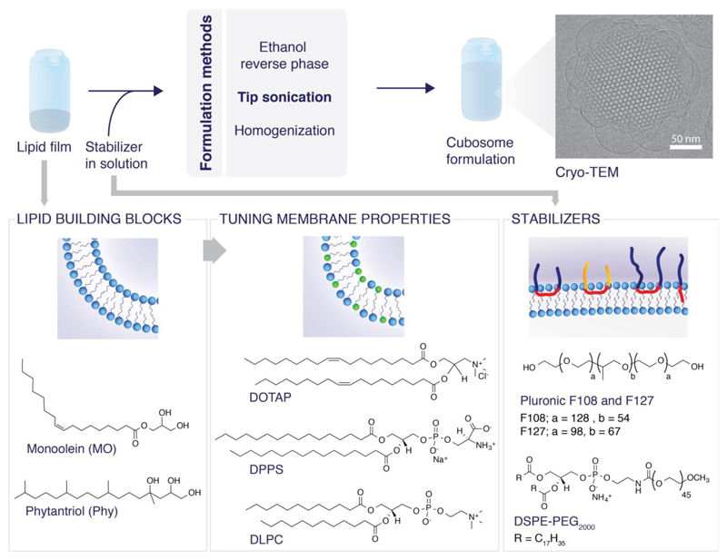
Cubosomes are highly stable nanoparticles formed from the lipid cubic phase and stabilized by a polymer based outer corona. Bicontinuous lipid cubic phases consist of a single lipid bilayer that forms a continuous periodic membrane lattice structure with pores formed by two interwoven water channels. Cubosome composition can be tuned to engineer pore sizes or include bioactive lipids, the polymer outer corona can be used for targeting and they are highly stable under physiological conditions. Compared to liposomes, the structure provides a significantly higher membrane surface area for loading of membrane proteins and small drug molecules. Owing to recent advances, they can be engineered in vitro in both bulk and nanoparticle formats with applications including drug delivery, membrane bioreactors, artificial cells, and biosensors. This review outlines recent advances in cubosome technology enabling their application and provides guidelines for the rational design of new systems for biomedical applications.

Keywords: cubosomes; drug delivery; lipids; nanoparticles; self-assembly.


Figures

Figure 1
Figure 1
Intelligent design of cubosomes for biomedical applications. Cryo-TEM tomography image reproduced with permission from D. Demurtas et al.[33]
Figure 2
Figure 2
Primitive and double diamond cubic phases commonly found in cubosomes. A, example Cryo TEM and Fourier transform of cubosomes. B & C, reconstructed 3D image of cubosome from cryo-TEM tomography data showing the lipid arrangement (B) and the water channels (C). D, primitive phase reduced SAXS scattering pattern.[58] E, 2D SAXS scattering pattern[58] corresponding to D and illustration of the primitive cubic phase[45] showing the lipid membrane (grey) and the water channels (blue, orange). F, double diamond cubic phase reduced SAXS scattering pattern.[58] G, 2D SAXS scattering pattern[58] corresponding to F and illustration of the double diamond bicontinuous cubic phase[45] showing the lipid membrane (grey) and the water channels (blue, orange). A, B and C are reproduced with permission from P. Demurtas et al.[33] The reduced SAXS scattering patterns in D & F and the 2D SAXS scattering patterns in E & G are adapted from Ref. [58] with permission from The Royal Society of Chemistry. Illustrations of the primitive and diamond cubic phases in E and G are adapted with permission from H. Kim, C. Leal, ACS Nano 2015, 9, 10214–10226. Copyright (2017) American Chemical Society.
Figure 3
Figure 3
Formulations of cubosomes and molecular structures of the common constituent lipids and stabilizers.
Figure 4
Figure 4
In vitro and in vivo examples of cubosome formulations in delivery and imaging. A: Photomicrographs of histopathological sections representing burned skin of rat groups following treatment using cubogels for 21 days. Reproduced with permission from Morsi et al.[137] B: i) Viability of HeLa cells after incubation with uncoated (RF) and coated (RFPεL) cubosomes (assayed by MTT). The error bar is standard error from three independent experiments done in triplicate. ii) Cellular uptake of Nile Red (NR) loaded cubosomes indicated by the shift in NR fluorescence intensity for the cell count due to the uptake of RFNR (red) and RFPεLNR (blue) compared to the control (gray). iii) Cellular uptake of NR loaded cubosomes observed by fluorescence microscopy. DAPI was used for counterstaining nucleus. Imaging was done at 63× magnification: (blue) DAPI, (red) cubosomes. Scale bar = 20 μm. Reproduced with permission from Deshpande et al.[35] C: In vivo MRI images of male C57Bl/6 mouse spleen and liver pre-injection (i and iii) and 30 minutes post-injection (ii and iv) of NIRF-MRI cubosomes. Enhanced MRI signals were observed from regions marked by dotted lines for the spleen and the liver. Reproduced with permission from Tran et al.[148] D: i) Viability of HeLa cells after incubating with Naproxen-loaded uncoated (RFNap) and coated (RFPεLNap) cubosomes for 24 hours. The error bar is standard error of three independent experiments performed in triplicate. Statistical significance is indicated by *** (p < 0.001). ii) Images of HeLa cells after incubation with Nile Red (NR) and Naproxen (Nap) loaded cubosomes. The live cells were stained with calcein AM. Red represents fluorescence due to NR. Imaging was done at 20× magnification: (green) calcein AM, (red) cubosomes. Scale bar = 100 μm. Reproduced with permission from Deshpande et al.[35]

References

    1. van Meer G, Voelker DR, Feigenson GW. Nat Rev Mol Cell Biol. 2008;9:112–24. - PMC - PubMed
    1. Stepanyants N, Macdonald PJ, Francy CA, Mears JA, Qi X, Ramachandrana R. Mol Biol Cell. 2015;26:3104–3116. - PMC - PubMed
    1. Simunovic M, Evergren E, Golushko I, Prévost C, Renard H-F, Johannes L, McMahon HT, Lorman V, Voth GA, Bassereau P. Proc Natl Acad Sci. 2016;113:11226–11231. - PMC - PubMed
    1. Schmidt A, Wolde M, Thiele C, Fest W, Kratzin H, Podtelejnikov AV, Witke W, Huttner WB, Söling H-D. Nature. 1999;401:133–141. - PubMed
    1. Deng Y, Marko M, Buttle KF, Leith A, Mieczkowski M, Mannella CA. J Struct Biol. 1999;127:231–239. - PubMed

Publication types