

Protective effects of gonadal hormones on spinal motoneurons following spinal cord injury
- PMID: 29926818
- PMCID: PMC6022470
- DOI: 10.4103/1673-5374.233434
Protective effects of gonadal hormones on spinal motoneurons following spinal cord injury
Abstract
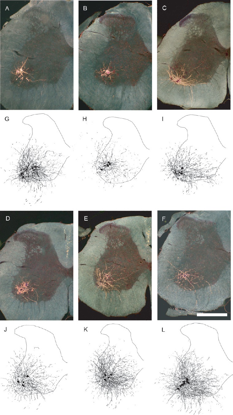
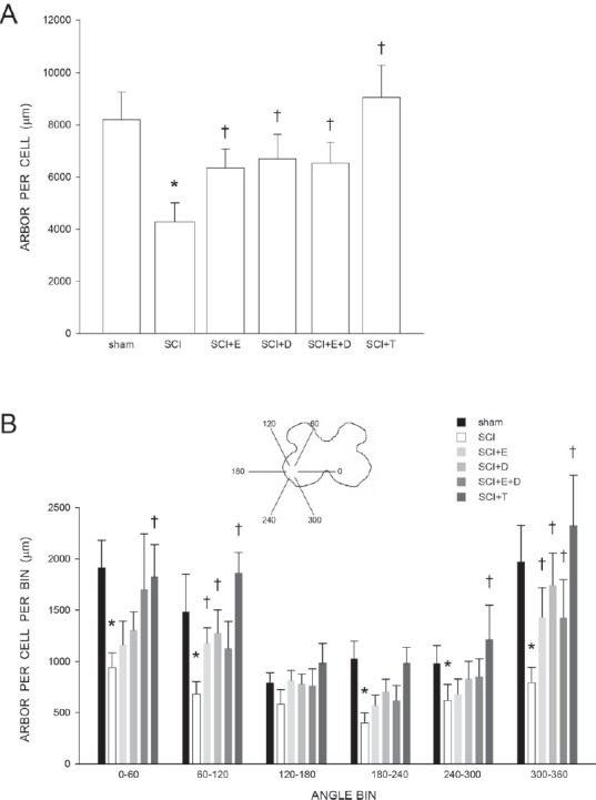
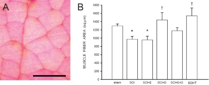
Spinal cord injury (SCI) results in lesions that destroy tissue and disrupt spinal tracts, producing deficits in locomotor and autonomic function. The majority of treatment strategies after SCI have concentrated on the damaged spinal cord, for example working to reduce lesion size or spread, or encouraging regrowth of severed descending axonal projections through the lesion, hoping to re-establish synaptic connectivity with caudal targets. In our work, we have focused on a novel target for treatment after SCI, surviving spinal motoneurons and their target musculature, with the hope of developing effective treatments to preserve or restore lost function following SCI. We previously demonstrated that motoneurons, and the muscles they innervate, show pronounced atrophy after SCI. Importantly, SCI-induced atrophy of motoneuron dendrites can be attenuated by treatment with gonadal hormones, testosterone and its active metabolites, estradiol and dihydrotestosterone. Similarly, SCI-induced reductions in muscle fiber cross-sectional areas can be prevented by treatment with androgens. Together, these findings suggest that regressive changes in motoneuron and muscle morphology seen after SCI can be ameliorated by treatment with gonadal hormones, further supporting a role for steroid hormones as neurotherapeutic agents in the injured nervous system.
Keywords: atrophy; dendrites; dihydrotestosterone; estradiol; morphology; muscle fibers; neuroprotection; retrograde labeling; steroids; testosterone.
Conflict of interest statement
None declared
Figures
Similar articles
-
Protective Effects of Estradiol and Dihydrotestosterone following Spinal Cord Injury.J Neurotrauma. 2018 Mar 15;35(6):825-841. doi: 10.1089/neu.2017.5329. Epub 2018 Jan 11. J Neurotrauma. 2018. PMID: 29132243 Free PMC article.
-
Differential effects of exercise and hormone treatment on spinal cord injury-induced changes in micturition and morphology of external urethral sphincter motoneurons.Restor Neurol Neurosci. 2024;42(2):151-165. doi: 10.3233/RNN-241385. Restor Neurol Neurosci. 2024. PMID: 39213108 Free PMC article.
-
Inhibition of Cytosolic Phospholipase A2 Has Neuroprotective Effects on Motoneuron and Muscle Atrophy after Spinal Cord Injury.J Neurotrauma. 2021 May 1;38(9):1327-1337. doi: 10.1089/neu.2014.3690. J Neurotrauma. 2021. PMID: 25386720 Free PMC article.
-
The role of embryonic motoneuron transplants to restore the lost motor function of the injured spinal cord.Ann Anat. 2011 Jul;193(4):362-70. doi: 10.1016/j.aanat.2011.04.001. Epub 2011 Apr 30. Ann Anat. 2011. PMID: 21600746 Review.
-
Intervention strategies to enhance anatomical plasticity and recovery of function after spinal cord injury.Adv Neurol. 1997;72:257-75. Adv Neurol. 1997. PMID: 8993704 Review.
Cited by
-
Exercise promotes recovery after motoneuron injury via hormonal mechanisms.Neural Regen Res. 2020 Aug;15(8):1373-1376. doi: 10.4103/1673-5374.274323. Neural Regen Res. 2020. PMID: 31997795 Free PMC article. Review.
-
Evaluating Sex Steroid Hormone Neuroprotection in Spinal Cord Injury in Animal Models: Is It Promising in the Clinic?Biomedicines. 2024 Jul 4;12(7):1478. doi: 10.3390/biomedicines12071478. Biomedicines. 2024. PMID: 39062051 Free PMC article. Review.
-
Diminished enteric neuromuscular transmission in the distal colon following experimental spinal cord injury.Exp Neurol. 2020 Sep;331:113377. doi: 10.1016/j.expneurol.2020.113377. Epub 2020 Jun 8. Exp Neurol. 2020. PMID: 32526238 Free PMC article.
-
Neuroprotective Effects of Testosterone in Male Wobbler Mouse, a Model of Amyotrophic Lateral Sclerosis.Mol Neurobiol. 2021 May;58(5):2088-2106. doi: 10.1007/s12035-020-02209-5. Epub 2021 Jan 7. Mol Neurobiol. 2021. PMID: 33411236
-
Sex Difference in Spinal Muscular Atrophy Patients - are Males More Vulnerable?J Neuromuscul Dis. 2023;10(5):847-867. doi: 10.3233/JND-230011. J Neuromuscul Dis. 2023. PMID: 37393514 Free PMC article.
References
-
- Ahlbom E, Prins G, Ceccatelli S. Testosterone protects cerebellar granule cells from oxidative stress-induced cell death through a receptor mediated mechanism. Brain Res. 2001;892:255–262. - PubMed
-
- Bernstein JJ, Standler N. Dendritic alteration of rat spinal motoneurons after dorsal horn mince: computer reconstruction of dendritic fields. Exp Neurol. 1983;82:532–540. - PubMed
-
- Bernstein JJ, Wacker W, Standler N. Spinal motoneuron dendritic alteration after spinal cord hemisection in the rat. Exp Neurol. 1984;83:548–554. - PubMed
-
- Brown TJ, Storer P, Oblinger M, Jones KJ. Androgenic enhancement of ßII-tubulin mRNA in spinal motoneurons following sciatic nerve injury. Rest Neurol Neurosci. 2001;18:191–198. - PubMed
Publication types
Grants and funding
LinkOut - more resources
Full Text Sources
Other Literature Sources
