. 2018 Aug;50(1):e56.
doi: 10.1002/cpmc.56. Epub 2018 May 18.

Salmonella Typhimurium Infection of Human Monocyte-Derived Macrophages

Affiliations

Salmonella Typhimurium Infection of Human Monocyte-Derived Macrophages

Stephanie K Lathrop et al. Curr Protoc Microbiol. 2018 Aug.

Abstract

The successful infection of macrophages by non-typhoidal serovars of Salmonella enterica is likely essential to the establishment of the systemic disease they sometimes cause in susceptible human populations. However, the interactions between Salmonella and human macrophages are not widely studied, with mouse macrophages being a much more common model system. Fundamental differences between mouse and human macrophages make this less than ideal. Additionally, the inability of human macrophage-like cell lines to replicate some properties of primary macrophages makes the use of primary cells desirable. Here we present protocols to study the infection of human monocyte-derived macrophages with Salmonella Typhimurium. These include a method for differentiating monocyte-derived macrophages in vitro and protocols for infecting them with Salmonella Typhimurium, as well as assays to measure the extent of infection, replication, and death. These protocols are useful for the investigation of both bacterial and host factors that determine the outcome of infection. © 2018 by John Wiley & Sons, Inc.

Keywords: bacteria; infection; intracellular pathogen; macrophages; monocyte-derived macrophage; salmonella.


Figures

Figure 1
Figure 1. Schematic overview of the protocols
Numbers refer to the protocol describing each procedure.
Figure 2
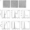
Figure 2. Phenotype of mature macrophages at day 7
A) Phase-contrast photos of macrophages after 7 days of differentiation as described in the protocol. Cells from 3 different donors show somewhat variable morphology. Bar = 100 μm B) Representative flow cytometry data of day 7 macrophages showing the expression of macrophage cell surface markers. Cells were removed from 6-well plates and stained as previously described. Cells were gated on CD33 and CD11b expression. The background autofluorescence level of unstained cells is shown in gray.
Figure 3
Figure 3. SPI1-expression of bacteria cultured in LB-Miller with aeration as described in Basic Protocol 3
Bacteria containing a plasmid-encoded fluorescent reporter for a SPI1 gene promoter, PprgH-gfp[LVA], were subcultured as described in the protocol, and samples removed at the indicated times. Bacteria were fixed for 15 min at R.T. with 2.5% PFA, and stained with the nucleic acid stain Syto 41 (ThermoFisher Scientific). The GFP fluorescence intensity of Syto 41+ events was detected by flow cytometry, and the percentage of bacteria expressing GFP was determined using FlowJo analysis software. Shown is the average percentage of GFP+ve bacteria (left axis, green circles) and the corresponding O.D. at 600 nm (right axis, black triangles) at each time point in two experiments, +/− the range.
Figure 4
Figure 4. The total percentage of cells infected directly correlates with the number of bacteria per cell
Data from 10 independent experiments/donors show that as the percentage of infected cells increases, so does the proportion of cells with 5 or more bacteria. The linear regression line is shown, r2 = 0.96.
Figure 5
Figure 5. Assessment of intracellular bacteria as recoverable colony-forming units (CFU) by gentamicin protection assay
(A) Macrophages grown as described in Basic Protocol 2 were infected with late log phase strain SL1344, and the recoverable CFU at 2, 8 and 18 h p.i. were determined as described in the protocol. Each symbol represents data from one independent experiment. Different donors were used in each experiment. Left, recoverable CFU per well. Right, the CFU values normalized to 2 h p.i., to facilitate comparisons between experiments. (B) Shown is the fold change in recoverable CFU between 2 and 18 h p.i. for cells infected with late log phase SL1344 in 15 independent experiments, using different donors. Note the wide range of replication between different experiments and donors. (C) Cells were infected with WT SL1344 or an isogenic strain lacking phoP. Recoverable CFU were determined at 2, 4, 8 and 18 h p.i., Each of 3 independent experiments using different donors is represented by a unique set of lines and symbols (circles, WT; squares, ΔphoP). Data are normalized to the 2 h value.
Figure 6
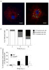
Figure 6. Immunofluorescence microscopy analysis showing increased number of intracellular bacteria per cell over time
Cells were infected with SPI1-induced SL1344 bacteria according to the protocol, fixed at 2, 8 and 18 h p.i., and stained for IF microscopy. (A) Representative images showing infected cells at 2 h and 18 h p.i. Red = LAMP1, Green = Salmonella (LPS), Blue = DAPI (DNA). Bar = 10 μm. B) The proportion of infected cells having 1 – 4, 5 – 9, and ≥10 bacteria is shown at the indicated time p.i. for a representative experiment. (C) Cells were infected with either WT SL1344 or an isogenic strain lacking phoP, and the percentage of infected cells containing ≥10 bacteria at 2, 8 and 18 h p.i. is shown. Data are the average of 3 independent experiments using cells from different donors. Copyright © American Society for Microbiology, Infection & Immunity, 2015, 83:2661–2671, DOI: 10.1128/IAI.00033-15
Figure 7
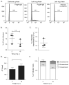
Figure 7. Infection of macrophages with stationary phase bacteria results in less replication than seen by late log phase bacteria
A) Bacteria containing the SPI1 reporter PprgH-gfp[LVA] were grown either to stationary phase (left) or late log phase (middle), as described in the protocols. The percentage of GFP+ve bacteria then detected by flow cytometry is shown. For determination of background fluorescence, WT bacteria lacking the PprgH-gfp[LVA] reporter are shown in the panel on the right. (B–E) Macrophages were infected with stationary phase bacteria (Alternative Protocol 1), followed by assessment of intracellular bacteria by gentamicin protection assay (Basic Protocol 4) and immunofluorescence microscopy (Basic Protocol 5) at 2 and 18 h p.i. Three independent experiments were done with different donors. All data are the mean +/− SD. (B) The percentage of total cells containing bacteria at 2 and 18 h p.i. was assessed by IF microscopy. (C) The efficiency of bacterial internalization was calculated by dividing the number of intracellular bacteria at 2 h p.i. by the number of live bacteria that was added to the cells during the infection. (D) The recoverable CFU at 2 h and 18 h p.i. E) The number of bacteria per infected cell as assessed by IF microscopy is represented by binning of the infected cells into 3 categories according to the number of bacteria they contain.

References

    1. Tsolis RM, Xavier MN, Santos RL, Baumler AJ. How to become a top model: impact of animal experimentation on human Salmonella disease research. Infect Immun. 2011;79:1806–1814. doi: 10.1128/IAI.01369-10. - DOI - PMC - PubMed
    1. Fields PI, Swanson RV, Haidaris CG, Heffron F. Mutants of Salmonella typhimurium that cannot survive within the macrophage are avirulent. Proc Natl Acad Sci U S A. 1986;83:5189–5193. - PMC - PubMed
    1. Gordon MA. Salmonella infections in immunocompromised adults. The Journal of infection. 2008;56:413–422. doi: 10.1016/j.jinf.2008.03.012. - DOI - PubMed
    1. Feasey NA, Dougan G, Kingsley RA, Heyderman RS, Gordon MA. Invasive non-typhoidal salmonella disease: an emerging and neglected tropical disease in Africa. Lancet. 2012;379:2489–2499. doi: 10.1016/S0140-6736(11)61752-2. - DOI - PMC - PubMed
    1. Schwan WR, Huang XZ, Hu L, Kopecko DJ. Differential bacterial survival, replication, and apoptosis-inducing ability of Salmonella serovars within human and murine macrophages. Infect Immun. 2000;68:1005–1013. - PMC - PubMed

Publication types

LinkOut - more resources