. 2018 Oct 1;315(4):G514-G528.
doi: 10.1152/ajpgi.00357.2017. Epub 2018 Jun 21.

Rho kinase inhibition maintains intestinal and vascular barrier function by upregulation of occludin in experimental necrotizing enterocolitis

Affiliations

Rho kinase inhibition maintains intestinal and vascular barrier function by upregulation of occludin in experimental necrotizing enterocolitis

Justyna S Grothaus et al. Am J Physiol Gastrointest Liver Physiol. .

Abstract

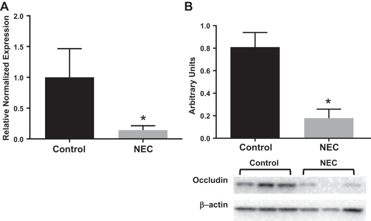
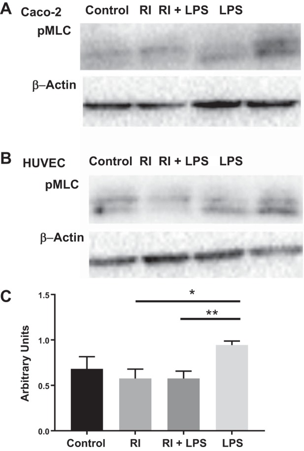
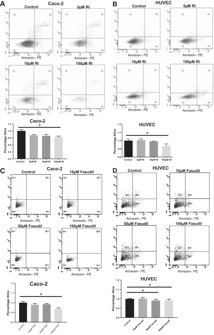
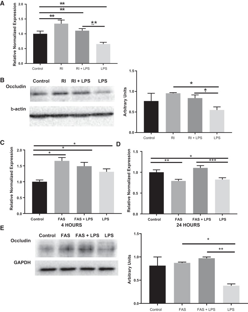
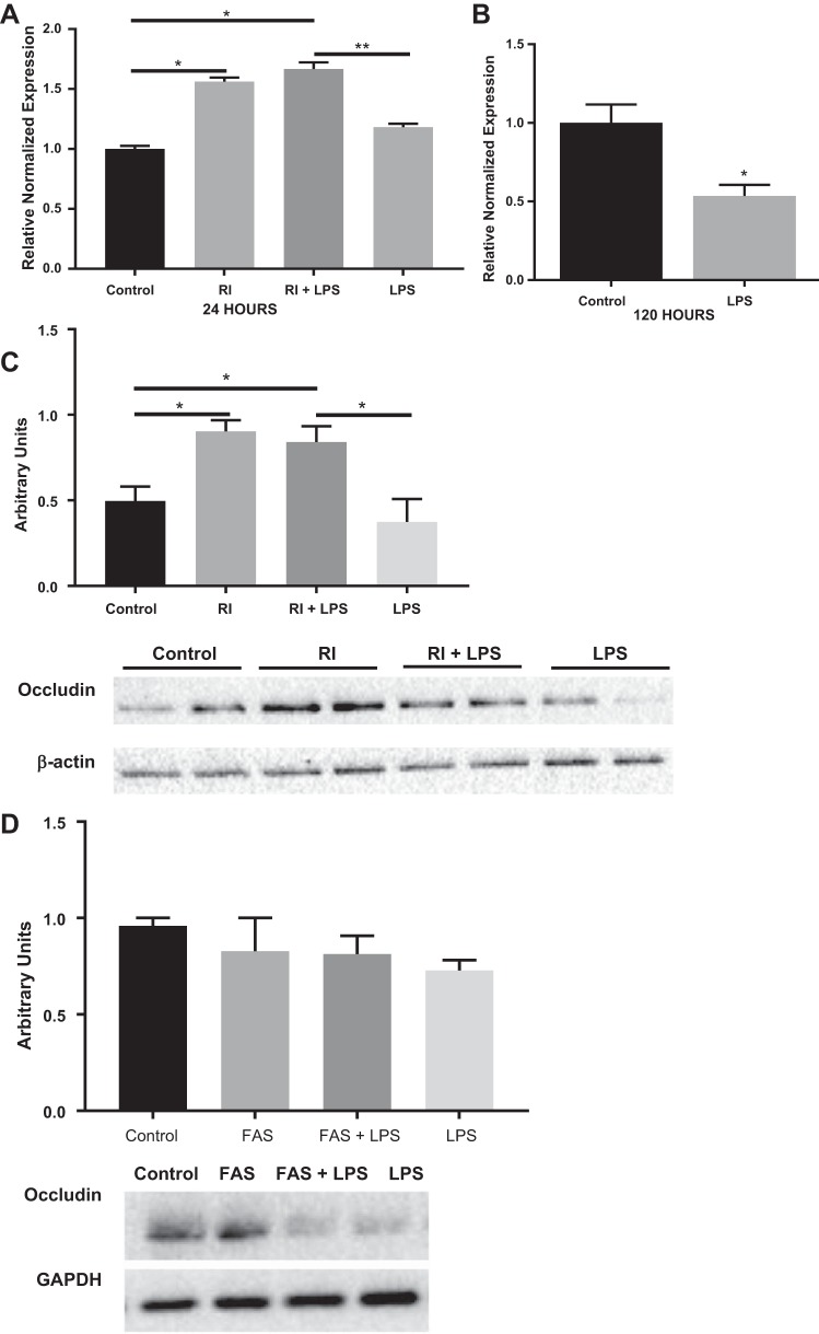
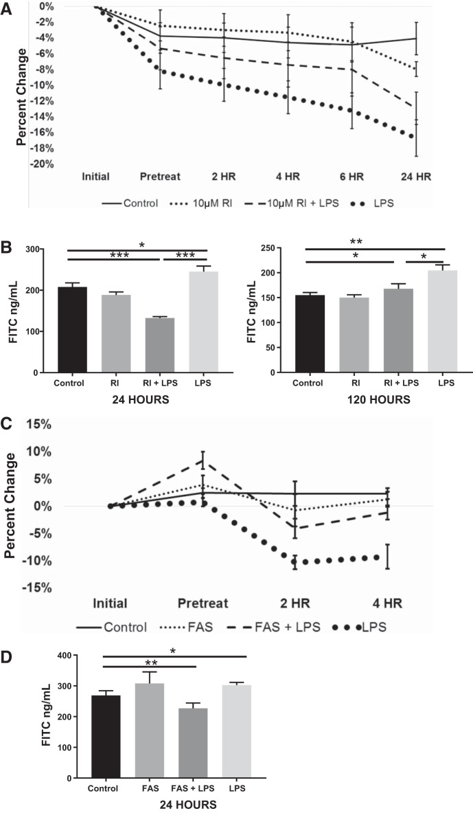
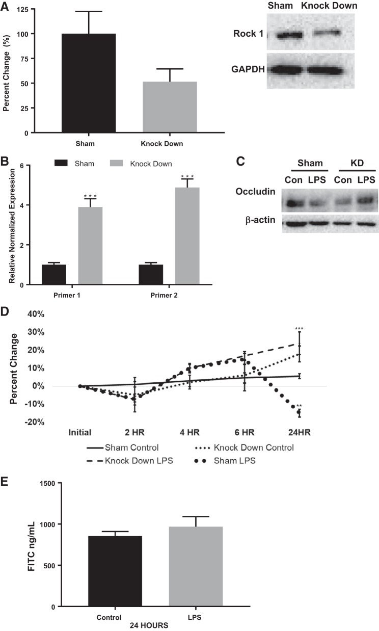
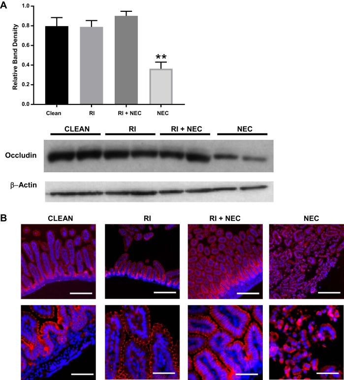
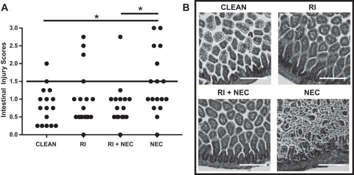
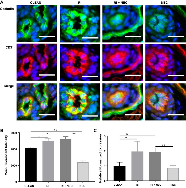
Necrotizing enterocolitis (NEC) is a deadly disease that occurs in 5-10% of neonates. Although NEC has been extensively studied, no single therapeutic target has been identified. Rho kinase (ROCK) is a serine/threonine kinase that affects multiple cellular processes, including tight junction (TJ) function, cellular permeability, and apoptosis. We hypothesized that ROCK inhibition would decrease cellular permeability, stabilize TJ proteins (occludin), and decrease the severity of NEC. To test this hypothesis, human colon epithelial cells (Caco-2) and human endothelial cells were studied. Cells were treated with lipopolysaccharide to simulate an in vitro model of NEC. The effect of ROCK inhibition was measured by transepithelial membrane resistance (TEER) and cellular permeability to FITC-dextran. The effects of ROCK inhibition in vivo were analyzed in the rat pup model of NEC. NEC was induced by feeding formula supplemented with Cronobacter sakazakii with or without gavaged ROCK inhibitor. Rat intestines were scored based on histological degree of injury. RNA and protein assays for occludin protein were performed for all models of NEC. Treatment with ROCK inhibitor significantly decreased cellular permeability in Caco-2 cells and increased TEER. Intestinal injury scoring revealed decreased scores in ROCK inhibitor-treated pups compared with NEC only. Both cell and rat pup models demonstrated an upregulation of occludin expression in the ROCK inhibitor-treated groups. Therefore, we conclude that ROCK inhibition protects against experimental NEC by strengthening barrier function via upregulation of occludin. These data suggest that ROCK may be a potential therapeutic target for patients with NEC. NEW & NOTEWORTHY These studies are the first to demonstrate an upregulation of occludin tight junction protein in response to Rho kinase (ROCK) inhibition. Furthermore, we have demonstrated that ROCK inhibition in experimental models of necrotizing enterocolitis (NEC) is protective against NEC in both in vitro and in vivo models of disease.

Keywords: Rho kinase inhibitors; necrotizing enterocolitis; occludin; tight junctions.


Figures

Fig. 1.
Fig. 1.
Occludin gene and protein expression is decreased in bowel samples of patients with NEC. A: occludin gene expression in human samples was assessed via quantitative PCR. Patients with NEC had significantly lower occludin expression compared with control samples (*P < 0.05 vs. control), n = 3 per group. B: occludin protein production in human samples was analyzed by Western blot. Samples obtained from patients with NEC had significantly lower occludin protein compared with control patients (*P < 0.05), n = 3 per group. Western blot shows increased band density of occludin in control group. NEC, necrotizing enterocolitis.
Fig. 2.
Fig. 2.
LPS increases production of phosphorylated myosin light chain (pMLC) in both HUVECs and Caco-2 cells (A and B). ROCK inhibition in the presence of LPS decreases pMLC production in both HUVECs and Caco-2 cells to values similar to that of the controls (C; **P < 0.005). Western blot of pMLC in both HUVEC and Caco-2 cells are shown with the corresponding β-actin blot. *P < 0.05. LPS, lipopolysaccharide; RI, ROCK inhibitor; ROCK, Rho kinase.
Fig. 3.
Fig. 3.
Increased concentrations of ROCK inhibitor are associated with more cellular death. A: flow cytometry performed on Caco-2 cells with different concentrations of ROCK inhibitor (RI). Q3 shows percentage of cells alive; Q2 shows percentage of cells in late stage apoptosis. Controls were untreated. Decreased percentage of cells alive with increasing concentration of RI, with 100 μM RI being significantly different from control (*P < 0.05 vs. control). B: a similar trend is seen in HUVECs (*P < 0.05 vs. control). C: flow cytometry on Caco-2 cells treated with fasudil hydrochloride (FAS) demonstrate increased apoptosis after treatment with 100 μM FAS vs. controls (*P < 0.05). D: HUVECs showed increased apoptosis after treatment with 50 μM FAS (*P < 0.05). Values are means ± SE; ROCK, Rho kinase.
Fig. 4.
Fig. 4.
ROCK inhibition increases occludin gene and protein expression in HUVECs. A: occludin gene expression in HUVECs was assessed by quantitative PCR. LPS had significantly less occludin expression compared with controls (**P < 0.005). RI was significantly higher than controls (**P < 0.005), and RI + LPS was the same as control. RI + LPS was significantly higher than LPS (**P < 0.005). B: occludin protein expression in HUVECs was assessed via Western blot. A representative Western blot shows that control, RI, and RI + LPS groups had more occludin than LPS-treated cells. C and D: occludin gene expression in HUVECs is increased early after 4 h of FAS administration compared with controls (*P < 0.05); LPS-treated group had decreased occludin gene expression by 24 h, whereas expression in FAS + LPS treated cells remains increased vs. LPS (*P < 0.0005). E: occludin protein expression using FAS instead of RI shows more occludin in FAS (*P < 0.05) and FAS + LPS (**P < 0.005) groups compared with LPS-treated cells. *P < 0.05, **P < 0.005, ***P < 0.0005. FAS, fasudil hydrochloride; LPS, lipopolysaccharide; RI, ROCK inhibitor; ROCK, Rho kinase.
Fig. 5.
Fig. 5.
ROCK inhibition increases gene and protein expression of occludin in Caco-2 cells. A: occludin gene expression in Caco-2 is increased in both RI groups compared with control (*P < 0.05). With the addition of RI to LPS-treated cells, occludin gene expression increases when compared with LPS alone (**P < 0.005). B: LPS-treated Caco-2 cells had significantly decreased occludin gene expression compared with controls at 120 h (*P < 0.05). C: representative Western blot shows that both RI groups had increased occludin compared with LPS-treated cells. D: Western blot shows a trend toward decreased occludin in LPS-treated cells compared with controls, whereas the addition of FAS increases occludin protein expression (P = 0.08). FAS, fasudil hydrochloride; LPS, lipopolysaccharide; RI, ROCK inhibitor; ROCK, Rho kinase.
Fig. 6.
Fig. 6.
Permeability and TEER in Caco-2 cells. A: TEER in Caco-2 cells over 24 h in the 4 experimental groups: control, RI, RI+ LPS, and LPS. LPS significantly decreases TEER at 24 h compared with control (**P < 0.005). Addition of RI to LPS-treated cells increases TEER significantly compared with LPS-treated cells alone (*P < 0.05). B: Caco-2 cells treated with LPS have increased permeability compared with controls at 24 and 120 h (*P < 0.05). Addition of RI to LPS-treated cells decreases cellular permeability compared with LPS only treated cells both at 24 and 120 h (***P < 0.0005 and *P < 0.05, respectively). C: Caco-2 cells treated with LPS had increased TEER compared with controls at 4 h. FAS and FAS + LPS groups had no differences in TEER compared with controls (***P < 0.0005). D: similarly, there was increased permeability in LPS-treated cells that was significantly diminished in FAS-treated cells (**P < 0.005). FAS, fasudil hydrochloride; LPS, lipopolysaccharide; RI, ROCK inhibitor; ROCK, Rho kinase; TEER, transendothelial and transepithelial electrical resistance.
Fig. 7.
Fig. 7.
Occludin is upregulated with genetic inhibition of ROCK in Caco-2 cells. A: ROCK1 protein expression in Caco-2 cells assayed with Western blot. Immunoblot density for sham shRNA cells and shRNA ROCK1 knockdown cells showed a 49% ROCK1 decrease in protein expression in knockdown cells compared with controls; representative immunoblots are shown for each group. B: occludin gene expression is increased fourfold following the knockdown of ROCK1 in Caco-2 cells compared with controls (n = 3; ***P < 0.0005). C: immunoblot density for occludin showed increased occludin expression in ROCK1 knockdown cells treated with LPS compared with untreated knockdown cells and sham controls. D: ROCK1 knockdown cells had increased TEER compared with sham controls (**P < 0.005). LPS-treated cells had significantly lower TEER compared with sham controls and with ROCK1 knockdown cells (***P < 0.0005). E: permeability to FITC-dextran was not different between ROCK1 knockdown Caco-2 cells treated with LPS vs. control. LPS, lipopolysaccharide; ROCK, Rho kinase; TEER, transendothelial and transepithelial electrical resistance.
Fig. 8.
Fig. 8.
Occludin is upregulated with RI and localizes to the cellular membrane in the rat pup intestine. A: occludin protein expression in rat pup intestine assessed via Western blot. Rat pups inflicted with NEC had lower amounts of occludin protein in the intestine compared with RI + NEC, RI, and clean groups; n = 4 per group. B: immunofluorescence imaging of occludin (red) in the rat pup intestine. Preserved occludin staining on cellular membrane seen in clean, RI, and RI + NEC groups in both high- and low-power views. In the NEC group, there is breakdown of the villus architecture, and occludin is no longer part of the cellular membrane. **P < 0.005. Scale bar = 200 μm for ×10 images. NEC, necrotizing enterocolitis; RI, ROCK inhibitor, ROCK, Rho kinase.
Fig. 9.
Fig. 9.
RI decreases intestinal injury in a rat pup model of NEC. A: intestinal injury scoring of rat pup intestine from hematoxylin-eosin slides. Clean pups had significantly lower intestinal injury scores compared with the NEC group (*P < 0.05). Addition of RI to the formula decreased average intestinal injury scores in the RI + NEC group vs. NEC only (*P < 0.05); n = 17–19 pups per group. B: hematoxylin-eosin slides of rat pup intestine. Loss of villus architecture and epithelial sloughing seen in the NEC group. Preservation of epithelial barrier seen in clean, RI, and RI + NEC group. Scale bar = 200 μm. NEC, necrotizing enterocolitis; RI, ROCK inhibitor, ROCK, Rho kinase.
Fig. 10.
Fig. 10.
Increased occludin gene expression and protein is found in the mesentery of intestines in a rat pup model of NEC. A: immunofluorescence imaging of mesentery in four treatment groups: clean, RI, RI + NEC and NEC groups. CD31 staining (red) was used as a marker for endothelial cells of arteries and veins of mesentery. Occludin staining (green) was brighter in RI groups indicating increased occludin, n = 3–6 per group. Scale bar = 20 μm. B: gene expression of occludin in rat pup mesentery. Clean vs. NEC (NS), clean vs. RI (**P < 0.005), clean vs. RI + NEC (**P < 0.005), RI + NEC vs. NEC (**P < 0.005); n = 4 pups per group. *P < 0.05. NEC, necrotizing enterocolitis; RI, ROCK inhibitor, ROCK, Rho kinase.

Similar articles

Cited by

References

    1. Al-Sadi R, Khatib K, Guo S, Ye D, Youssef M, Ma T. Occludin regulates macromolecule flux across the intestinal epithelial tight junction barrier. Am J Physiol Gastrointest Liver Physiol 300: G1054–G1064, 2011. doi:10.1152/ajpgi.00055.2011. - DOI - PMC - PubMed
    1. Almajed FS, Forsythe SJ. Cronobacter sakazakii clinical isolates overcome host barriers and evade the immune response. Microb Pathog 90: 55–63, 2016. doi:10.1016/j.micpath.2015.11.014. - DOI - PubMed
    1. Amano M, Nakayama M, Kaibuchi K. Rho-kinase/ROCK: a key regulator of the cytoskeleton and cell polarity. Cytoskeleton 67: 545–554, 2010. doi:10.1002/cm.20472. - DOI - PMC - PubMed
    1. Antonetti DA, Barber AJ, Khin S, Lieth E, Tarbell JM, Gardner TW; Penn State Retina Research Group . Vascular permeability in experimental diabetes is associated with reduced endothelial occludin content: vascular endothelial growth factor decreases occludin in retinal endothelial cells. Diabetes 47: 1953–1959, 1998. doi:10.2337/diabetes.47.12.1953. - DOI - PubMed
    1. Balda MS, Flores-Maldonado C, Cereijido M, Matter K. Multiple domains of occludin are involved in the regulation of paracellular permeability. J Cell Biochem 78: 85–96, 2000. doi:10.1002/(SICI)1097-4644(20000701)78:1<85::AID-JCB8>3.0.CO;2-F. - DOI - PubMed

Publication types

MeSH terms

LinkOut - more resources