. 2018 Jul 10;18(14):2047-2054.
doi: 10.1039/c8lc00202a.

Real-time observation of leukocyte-endothelium interactions in tissue-engineered blood vessel

Affiliations

Real-time observation of leukocyte-endothelium interactions in tissue-engineered blood vessel

Z Chen et al. Lab Chip. .

Abstract

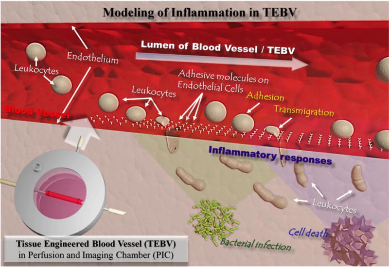
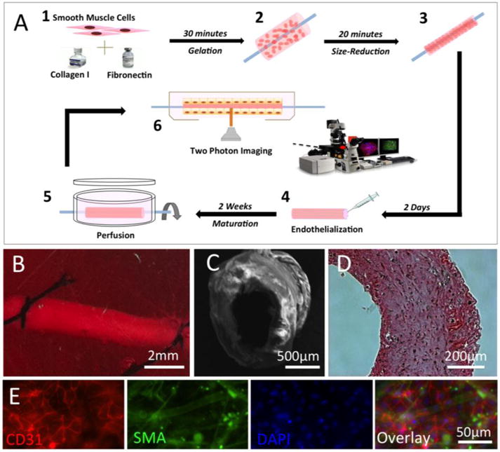
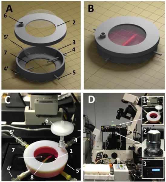
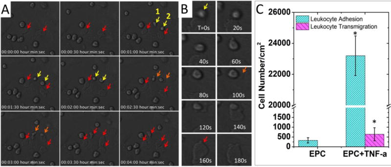
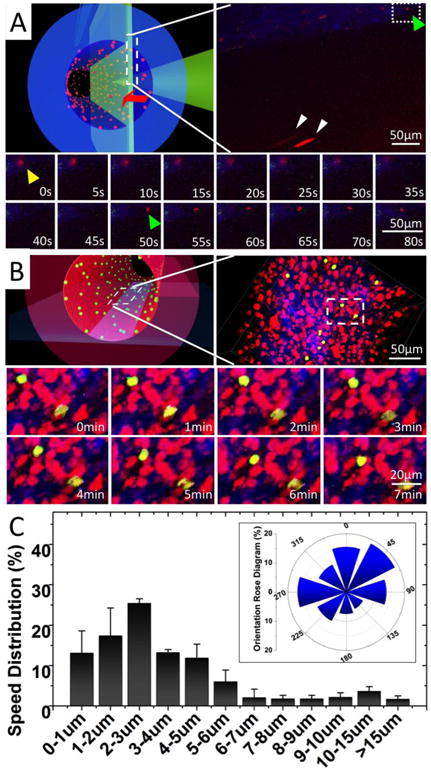
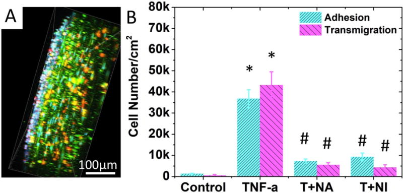
Human cell-based 3D tissue constructs play an increasing role in disease modeling and drug screening. Inflammation, atherosclerosis, and many autoimmune disorders involve the interactions between immune cells and blood vessels. However, it has been difficult to image and model these interactions under realistic conditions. In this study, we fabricated a perfusion and imaging chamber to allow the real-time visualization of leukocyte perfusion, adhesion, and migration inside a tissue-engineered blood vessel (TEBV). We monitored the elevated monocyte adhesion to the TEBV wall and transendothelial migration (TEM) as the TEBV endothelium was activated by the inflammatory cytokine TNF-α. We demonstrated that treatment with anti-TNF-α or an NF-kB signaling pathway inhibitor would attenuate the endothelium activation and reduce the number of leukocyte adhesion (>74%) and TEM events (>87%) close to the control. As the first demonstration of real-time imaging of dynamic cellular events within a TEBV, this work paves the way for drug screening and disease modeling in TEBV-associated microphysiological systems.


Conflict of interest statement

Conflicts of interest

There are no conflicts to declare

Figures

Figure 1
Figure 1
Schematic illustration of immune cell interactions with endothelium in blood vessels during inflammation caused by stimuli such as external bacterial infection or internal cell necrosis. Human cell-based tissue-engineered blood vessel (TEBV) is proposed as a model for disease modelling or drug screening in the vascular system.
Figure 2
Figure 2
Fabrication of human cell-based tissue-engineered blood vessel (TEBV). (A) Schematic illustration of the fabrication process of a TEBV. (B) A TEBV in perfusion inside of a bioreactor. (C) Image of TEBV with Scanning Electron Microscopy (SEM). (D) Image of TEBV with H&E staining. (E) Immunostaining of TEBV showing the blood vessel wall with an intact endothelium (Red: CD31 as a marker for endothelial cells) and vascular smooth muscle cells (Green: smooth muscle actin as a marker for smooth muscle cells). Scale bars are indicated in each panel.
Figure 3
Figure 3
The PIC chamber and the imaging system setup. (A) Components of a PIC chamber; numbers described in (C). (B) Illustration of a TEBV inside a PIC chamber. (C) The TEBV-PIC chamber in actual imaging condition. Major components are labelled: top observation window (1), cover (2), chamber (3), TEBV perfusion inlet/outlet (4/4′), chamber medium perfusion inlet/outlet (5′), connector for air filter (6), bottom observation window (7), TEBV (8). (D) The setup of PIC perfusion on the confocal microscope. Inserted figures are the key components for TEBV imaging: PIC chamber (1), NIKON 25× LWD (2.0mm) objective (2), two-photon laser (3).
Figure 4
Figure 4
Imaging of monocyte adhesion and transmigration on 2D-cultured endothelial cells. (A) Time-lapse imaging of monocyte-like HL-60 cells attached to TNF-α-activated endothelial progenitor cells (EPC). Arrows indicate monocytes attached to ECs. Yellow arrow: attached monocytes. Orange arrow: monocytes undergoing transendothelial migration. Red arrow: monocytes after transendothelial migration. Cell morphologies changed after transmigration, as they were squeezed and trapped between ECs and the substrate. (B) A montage of monocytes undergoing trans-endothelial migration. The transmigration event happened during 100s-160s. (C) Comparison of the monocytes interaction with EPCs with or without TNF-α treatment. Adhesion: control (330±133/cm2) vs. TNF-α (23200±1276/cm2); transmigration: control (not observed) vs. TNF-α (640±332/cm2). In each experiment, the cell number was counted and averaged from 3 microscopic views. Data are shown as mean ± SEM from four independent experiments. Significance was determined by student t-test; * p<0.05.
Figure 5
Figure 5
Real-time imaging of monocyte perfusion, adhesion and migration inside TEBV. (A) Monocyte perfusion and adhesion in TEBV. Upper left panel, a schematic illustration of the imaging focal-plane. Upper right panel, a full view of monocyte perfusion in TEBV. Yellow arrow: a monocyte adhered to endothelium during its transmigration. Green arrow: a new monocyte attachment event. White arrow: monocyte in perfusion. Lower panel, montage of the monocyte attachment and transmigration events (Supplemental Movie 2). (B) Monocyte migration on the TEBV endothelium. Upper left panel, a schematic illustration of the imaging focal-plane. Upper-right panel, a full view of monocyte migration on TEBV endothelium. Lower panel, montage of leukocyte migration on TEBV endothelium (Supplemental Movie 3). (C) Quantification of leukocyte migration speed distribution on TEBV endothelium and their migration directionality with Rose Diagram (inserted panel). The flow direction is set as degree zero. Results were calculated from 60 leukocytes in three experiments. Data are shown as mean ± SEM.
Figure 6
Figure 6
Effect of inflammatory cytokine and anti-inflammatory drugs on leukocyte-endothelium interactions. (A) A side view of the 3D-reconstructed two-photon confocal images in which monocytes attached to or transmigrated into activated TEBV endothelium in a TNF-α activated TEBV (perfused with medium containing TNF-α, 200U/mL, 4 hrs). B) Quantification of cell attachment and transmigration in TEBV with different treatments. TEBV was treated with i) Null, ii) 200U/mL TNF-α, iii) 200U/mL TNF-α + 20ng/mL TNF-α neutralizing antibody, and iv) 200U/mL TNF-α + 1μM Bay117082. N=4 for each group. Data are shown as mean ± SEM. Significance was determined by one-way ANOVA and Tukey’s post hoc test; * p<0.05 vs. number of cells adhered or transmigrated in control group, respectively; # p<0.05 vs. number of cells adhered or transmigrated in TNF-α treated group.

References

    1. Vestweber D, Wessel F, Nottebaum AF. Semin Immunopathol. 2014;36:177. - PubMed
    1. Tabas I, García-Cardeña G, Owens GK. J Cell Biol. 2015;209:13. - PMC - PubMed
    1. Steyers CM, Miller FJ. Int J Mol Sci. 2014;15:11324. - PMC - PubMed
    1. Cedervall J, Dimberg A, Olsson AK. Mediators Inflamm. 2015:418290. - PMC - PubMed
    1. Machida H, Tanaka I, Fukui R, Shen Y, Ishikawa T, Tate E, Ueno E. RadioGraphics. 2015;35:991. - PubMed

Publication types