Review
. 2018 Jul 25;118(14):6656-6705.
doi: 10.1021/acs.chemrev.8b00008. Epub 2018 Jun 21.

Chemical and Biochemical Perspectives of Protein Lysine Methylation

Affiliations
Review

Chemical and Biochemical Perspectives of Protein Lysine Methylation

Minkui Luo. Chem Rev. .

Abstract

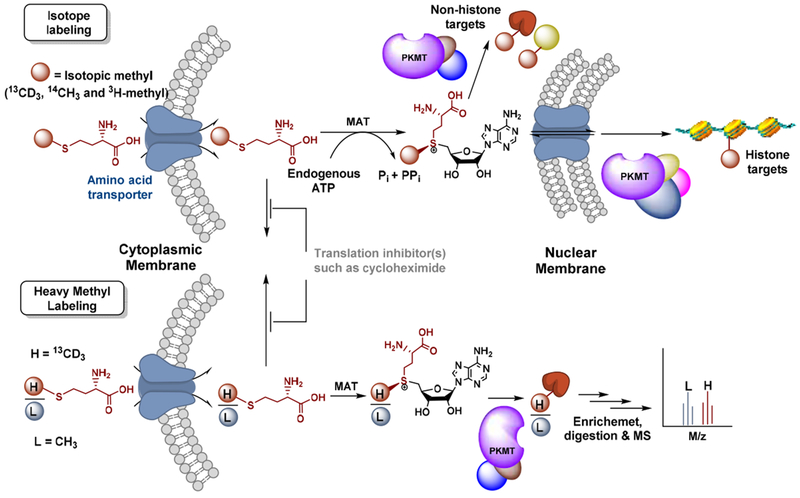
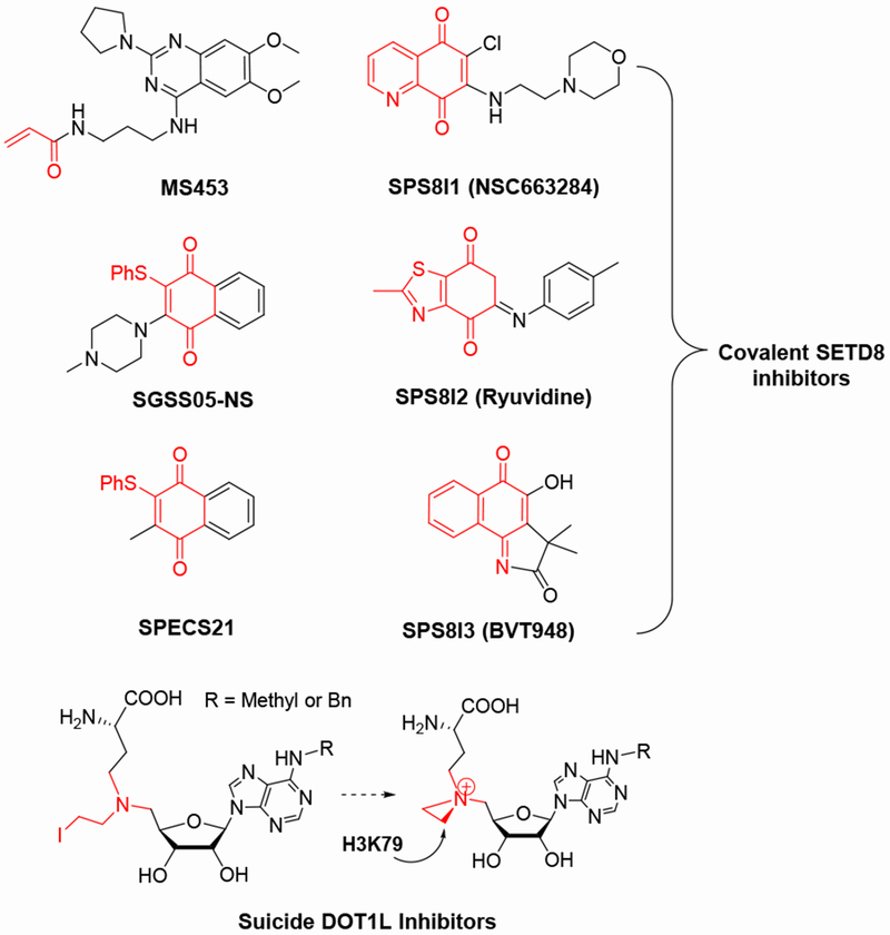
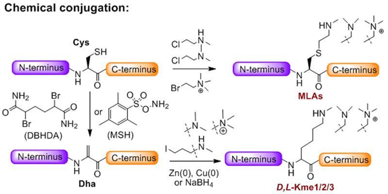
Protein lysine methylation is a distinct posttranslational modification that causes minimal changes in the size and electrostatic status of lysine residues. Lysine methylation plays essential roles in regulating fates and functions of target proteins in an epigenetic manner. As a result, substrates and degrees (free versus mono/di/tri) of protein lysine methylation are orchestrated within cells by balanced activities of protein lysine methyltransferases (PKMTs) and demethylases (KDMs). Their dysregulation is often associated with neurological disorders, developmental abnormalities, or cancer. Methyllysine-containing proteins can be recognized by downstream effector proteins, which contain methyllysine reader domains, to relay their biological functions. While numerous efforts have been made to annotate biological roles of protein lysine methylation, limited work has been done to uncover mechanisms associated with this modification at a molecular or atomic level. Given distinct biophysical and biochemical properties of methyllysine, this review will focus on chemical and biochemical aspects in addition, recognition, and removal of this posttranslational mark. Chemical and biophysical methods to profile PKMT substrates will be discussed along with classification of PKMT inhibitors for accurate perturbation of methyltransferase activities. Semisynthesis of methyllysine-containing proteins will also be covered given the critical need for these reagents to unambiguously define functional roles of protein lysine methylation.


Conflict of interest statement

The author declares no competing financial interest.

Figures

Figure 1.
Figure 1.
Biosynthesis and bio-consumption of SAM. In general, SAM’s biosynthesis is carried out by L-methionine adenosyltransferases (MATs or SAM synthetases) with L-methionine and ATP as substrates. Occasionally, bacterial enzymes such as SalL and FDAS can generate SAM with 5′-Cl/F-5′-deoxyadenosine and L-methionine as substrates. Bio-consumption of SAM can be classified as (i) homolytic, (ii) intramolecular heterolytic, (iii) intermolecular heterolytic cleavage of two CH2-sulfonium bonds, and (iv) intermolecular heterolytic cleavage of the methyl-sulfonium bond. The latter accounts for methylation with DNA, RNA, proteins and small-molecule metabolites as substrates.
Figure 2.
Figure 2.
Biophysical and biochemical properties of free lysine (Lys) and methyllysine (Kme1/2/3). The side chains of Lys and Kme1/2/3 are shown in a space-filling model and with electrostatic potential surface. Their pKa values and capability to form a salt bridge and hydrogen bonds at physiological pH of 7.4 are compared.
Figure 3.
Figure 3.
Structures and formal charges of posttranslationally modified Lys residues. Different from other posttranslational modifications, lysine methylation is characterized by unaltered charge (+1) at physiological pH and a minimal change of size relative to an unmodified lysine. Relative sizes of the posttranslational modifications except ubiquitin, SUMO and Nedd8 are compared in a space-filling model.
Figure 4.
Figure 4.
Structures and topology of PKMTs. The cartoon 3-D structures and 2–D topology of G9a (PDB 2O8J) and DOT1L (PDB 1NW3) are shown as representative examples of SET domain-containing PKMTs and Rossmann-fold-like (non-SET-domain) PKMTs, respectively. The SAM binding of the two PKMTs and the “pseudoknot” fold of G9a in red are highlighted.
Figure 5.
Figure 5.
Phylogenic trees of SET domain-containing PKMTs and Rossmann-fold-like PKMTs. Classification and relative positions of PKMTs were referred in a previous report.(28, 73)
Figure 6.
Figure 6.
Representative PKMT complexes. Here shown are key components of catalytically active MLL1-4 and EZH1/2 complexes.(90, 91) MLL1/2 contain cleavage sites for the threonine aspartase Taspase1. (92) The relative topology of individual subunits was presented on the basis of homogenous X-ray structures as reported.(90, 91, 93, 94)
Figure 7.
Figure 7.
Reaction path of PKMT-catalyzed lysine monomethylation and relevant biochemical methods to examine this process. PKMT first recognizes the SAM cofactor and its substrate. The ε-amine of lysine is then subjected to enzyme-mediated deprotonation. The overall rate-limitation step is expected to be the assembling of a SN2 transition state, followed by releasing methylated lysine and SAH as a product and a byproduct, respectively. Reproduced with permission from Ref. (98) Copyright 2016 PNAS.
Figure 8.
Figure 8.
SAM-binding modes of PKMTs. The interacting networks of SAM in SETD8 (PDB 3F9W) and DOT1L (PDB 1NW3) are shown as the representative examples of SET domain-containing PKMTs and Rossmann-fold-like (non-SET-domain) PKMTs, respectively. Both the stick mode generated with PyMOL and structural details of SAM binding are shown for clarity.
Figure 9.
Figure 9.
Noncanonical carbon-oxygen (CH•••O) interaction for SAM engagement. SET7/9 was used as an example to illustrate the noncanonical CH•••O interaction. The hypothetic transition state structure was generated upon aligning two PDB files 1XQH and 1N6C. Both the stick mode generated with PyMOL and a chemical structure model are shown for clarity.
Figure 10.
Figure 10.
Inverse BIEs of CD3-SAM associated with SAM binding. Inverse BIEs of CD3-SAM were reported for SETD8 and NSD2. The noncanonical CH…O interactions were highlighted with the stick mode generated with PyMOL (PDB 3F9W for SETD8 and 5LSU for NSD2) and with the chemical structures at proposed transition states. SAM in the NSD2-SAM complex (PDB 5LSU) was used to depict the interactions of SETD8 and NSD2 at the catalytic sites.
Figure 11.
Figure 11.
Noncanonical sulfur-oxygen (S•••O) interaction for SAM engagement. SET7/9 was used as an example to illustrate the noncanonical chalcogen-oxygen interaction. The hypothetic transition state structure was generated upon aligning two PDB files 1XQH and 1N6C. Both the stick mode generated with PyMOL and the chemical structure model at a proposed transition state are shown for clarity.
Figure 12.
Figure 12.
Substrate-binding mode of SET domain-containing PKMTs. With G9a as an example (PDB 2O8J), its lysine binding pocket consists of multiple aromatic residues to engage in cation-π interactions with positively charged lysine or methyllysine residues. The hydrophobic pocket positions the side chains of these residues in a ready linear trajectory for a SN2 transition state, partially through a hydrogen bond between their ε-amine moieties and G9a’s Y1154 residue.
Figure 13.
Figure 13.
Transition states of NSD2 and SETD8. A late SN2 transition state of NSD2 and an early SN2 transition state of SETD8 were solved with their KIEs as geometrical constraints.
Figure 14.
Figure 14.
Summary of the reported transition states of PKMTs. Described here are the PKMTs, their substrates, and characteristic C-S and C-N distances at the solved transition states.
Figure 15.
Figure 15.
Phe-Tyr switch associated with product specificity of PKMTs. G9a can carry out dimethylation. In contrast, SETD8 and SET7/9 mainly monomethylate their substrates. Such product specificity is expected to be controlled by a characteristic Phe-Tyr switch and the associated water molecule. The Phe residue allows a vacant space for dimethylation. The Tyr residue binds a water molecule, which occupies the space otherwise for a monomethylated substrate, and thus prevents further methylation at their transition states. PDB files of 2O8J, 2F9W and 1XQH for G9a, SETD8 and SET7/9, respectively.
Figure 16.
Figure 16.
Recognition of Kme3 by diverse reader domains. Shown here are a collection of reader domains, their preferential Kme3 ligands and the associated PDB files. Both the stick mode generated by PyMOL and chemical structures are shown for clarity.
Figure 17.
Figure 17.
Recognition of Kme2 by diverse reader domains. Shown here are a collection of reader domains, their preferential Kme2 ligands and the associated PDB files. Both the stick mode generated by PyMOL and chemical structures are shown for clarity.
Figure 18.
Figure 18.
Promiscuous recognition of methyllysine by diverse reader domains. Shown here are a collection of reader domains, their promiscuous methyllysine ligands and PDB files. Both the stick mode generated with PyMOL and chemical structures are shown for clarity.
Figure 19.
Figure 19.
Recognition of unmodified Lys and Kme1 by reader domains. Shown here are a collection of reader domains, their preferential low states of lysine methylation and the associated PDB files. Both the stick mode generated with PyMOL and chemical structures are shown for clarity.
Figure 20.
Figure 20.
Structure and topology of human BPTF. Human BPTF (PDB: 2F6J) contains two reader domains to recognize H3K4me3 and H4K16ac in a synergistic manner. The 2–D topology and 3-D cartoon structure of human BPTF are shown.
Figure 21.
Figure 21.
Modules to agonize or antagonize methyllysine reader domains. The interaction of methyllysine reader domains and their ligands can be strengthened in the presence of locally high concentrations of these ligands or through additional interactions of reader domains with other posttranslational marks. In contrast, such interactions can be antagonized in the presence of unmatched ligands.
Figure 22.
Figure 22.
Structure and topology of human ZMET2. ZMET2 (PDB 4FT2) contains BAH and chromo domains, and is expected to recognize H3K9me2-containing nucleosomes through binding two H3K9me2 simultaneously. Such multivalent interactions positions the DNA methyltransferase domain of ZMET2 to methylate nearby nucleosome DNA. The 2–D topology and 3-D cartoon structure of ZMET2 are shown.
Figure 23.
Figure 23.
Structure of the PHD finger of ING2 and its preferential ligand. The PHD finger of ING2 (PDB 2G6Q) recognizes H3K4me3, which is separated from H3R2 by Thr3. In contrast, this PHD finger disfavors H3K9me3 and H3K27me3, which are adjacent to an Arg residue (R8/R26).
Figure 24.
Figure 24.
Topology and 3-D structures of LSD1 and LSD2. The structures of LSD1/2 were generated on the basis of PDB files 2H94 and 4GU0 with PyMOL, respectively.(237, 238)
Figure 25.
Figure 25.
Phylogenic tree of JmjC-domain-containing human KDMs. Classification and relative positions of KDMs were referred in a previous report.(–35)
Figure 26.
Figure 26.
Representative structure and catalytic site of JmjC domain-containing KDMs. KDM4A (PDB 2OQ6) is shown here as an example.
Figure 27.
Figure 27.
Chemical mechanism of demethylation reaction catalyzed by LSDs.
Figure 28.
Figure 28.
Proposed chemical mechanism of a demethylation reaction catalyzed by JmjC-domain-containing KDMs. Two possible mechanisms: a stepwise radical reaction versus a concerted reaction. The stepwise radical mechanism is consistent with a model system.(242)
Figure 29.
Figure 29.
Structure and distinct catalytic site of the H3K9me2 demethylase PHF2. PHF2 appears to use a Tyr (Y321) instead of the conserved His of the facial triad in other KDMs to coordinate the Fe cofactor.
Figure 30.
Figure 30.
Proposed catalytic cycle of a LOXL2-catayzled deamination reaction.
Figure 31.
Figure 31.
Structure, specific long-range substrate recognition and promiscuous substrate sequences of LSD1.
Figure 32.
Figure 32.
Isotopical labeling of methylomes within living cells. To label PKMT targets with isotopic SAM cofactors in living cells, S-[heavy-methyl] methionine can be used as the substrates of MAT. The resultant biosynthesized SAM will be used as cofactors by native PMTs to label their substrates. Alternatively, a mixture of S-[heavy-methyl] L-methionine and unlabeled L-methionine with known ratios can be used. The resultant labeled PKMT targets can be identified upon detecting the light/heavy pair of labeled products in a quantitaive manner. Adapted from Epigenetic Technological Applications, Luo M., “Chapter 10: Current Methods for Methylome Profiling” 187-212, Copyright 2015 with permission from Elsevier.
Figure 33.
Figure 33.
Methylome derivatization with isotopic bar codes. Characteristic light-to-heavy isotopic mass shifts can be introduced at various stages of sample processing. Their relative MS ratios will be used for MS quantification. Free amine group can be labeled with isotopic propionic anhydride or formaldehyde. Free carboxylic group can be labeled with isotopic methanol. *Positions for isotopic labeling. Adapted from Epigenetic Technological Applications, Luo M., “Chapter 10: Current Methods for Methylome Profiling” 187-212, Copyright 2015 with permission from Elsevier.
Figure 34.
Figure 34.
Methylome chemical labeling with SAM analogs. SAM analog cofactors containing clickable sulfonium-alkyl moieties such as terminal alkyne or azide groups can be processed by some native PKMTs for substrate labeling. The terminal alkyne or azide groups feature their ready conjugation with other probes (e. g. dye and biotin) via the well-established Huisgen cycloaddition reaction (the click chemistry).
Figure 35.
Figure 35.
Bioorthogonal Profiling of Protein Methylation (BPPM). For BPPM, SAM-binding pockets of PKMTs are engineered to accommodate S-alkyl SAM analogs, which are otherwise too bulky to serve as cofactors of native PKMTs. The engineered PKMTs can then transfer the distinct alkyl moieties to the substrates of native PKMTs. The BPPM approach allows the distinctly-labeled targets to be assigned to individual (engineered) PKMTs in an unambiguous manner. Reproduced from ACS Chem. Biol. 2012, 7, 443-463, Copyright 2012 American Chemical Society.
Figure 36.
Figure 36.
Workflow of methylome profiling by BPPM. Cells were transfected with a PKMT mutant plasmid and then lysed, followed by treatment with SAM analog cofactors. BPPM-labeled targets were then conjugated with fluorescent dyes for in-gel fluorescence or with cleavable azido-azo-biotin probes for target enrichment. Adapted from Epigenetic Technological Applications, Luo M., “Chapter 10: Current Methods for Methylome Profiling” 187-212, Copyright 2015 with permission from Elsevier.
Figure 37.
Figure 37.
Structure-activity-relationship for efficient BPPM. Engineered G9a was used as an example to rationalize how structurally matched G9a variants and SAM analog cofactors facilitate target labeling by increasing kcat rather than decreasing Km. Such an observation is expected to associate with more readily assembled transition states for structurally matched enzyme-cofactor pairs. Adapted with permission from Ref. (99) Copyright 2013 PNAS.
Figure 38.
Figure 38.
BPPM in living cells. SAM biosynthetic pathway is hijacked by engineered MATs to process membrane-permeable S-alkyl methionine analogs for in situ production of the corresponding S-alkyl SAM analogs. The three-step BPPM within living cells consist of the biosynthesis of SAM analogs from methionine analog precursors by engineered MATs, in situ target labeling by engineered PKMTs, and subsequent enrichment of the distinct modified targets via the click chemistry. Adapted from Epigenetic Technological Applications, Luo M., “Chapter 10: Current Methods for Methylome Profiling” 187-212, Copyright 2015 with permission from Elsevier.
Figure 39.
Figure 39.
MOA and representative structures of SAM-competitive PKMT inhibitors. The PKMT targets of these inhibitor are shown in parenthesis. Partially adapted with permission from Ref. (38) Copyright 2015 Future Medicine Ltd.
Figure 40.
Figure 40.
MOA and representative structures of substrate-competitive inhibitors. These inhibitors can be further classified as SAM-noncompetitive or SAM-dependent inhibitors on the basis of their ability to form more stable PKMT-SAM-inhibitor ternary complexes. Partially adapted with permission from Ref. (38) Copyright 2015 Future Medicine Ltd.
Figure 41.
Figure 41.
Structures of SMYD3 in complex with its substrate and inhibitors. The overall structure of SMYD3 (green) is displayed with its substrate MAP3K2 (blue) and the SMYD3 inhibitors EPZ030456 (pink in left) and GSK2807 (pink in right). The two inhibitors show potential steric clashes with the substrate.
Figure 42.
Figure 42.
MOA of allosteric inhibitors of PKMTs and their representative structures. Partially adapted with permission from Ref. (38) Copyright 2015 Future Medicine Ltd.
Figure 43.
Figure 43.
Representative structures of covalent inhibitors of SETD8 and a suicide inhibitor of DOT1L.
Figure 44.
Figure 44.
Semi-synthesis of proteins containing site-specific methyllysine analogs via direct chemical conjugation. Cys and Dha are the sites allowing chemical incorporation of methyllysine side chains into proteins.
Figure 45.
Figure 45.
Biosynthesis of proteins containing site-specific methyllysine residues or methyllysine analogs via nonsense-suppression mutagenesis. A dozen of nonnatural methyllysine precursors can be incorporated into proteins through nonsense-suppression mutagenesis and then converted into corresponding methyllysine or methyllysine analogs.
Figure 46.
Figure 46.
Biosynthesis of proteins containing site-specific methyllysine residues via diverse chemical ligation strategies. Native chemical ligation, expressed protein ligation and ultrafast trans-splicing ligation can be implemented to incorporate methyllysine-containing truncated peptides into full-length products.

References

    1. Reid MA, Dai Z, and Locasale JW The Impact of Cellular Metabolism on Chromatin Dynamics and Epigenetics. Nat. Cell Biol. 2017, 19, 1298–1306. - PMC - PubMed
    1. Allis CD, and Jenuwein T The Molecular Hallmarks of Epigenetic Control. Nat. Rev. Genet 2016, 17, 487–500. - PubMed
    1. Jakovcevski M, and Akbarian S Epigenetic Mechanisms in Neurological Disease. Nat. Med 2012, 18, 1194–1204. - PMC - PubMed
    1. Han S, and Brunet A Histone Methylation Makes its Mark on Longevity. Trends Cell Biol. 2012, 22, 42–49. - PMC - PubMed
    1. Flavahan WA, Gaskell E, and Bernstein BE Epigenetic Plasticity and the Hallmarks of Cancer. Science 2017, 357, eaal2380. - PMC - PubMed

Publication types

MeSH terms