. 2018 Jul 24;57(29):4338-4348.
doi: 10.1021/acs.biochem.8b00536. Epub 2018 Jul 10.

Primary Deuterium Kinetic Isotope Effects: A Probe for the Origin of the Rate Acceleration for Hydride Transfer Catalyzed by Glycerol-3-Phosphate Dehydrogenase

Affiliations

Primary Deuterium Kinetic Isotope Effects: A Probe for the Origin of the Rate Acceleration for Hydride Transfer Catalyzed by Glycerol-3-Phosphate Dehydrogenase

Archie C Reyes et al. Biochemistry. .

Abstract

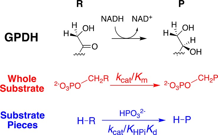
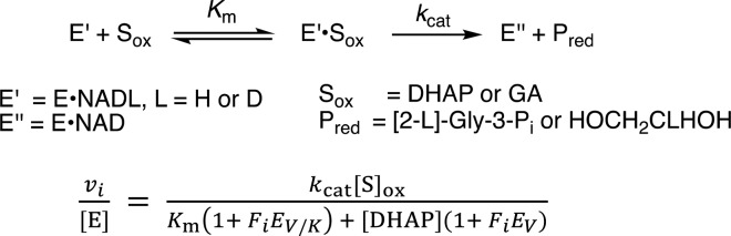
Large primary deuterium kinetic isotope effects (1° DKIEs) on enzyme-catalyzed hydride transfer may be observed when the transferred hydride tunnels through the energy barrier. The following 1° DKIEs on kcat/ Km and relative reaction driving force are reported for wild-type and mutant glycerol-3-phosphate dehydrogenase (GPDH)-catalyzed reactions of NADL (L = H, D): wild-type GPDH, ΔΔ G = 0 kcal/mol, 1° DKIE = 1.5; N270A, 5.6 kcal/mol, 3.1; R269A, 9.1 kcal/mol, 2.8; R269A + 1.0 M guanidine, 2.4 kcal/mol, 2.7; R269A/N270A, 11.5 kcal/mol, 2.4. Similar 1° DKIEs were observed on kcat. The narrow range of 1° DKIEs (2.4-3.1) observed for a 9.1 kcal/mol change in reaction driving force provides strong evidence that these are intrinsic 1° DKIEs on rate-determining hydride transfer. Evidence is presented that the intrinsic DKIE on wild-type GPDH-catalyzed reduction of DHAP lies in this range. A similar range of 1° DKIEs (2.4-2.9) on ( kcat/ KGA, M-1 s-1) was reported for dianion-activated hydride transfer from NADL to glycolaldehyde (GA) [Reyes, A. C.; Amyes, T. L.; Richard, J. P. J. Am. Chem. Soc. 2016, 138, 14526-14529]. These 1° DKIEs are much smaller than those observed for enzyme-catalyzed hydrogen transfer that occurs mainly by quantum mechanical tunneling. These results support the conclusion that the rate acceleration for GPDH-catalyzed reactions is due to the stabilization of the transition state for hydride transfer by interactions with the protein catalyst. The small 1° DKIEs reported for mutant GPDH-catalyzed and for wild-type dianion-activated reactions are inconsistent with a model where the dianion binding energy is utilized in the stabilization of a tunneling ready state.


Conflict of interest statement

The authors declare no competing financial interest.

Figures

Figure 1
Figure 1
Free-energy reaction profiles that illustrate different strategies for enzymes to reduce the effective activation barrier. (A) The deprotonation of DHAP to form an enediolate phosphate intermediate catalyzed by triosephosphate isomerase. The activation barrier to the enzyme-catalyzed reaction is reduced by protein–ligand interactions that result in similar stabilization of the enediolate intermediate and the enediolate-like transition state relative to the bound substrate. (B) A reaction coordinate profile for hydride transfer from NADH to the carbonyl group catalyzed by a representative dehydrogenase. The apparent activation barrier may be reduced below that for the formation of the transition state by quantum mechanical tunneling through the energy surface.
Scheme 1
Scheme 1. GPDH-Catalyzed Reduction of the Whole Substrate DHAP and the Dianion-Activated Reduction of Substrate Piece Glycolaldehyde
Scheme 2
Scheme 2. Minimal Kinetic Mechanism for GPDH-Catalyzed Reactions
Figure 2
Figure 2
Dependence of v/[E] (s–1) on [DHAP] for the mutant hlGPDH-catalyzed reduction of DHAP by NADH or NADD (0.2 mM) at pH 7.5, 25 °C, and I = 0.12 (NaCl): (A) R269A mutant, (B) N270A mutant, (C) R269A/N270A mutant.
Figure 3
Figure 3
Dependence of v/[E] (s–1) on [GA] for the N270A mutant hlGPDH-catalyzed reduction of DHAP by NADH or NADD (0.2 mM) at pH 7.5, 25 °C, and I = 0.12 (NaCl).
Scheme 3
Scheme 3. Activation of the R269A Mutant hlGPDH-Catalyzed Reduction of S = DHAP by NADL
Figure 4
Figure 4
Effect of the guanidine cation on the R269A mutant hlGPDH-catalyzed reduction of DHAP by NADH or NADD for reactions at pH 7.5 (20 mM TEA buffer), 25 °C, 0.2 mM NADL, and I = 0.12 (NaCl): (⧫) 2 mM Gua+, (▼) 5 mM Gua+, (▲) 10 mM Gua+, (●) 15 mM Gua+, and (○) 20 mM Gua+.
Figure 5
Figure 5
Effect of increasing [Gua+] on the values of (kcat/Km)obs determined as the slopes of correlations from Figure 4 for the R269A mutant hlGPDH-catalyzed reduction of DHAP by NADH and NADD at pH 7.5 (20 mM TEA buffer), 25 °C, 0.2 mM NADL, and I = 0.12 (NaCl).
Scheme 4
Scheme 4. Kinetic Mechanism for the hlGPDH-Catalyzed Reduction of GA at Saturating Concentrations of NADH or NADD
Figure 6
Figure 6
Representation of the X-ray crystal structure of the nonproductive ternary complex between hlGPDH, DHAP, and NAD+ (PDB entry 1WPQ) showing the side chains for Arg-269 and Asn-270 that interact with the substrate phosphodianion. Also shown are loop residues 292–297 (green) that fold over DHAP and the hydrogen-bonded side chains [Asn-270, Thr-264, Asn-205, Lys-204, Asp-260, and Lys-120] that connect the catalytic and dianion activation sites.
Scheme 5
Scheme 5. Cycle That Shows the Effect of Consecutive R269A and N270A Mutations on the Activation Barrier ΔGDHAP for the Wild-Type hlGPDH-Catalyzed Reduction of DHAP by NADH
Figure 7
Figure 7
Hypothetical reaction coordinates which show the barriers for the microscopic rate constants that control the rate-determining step for the hlGPDH-catalyzed reduction of Sox = GA or DHAP: (k–d)S for the release of substrate Sox; kchem for hydride transfer; and (k–d)P for the release of product Pred. Hydride transfer is only partially rate-determining for the reaction catalyzed by wild-type hlGPDH (black lines). The observation of intrinsic kinetic isotope effects on the hlGPDH-catalyzed reactions of the substrate in pieces or the mutant hlGPDH-catalyzed reactions reflects the decreasing barriers to (k–d)S and (k–d)P and the increasing barrier to kchem (blue lines), as described in the text.

References

    1. Richard J. P. (1998) The Enhancement of Enzymic Rate Accelerations by Bronsted Acid-Base Catalysis. Biochemistry 37, 4305–4309. 10.1021/bi972655r. - DOI - PubMed
    1. Richard J. P.; Huber R. E.; Lin S.; Heo C.; Amyes T. L. (1996) Structure-reactivity relationships for β-galactosidase (Escherichia coli, lac Z). 3. Evidence that Glu-461 participates in Bronsted acid-base catalysis of β-D-galactopyranosyl group transfer. Biochemistry 35, 12377–12386. 10.1021/bi961028j. - DOI - PubMed
    1. Richard J. P.; Huber R. E.; Heo C.; Amyes T. L.; Lin S. (1996) Structure-reactivity relationships for β-galactosidase (Escherichia coli, lac Z). 4. Mechanism for reaction of nucleophiles with the galactosyl-enzyme intermediates of E461G and E461Q β-galactosidases. Biochemistry 35, 12387–12401. 10.1021/bi961029b. - DOI - PubMed
    1. Richard J. P. (2012) A Paradigm for Enzyme-Catalyzed Proton Transfer at Carbon: Triosephosphate Isomerase. Biochemistry 51, 2652–2661. 10.1021/bi300195b. - DOI - PMC - PubMed
    1. Jonnalagadda V.; Toth K.; Richard J. P. (2012) Isopentenyl diphosphate isomerase catalyzed reactions in D2O: Product release limits the rate of this sluggish enzyme-catalyzed reaction. J. Am. Chem. Soc. 134, 6568–6570. 10.1021/ja302154k. - DOI - PMC - PubMed

Publication types

LinkOut - more resources