Review
. 2018 Nov;91(1091):20180142.
doi: 10.1259/bjr.20180142. Epub 2018 Jun 27.

Peripheral vision: abdominal pathology missed outside the centre of gaze

Affiliations
Review

Peripheral vision: abdominal pathology missed outside the centre of gaze

Catalin Vasile Ivan et al. Br J Radiol. 2018 Nov.

Abstract

Radiology misses have been the subject of much debate on both sides of the Atlantic in recent years. There is now greater focus in trying to reduce radiology errors by continuous education and changing the working environment to try and protect the radiologist, and ultimately the patient from potential harm. Duty of candour is a relevant and sensitive area. Developing robust validated reporting pathways within the healthcare structure is very important so as to encourage a "learning from discrepancies" culture and to put the patient and their families at the center of reporting and acknowledging errors in radiology. Having reflected in our daily practice and while writing this pictorial review, we have concluded that during reporting MRI scans, routine assessment of the localizer images, focusing outside the area of interest and having a more structured approach to image interrogation are key actions which may help reduce the number of omissions. We present a myriad of cases where pathology was "missed" outside the center of gaze in relation to the abdomen or outside the abdomen on abdominal MRI, and suggest key high yield sequence related review areas to minimize the chance of missing potentially significant pathology.


Figures

Figure 1.
Figure 1.
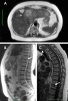
Ascending colon cancer with spinal metastases on MRI liver. Large ascending colon tumour and left pedicle metastatic lesion with a soft tissue component narrowing the spinal canal. Both the primary and metastatic lesions demonstrate restricted diffusion and enhance on the Fat Sat T1 C + sequence.
Figure 2.
Figure 2.
Large synchronous primary bowel cancer within the caecum on localiser for staging MR rectum. Large shouldered caecal tumour within the caecum on T2 weighted coronal localiser. This was subsequently identified on the subsequent staging CT scan. Sagittal T2 weighted image of a large rectal tumour (right).
Figure 3.
Figure 3.
Lesion within gastric antrum on MRCP. Coronal heavily weighted T 2 imaging shows high T 2 signal thickening of the gastric antrum (yellow arrows). Axial T2 weighted image showing the circumferential antral thickening (yellow circle). Histology confirmed adenocarcinoma. Note the distended gallbladder and gallstone. MRCP, magnetic resonance cholangiopancreatography.
Figure 4.
Figure 4.
Renal lesion on MRI liver. Axial T 1 C+ (GD) and following portal venous phase CT reveal a left anterior cortical renal lesion with thick, nodular septal enhancement.
Figure 5.
Figure 5.
Right ovarian mature (cystic) teratoma. Large, fat containing right ovarian mass seen on the coronal T2 weighted localizer of an MRCP study also subsequently demonstrated on a CT scan. MRCP, magnetic resonance cholangiopancreatography.
Figure 6.
Figure 6.
Haemangioma on coronal localiser for MR spine. Single image T2 weighted localiser of MR spine shows a focal high T 2 lesion in Segment 7 of the liver (yellow arrow). Subsequent CT showed peripheral nodular early arterial enhancement in keeping with haemangioma (arrows).
Figure 7.
Figure 7.
Splenic lesion on MR lumbar spine. Coronal localiser image showing focal lesion at the splenic hilum (arrow). Subsequent ultrasound shows a poorly echogenic lobulated lesion which was proven to be a metastatic deposit.
Figure 8.
Figure 8.
Right renal mass on MR spine. (A) Axial and coronal localisers showing a heterogeneous mass in the interpolar region of the right kidney. (B) Coronal portal venous phase CT confirming the heterogeneous mass. (C) Renal mass on coronal contrast enhanced CT images. (D) Renal mass on ultrasound.
Figure 9.
Figure 9.
Large retroperitoneal mass on MR spine showing multiple spinal metastases. (A) Sagittal T2 weighted localiser showing lobulated large retroperitoneal mass and focal high T 2 spinal lesions. (B, C) T 1 and T2 weighted axial images showing spinal metastasis and retroperitoneal mass (star). Note the mass is partially obscured by saturation bands but still visible (arrow).
Figure 10.
Figure 10.
Sigmoid tumour on MR hip. (A, B) Sagittal and coronal localisers showing abnormal sigmoid colon. (C) Coronal fat saturated planning scan shows high signal tumour in the sigmoid. (D) T1 weighted axial image showing circumferential thickening with local fat stranding.
Figure 11.
Figure 11.
Focal spinal lesion on MRCP. (A) Axial T2 weighted sequence showing multiple liver metastases and a focal lesion in the superior endplate of a lower thoracic vertebral body. (B) Sagittal localiser from the same study confirmed the lesion is in the superior endplate of T12 (arrow). (C) Subsequent T 1 weighted sagittal image from a dedicated MR showing metastatic lesions at T12 and T 1 (arrows). MRCP, magnetic resonance cholangiopancreatography.
Figure 12.
Figure 12.
AVN in the right femoral head on MR small bowel. (A) T2 weighted coronal image from MR small bowel showing the classic “Double-line” sign of AVN on the right femoral head. (B) Axial CT shows sclerosis of the right femoral head. AVN, avascular necrosis.
Figure 13.
Figure 13.
External iliac artery aneurysm on MR prostate. (A) Coronal localiser showing large rounded lesion in the right pelvis. (B) On axial T1 weighted images, the lesion was continuous with the external iliac artery and later confirmed as an aneurysm (arrows).
Figure 14.
Figure 14.
Malignant lung nodule on MRCP. Sagittal T 2 W localizer showing a posterior basal left lung nodule. CT scan after 1 year, demonstrates a necrotic lung mass invading through the diaphragm, into the spleen.
Figure 15.
Figure 15.
Pleural effusion and pericardiophrenic angle lung nodule on MRCP. Sagittal and axial T 2 W localizers and the accompanying CT scan demonstrating a nodule at the posterior cardiophrenic angle (arrows). MRCP, magnetic resonance cholangiopancreatography.

References

    1. Kim YW, Mansfield LT. Fool me twice: delayed diagnoses in radiology with emphasis on perpetuated errors. AJR Am J Roentgenol 2014; 202: 465–70. doi: 10.2214/AJR.13.11493 - DOI - PubMed
    1. Berlin L. Reviewing the CT scout view: medicolegal and ethical considerations. AJR Am J Roentgenol 2014; 202: 1264–6. doi: 10.2214/AJR.12.10444 - DOI - PubMed
    1. Daffner RH. Reviewing CT scout images: observations of an expert witness. AJR Am J Roentgenol 2015; 205: 589–91. doi: 10.2214/AJR.15.14405 - DOI - PubMed
    1. Cunliffe WJ, Hasleton PS, Tweedle DE, Schofield PF. Incidence of synchronous and metachronous colorectal carcinoma. Br J Surg 1984; 71: 941–3. doi: 10.1002/bjs.1800711210 - DOI - PubMed
    1. Lam AK, Carmichael R, Gertraud Buettner P, Gopalan V, Ho YH, Siu S, et al. . Clinicopathological significance of synchronous carcinoma in colorectal cancer. Am J Surg 2011; 202: 39–44. doi: 10.1016/j.amjsurg.2010.05.012 - DOI - PubMed

MeSH terms

LinkOut - more resources