. 2018 Jun 21;14(6):e1007100.
doi: 10.1371/journal.ppat.1007100. eCollection 2018 Jun.

PPARγ is critical for Mycobacterium tuberculosis induction of Mcl-1 and limitation of human macrophage apoptosis

Affiliations

PPARγ is critical for Mycobacterium tuberculosis induction of Mcl-1 and limitation of human macrophage apoptosis

Eusondia Arnett et al. PLoS Pathog. .

Abstract

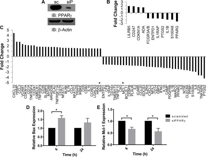
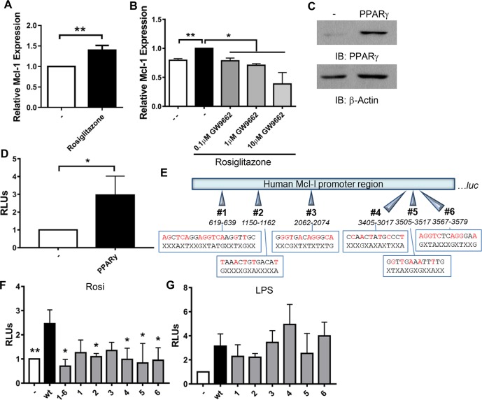
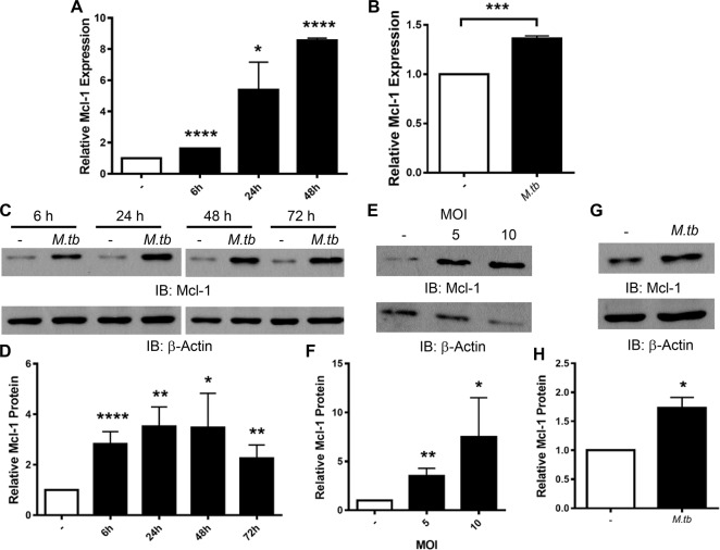
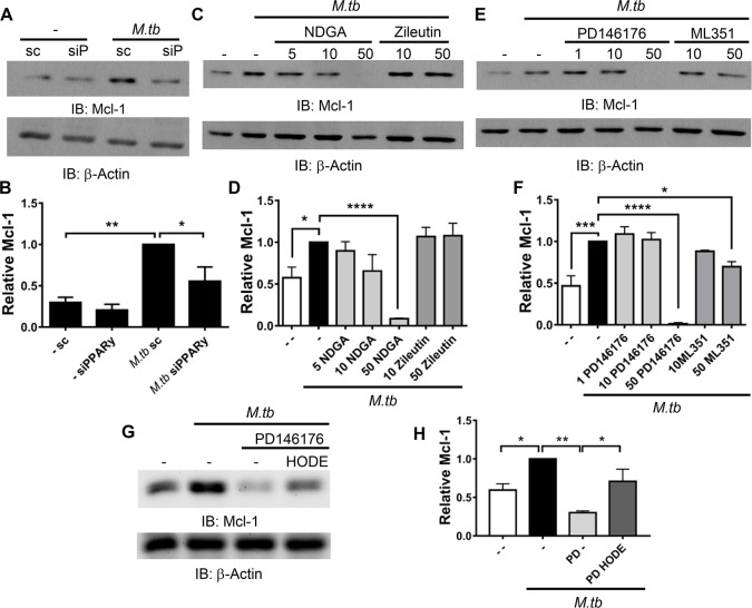
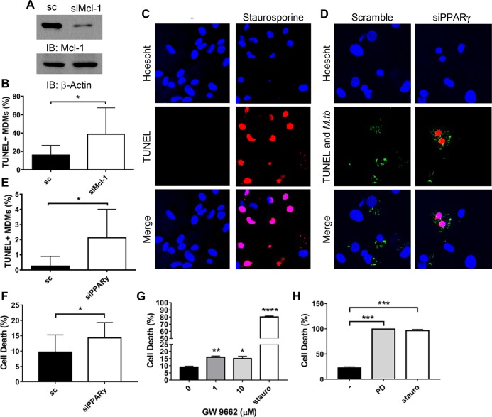
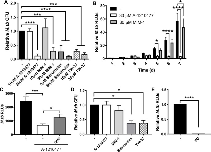
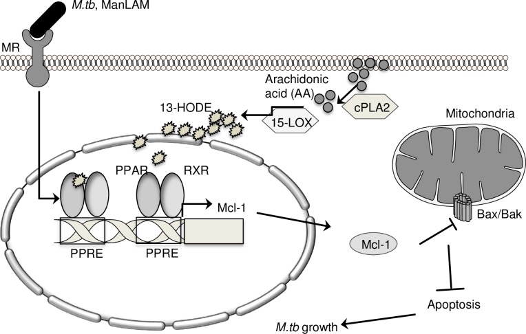
Peroxisome proliferator-activated receptor (PPAR)γ is a global transcriptional regulator associated with anti-inflammatory actions. It is highly expressed in alveolar macrophages (AMs), which are unable to clear the intracellular pathogen Mycobacterium tuberculosis (M.tb). Although M.tb infection induces PPARγ in human macrophages, which contributes to M.tb growth, the mechanisms underlying this are largely unknown. We undertook NanoString gene expression analysis to identify novel PPARγ effectors that condition macrophages to be more susceptible to M.tb infection. This revealed several genes that are differentially regulated in response to PPARγ silencing during M.tb infection, including the Bcl-2 family members Bax (pro-apoptotic) and Mcl-1 (pro-survival). Apoptosis is an important defense mechanism that prevents the growth of intracellular microbes, including M.tb, but is limited by virulent M.tb. This suggested that M.tb differentially regulates Mcl-1 and Bax expression through PPARγ to limit apoptosis. In support of this, gene and protein expression analysis revealed that Mcl-1 expression is driven by PPARγ during M.tb infection in human macrophages. Further, 15-lipoxygenase (15-LOX) is critical for PPARγ activity and Mcl-1 expression. We also determined that PPARγ and 15-LOX regulate macrophage apoptosis during M.tb infection, and that pre-clinical therapeutics that inhibit Mcl-1 activity significantly limit M.tb intracellular growth in both human macrophages and an in vitro TB granuloma model. In conclusion, identification of the novel PPARγ effector Mcl-1 has determined PPARγ and 15-LOX are critical regulators of apoptosis during M.tb infection and new potential targets for host-directed therapy for M.tb.


Conflict of interest statement

The authors have declared that no competing interests exist.

Figures

Fig 1
Fig 1. Identification of novel PPARγ effectors with NanoString.
MDMs were transfected with PPARγ (siP) or scrambled control (sc) siRNA, then infected with M.tb at MOI 5 for 6 and 24 h. A) Representative Western blot showing knockdown efficiency, mean knockdown efficiency was 81.7 ± 5.5% (N = 3). B and C) Total RNA was extracted and NanoString analysis was performed with a Human Immunology Panel. Shown are genes that displayed a significant (p < 0.05) mean fold change of at least 1.5 after PPARγ knockdown and 6 (B) or 24 (C) h of infection. Asterisks in C indicate Bax and Mcl-1. N = 3. D and E) Total RNA was collected and gene expression analyzed by qRT-PCR. Results are expressed as Bax (D) and Mcl-1 (E) expression relative to the scrambled control cells and are the mean ± SEM of 3, in triplicate, * p < 0.05.
Fig 2
Fig 2. PPARγ induces Mcl-1 expression.
A and B) MDMs were treated with 100 nM rosiglitazone without (A) or with (B) 1h pre-treatment with GW9662. After 24 h, total RNA was collected and Mcl-1 gene expression analyzed by qRT-PCR. Results are the mean ± SEM of N = 5 (A) or 2 (B). C, D, F) RAW cells were transfected with wild-type (wt; C, D, F) and mutated (F) Mcl-1 promoter luciferase reporter constructs, with and without PPARγ expression plasmids. C) Western blot was performed to confirm PPARγ expression. D and F) After transfection, cells were stimulated with 100 nM rosiglitazone. After 24 h, luciferase activity was determined and normalized to total protein. Results are expressed as (RLUs) normalized to cells not expressing PPARγ and are the mean ± SEM of N = 14 (D) or 2–4 (F), * indicate a significant difference between wt reporter and the indicated condition. E) Schematic of Mcl-1 promoter region upstream of the luciferase gene (luc) in pGL3. The six putative PPREs are indicated, wt sequences are shown on the top with red indicating consensus to PPRE (AGGTCAnAGGTCA), with mutated sequences below and X indicating no change. G) RAW cells were transfected with wt or mutated Mcl-1 promoter luciferase reporter constructs. After transfection, cells were stimulated with 1 μg/ml LPS. After 24 h, luciferase activity was determined and normalized to total protein. Results are expressed as relative luminescence units (RLUs) normalized to cells not stimulated with LPS and are the mean ± SEM of N = 2–3. A-F) * p < 0.05, ** p < 0.01.
Fig 3
Fig 3. M.tb induces Mcl-1 expression in human macrophages.
MDMs (A, C-F) and HAMs (B, G, H) were infected with M.tb at MOI 5 for the indicated times (A, C, D), at the indicated MOI for 24 h (E and F), or at MOI 5 for 24 h (B, G, H). A and B) Total RNA was collected and gene expression of Mcl-1 analyzed by qRT-PCR. Results are expressed as Mcl-1 expression relative to uninfected cells and are the mean ± SEM of N = 2–3. C-H) Protein lysates were collected and analyzed by Western blot (C, E, G). Densitometry analysis of Western blots was conducted with Image J (D, F, H). Data are expressed as amount of Mcl-1 protein relative to uninfected macrophages and are the mean ± SEM of at least 3 experiments. A-H) * p < 0.05, ** p < 0.01,*** p < 0.001, **** p < 0.0001.
Fig 4
Fig 4. M.tb induces Mcl-1 in a PPARγ- and 15-LOX-dependent manner.
A and B) MDMs were transfected with PPARγ (siP) or scrambled control (sc) siRNA then infected with M.tb at MOI 5 for 24 h. C-F) MDMs were treated with the indicated LOX inhibitors (μM) 1 h before, and during, M.tb infection (MOI 5 for 6 h). G and H) MDMs were treated with PD146176 (25 μM) 1 h before, and during, M.tb infection (MOI 5 for 6 h); along with 13-HODE (30 μM). A-H) MDMs were lysed and protein analyzed by Western blot, and densitometry analysis was conducted with Image J. Data are expressed as amount of Mcl-1 relative to M.tb infected scrambled control (B) or infected no inhibitor control (D, F, H) and are the cumulative mean ± SEM of at least 3 experiments, * p < 0.05, ** p < 0.01, *** p < 0.001, **** p < 0.0001.
Fig 5
Fig 5. PPARγ and Mcl-1 limit apoptosis during M.tb infection.
A, B, D, E) MDMs were transfected with Mcl-1 (A and B), PPARγ (D and E), or scrambled control (sc) siRNA then infected with fluorescent M.tb at MOI 50 for 24 h (B), or MOI 5 for 48 h (D and E). Due to the variability amongst donors, these different conditions were necessary to see low levels of apoptosis in the scrambled control cells, mean 8.66 ± 3.69% (B) and 2.30 ± 1.69% (E). A) Western blot showing Mcl-1 knockdown efficiency, mean knockdown efficiency was 76.6 ± 5.47% (N = 5). B and E) Data are representative of 3 experiments and are expressed as percentage of TUNEL+ MDMs and are the mean ± SD. The cumulative increase in TUNEL+ MDMs following knockdown (N = 3) is shown in S4A and S4B Fig. C) MDMs were treated with 5 μM staurosporine overnight then fixed and TUNEL staining performed. C and D) Representative images of TUNEL staining, with TUNEL staining indicated in red and fluorescent M.tb in green. F, G, H) MDMs were transfected with PPARγ or scrambled control (sc) siRNA (F) or pre-treated with GW9662 (G) or PD146176 (H) for 1 h, then infected with M.tb at MOI 5 for 24 h. MDMs were also treated with 5 μM staurosporine for 24 h. Cell death was determined with the CellTiter Glo Assay, data are expressed as % cell death, with uninfected cells set to 0%. Results are mean ± SEM of N = 3 (F) or 2 (H), or representative of n = 5 (G). A-H) * p < 0.05, ** p < 0.01, *** p < 0.001, **** p < 0.0001.
Fig 6
Fig 6. Mcl-1 inhibition limits M.tb growth.
A) MDMs were infected with M.tb at MOI 1, then treated with the indicated Mcl-1 inhibitors. After 4 d, cells were lysed and CFU enumerated. B) MDMs were infected with M.tb-lux at MOI 1, then treated with the indicated Mcl-1 specific inhibitors. M.tb luciferase activity was measured over time. C) MDMs were infected with M.tb-lux at MOI 1, treated with 100 nM Q-VD-OPH (QVD) for 1 h, then 30 μm A-1210477 and M.tb luciferase activity was measured after 3 d. Results are the mean ± SD of a representative experiment of 2, in triplicate. D) Human PBMCs were infected with M.tb at MOI 1 to generate in vitro TB granulomas, and after 1 day, treated with the indicated Mcl-1 inhibitors (30 μM). After 6 d with inhibitor, cells were lysed and CFU enumerated. E) MDMs were infected with M.tb-lux at MOI 1, then treated with PD146176 (50 μm). After 4 d, M.tb luciferase activity was measured. A-E) Results are the mean ± SEM of N = 3 unless indicated otherwise, * p < 0.05, *** p < 0.001, **** p < 0.0001.
Fig 7
Fig 7. Model.
In human macrophages, M.tb induces Mcl-1 expression through PPARγ, which requires 15-LOX, cPLA2, and the mannose receptor (MR) [4]. 15-LOX and PPARγ, through regulation of Mcl-1, contribute to M.tb’s ability to limit apoptosis and grow in human macrophages.

Comment in

References

    1. Glass CK, Saijo K. Nuclear receptor transrepressionpathways that regulate inflammationin macrophages and T cells. Nat Rev Immunol. Nature Publishing Group; 2010. May 1;10(5):365–76. doi: 10.1038/nri2748 - DOI - PubMed
    1. Ahmadian M, Suh JM, Hah N, Liddle C, Atkins AR, Downes M, et al. PPARγ signaling and metabolism: the good, the bad and the future. Nat Med. 2013. May;19(5):557–66. doi: 10.1038/nm.3159 - DOI - PMC - PubMed
    1. Almeida PE, Carneiro AB, Silva AR, Bozza PT. PPARγ Expression and Function in Mycobacterial Infection: Roles in Lipid Metabolism, Immunity, and Bacterial Killing. PPAR Res. 2012;2012:1–7. - PMC - PubMed
    1. Rajaram MVS, Brooks MN, Morris JD, Torrelles JB, Azad AK, Schlesinger LS. Mycobacterium tuberculosis activates human macrophage peroxisome proliferator-activated receptor gamma linking mannose receptor recognition to regulation of immune responses. J Immunol. 2010. July 15;185(2):929–42. doi: 10.4049/jimmunol.1000866 - DOI - PMC - PubMed
    1. Mahajan S, Dkhar HK, Chandra V, Dave S, Nanduri R, Janmeja AK, et al. Mycobacterium tuberculosis modulates macrophage lipid-sensing nuclear receptors PPARγ and TR4 for survival. J Immunol. 2012. June 1;188(11):5593–603. doi: 10.4049/jimmunol.1103038 - DOI - PubMed

Publication types

MeSH terms