

A case report on rare occurrence of periosteal ganglion cyst in femoral intercondylar region
- PMID: 29928104
- PMCID: PMC6008608
- DOI: 10.1016/j.jcot.2017.09.019
A case report on rare occurrence of periosteal ganglion cyst in femoral intercondylar region
Abstract
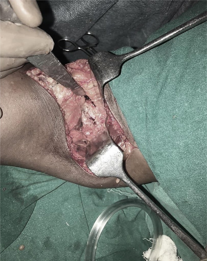
Ganglions are the cysts derived from myxomatous degeneration of periarticular tissue, commonly found around wrist and foot. Ganglion cysts are uncommon in Knee region and if present their occurrence is mostly incidental and benign. Based on their location, this could be extraarticular or intraarticular. Lesions with similar pathology in subchondral region around joints are lesser common entity called Intraosseous ganglions. Rarer still is the lesion produced by myxomatous degeneration of periosteum of long bone, called periosteal ganglion. We here present a case of 35 year old Male with gradual onset, poorly localised pain behind left knee which was radiologically suggestive of periosteal ganglion, eroding the posterior aspect of femur and confirmed histopathologically following the excision of a lesion from posterior aspect of femoral condyles.
Keywords: Benign cysts around knee; Intercondylar cyst; Periosteal ganglion.
Figures


References
-
- McCarthy E.F., Matz S., Steiner G.C., Dorfman H.D. Periosteal ganglion: a cause of cortical bone erosion. Skeletal Radiol. 1983;10:243–246. - PubMed
-
- Kobayashi H., Kotoura Y., Hosono M., Tsuboyama T., Sakahara H., Koinishi J. Periosteal ganglion of the tibia. Skeletal Radiol. 1996;25:381–383. - PubMed
-
- Valls R., Melloni P., Darnell A., Munoz J., Canalies J. Diagnostic imaging of tibial periosteal ganglion. Eur Radiol. 1997;7:70–72. - PubMed
-
- Ferguson N.N., Asarch A., Tschetter A.J., Stone M. Periosteal ganglia presenting as subcutaneous nodules on the tibia. JAMA Dermatol. 2014;150:663–664. - PubMed
Publication types
LinkOut - more resources
Full Text Sources
Other Literature Sources
