. 2018 Jun 6:9:1183.
doi: 10.3389/fimmu.2018.01183. eCollection 2018.

Sialylated Autoantigen-Reactive IgG Antibodies Attenuate Disease Development in Autoimmune Mouse Models of Lupus Nephritis and Rheumatoid Arthritis

Affiliations

Sialylated Autoantigen-Reactive IgG Antibodies Attenuate Disease Development in Autoimmune Mouse Models of Lupus Nephritis and Rheumatoid Arthritis

Yannic C Bartsch et al. Front Immunol. .

Abstract

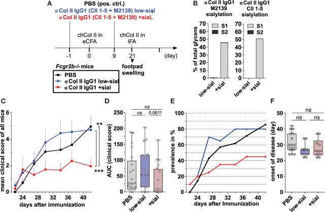
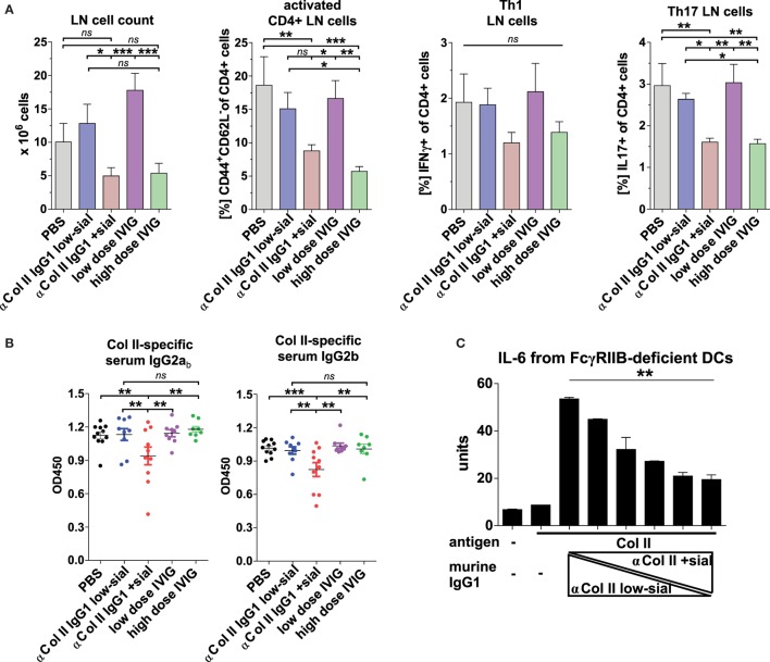
Pro- and anti-inflammatory effector functions of IgG antibodies (Abs) depend on their subclass and Fc glycosylation pattern. Accumulation of non-galactosylated (agalactosylated; G0) IgG Abs in the serum of rheumatoid arthritis and systemic lupus erythematosus (SLE) patients reflects severity of the diseases. In contrast, sialylated IgG Abs are responsible for anti-inflammatory effects of the intravenous immunoglobulin (pooled human serum IgG from healthy donors), administered in high doses (2 g/kg) to treat autoimmune patients. However, whether low amounts of sialylated autoantigen-reactive IgG Abs can also inhibit autoimmune diseases is hardly investigated. Here, we explore whether sialylated autoantigen-reactive IgG Abs can inhibit autoimmune pathology in different mouse models. We found that sialylated IgG auto-Abs fail to induce inflammation and lupus nephritis in a B cell receptor (BCR) transgenic lupus model, but instead are associated with lower frequencies of pathogenic Th1, Th17 and B cell responses. In accordance, the transfer of small amounts of immune complexes containing sialylated IgG Abs was sufficient to attenuate the development of nephritis. We further showed that administration of sialylated collagen type II (Col II)-specific IgG Abs attenuated the disease symptoms in a model of Col II-induced arthritis and reduced pathogenic Th17 cell and autoantigen-specific IgG Ab responses. We conclude that sialylated autoantigen-specific IgG Abs may represent a promising tool for treating pathogenic T and B cell immune responses in autoimmune diseases.

Keywords: IgG glycosylation; ST6gal1; Th17; autoimmunity; immunosuppression; rheumatoid arthritis; sialylation; systemic lupus erythematosus.


Figures

Figure 1
Figure 1
56R+/−Fcgr2b−/− mice develop self- and polyreactive IgG2a and IgG2b autoantibodies (autoAbs) earlier than Fcgr2b−/− but show no signs of renal inflammation. (A) Indirect immunofluorescence analysis of IgG2a immune complex (IC) depositions and macrophage infiltration in kidney sections from 6-month-old wt mice, IgG autoAb (AB)-negative and proteinuria (PU)-negative (ABPU−), AB+PU− and AB+PU+Fcgr2b−/− mice (all IgG2ab) and 56R+/−Fcgr2b−/− mice (IgG2aa). The presented immunofluorescence images are representative of at least five mice per group. (B) PU scores for 5–6-month-old wt (n = 30), Fcgr2b−/− (n = 39) and 56R+/−Fcgr2b−/− (n = 30) mice. Each data point represents an individual animal; horizontal lines represent mean values. (C) Schematic representation of the self- and polyreactive 56R VDJ4 knock-in (haplotype a) and the wt (haplotype b) heavy chain loci. The 56R VDJ4 knock-in replaced the endogenous IgH Js and can class-switch to all Ab isotypes (–46). (D) The frequencies of B220+ B cells expressing the knock-in allele (a) or the wt allele (b) were analyzed by flow cytometry of splenocytes from 2-month-old 56R−/−, 56R+/− or 56R+/+Fcgr2b−/− mice. About 5–10% of B cells in 56R+/−Fcgr2b−/− mice expressed the wt (IgMb+) BCR. (E) Anti-DNA IgG2ab, IgG2b or IgG2aa subclass Abs in sera of 5–7-month-old wt and Fcgr2b−/− mice and 2- or 6-month-old 56R+/−Fcgr2b−/− mice were determined via enzyme-linked immunofluorescence assay. Each data point represents an individual animal. Bars indicate mean values. (F) IgG2ab and IgG2aa Ab binding to HEp-2 slides from sera of 6-month-old Fcgr2b−/− and 56R+/−Fcgr2b−/− mice, respectively.
Figure 2
Figure 2
Absence of splenomegaly and no accumulation of Th1, Th17 and plasma cells (PCs) in 56R+/−Fcgr2b−/− mice. (A) Spleen sizes of 5–7-month-old wt mice, Fcgr2b−/− mice in the following disease states: IgG autoAb (AB)-negative and proteinuria (PU)-negative (ABPU), AB+PU and AB+PU+, and 56R+/−Fcgr2b−/− mice. The presented organ sizes are representative of a minimum of five mice per group. (B) The frequency of mesenteric lymph node (mLN) CD4+ IFNγ+ Th1 cells, splenic and mLN CD138+PCs and mLN and blood CD4+ IL-17+ Th17 cells of 5–7-month-old wt mice (Th1 cells, mLNs, n = 8/PCs, spleen, n = 3/PCs, mLNs, n = 3/Th17 cells, mLNs, n = 8/Th17 cells, blood, n = 15), Fcgr2b−/− mice [ABPU (n = 12/3/3/12/10), AB+PU (n = 12/3/3/21/17) and AB+PU+ (n = 11/3/3/13/7)] and 56R+/−Fcgr2b−/− mice (n = 12/12/3/12/26), as measured by flow cytometry. The bars indicate the mean values.
Figure 3
Figure 3
Self- and polyreactive IgG2a and IgG2b autoantibodies (autoAbs) in 56R+/−Fcgr2b−/− mice develop T cell independently and are sialylated. (A,B) AutoAb-positive 5-month-old Fcgr2b−/− mice or 2.5-month-old 56R+/−Fcgr2b−/− mice received i.p. injections of anti-mouse CD4 (GK1.5) every 4 days for 25 days to deplete CD4+ T cells (Figure S2 in Supplementary Material). (A) Serum anti-DNA IgG2ab, IgG2b and IgG2aa levels before and after CD4 depletion. (B) The frequencies of CD138+ PCs in the spleen and bone marrow (BM) of untreated (n = 6) versus CD4-depleted (n = 4) Fcgr2b−/− mice, and untreated (spleen, n = 13; BM, n = 3) versus CD4 depleted (n = 4) 56R+/−Fcgr2b−/− mice were analyzed via FACS. One representative experiment is shown. (C) The biantennary core of the glycan structure linked to Asn 297 in the Fc region of IgG Abs consists of four N-acetylglucosamines (GlcNAc; blue) and three mannoses (Man), which can be further modified with fucose, bisecting GlcNAc and terminal galactose (G) and sialic acid (S) residues. (D) The sialic acid content in serum IgG Abs of 5–6-month-old autoAb (AB)-negative and proteinuria (PU)-negative [ABPU (n = 8), AB+PU (n = 6) and AB+PU+ (n = 4)] Fcgr2b−/− mice and 56R+/−Fcgr2b−/− mice (n = 11) compared to wt mice (n = 10) were analyzed with the GlykoScreen™ Sialic Acid Quantification Kit (Prozyme) (Efl: fluorescence emission at 590 nm). (E,F) St6gal1 protein expression in splenic total and IgG+ PCs of 6–7-month-old ABPU (n = 4), AB+PU (n = 5) and AB+PU+ (n = 9) Fcgr2b−/− mice and 56R+/−Fcgr2b−/− mice (n = 6), compared to wt mice (n = 5). (E) Representative intracellular staining of St6gal1 protein expression levels in splenic PCs of wt, AB+PU+Fcgr2b−/− and 56R+/−Fcgr2b−/− mice measured by flow cytometry. (F) Relative median fluorescence levels of St6gal1 protein expression. Median St6gal1 protein expression levels in total or IgG+ PCs of three independent experiments were measured by flow cytometry and normalized (fold change) to the expression in wt controls (=1) in the respective experiments. The normalized data of three independent experiments were summarized (mean) in the graphs.
Figure 4
Figure 4
Transfer of sialylated polyreactive IgG antibodies (Abs) derived from 56R+/−Fcgr2b−/− mice or administration of sialylated antigen-specific monoclonal IgG Abs reduces nephritis-induced mortality in Fcgr2b−/− mice. (A–C) Transfer of sialylated polyreactive IgG Abs from 56R+/−Fcgr2b−/− mice reduces nephritis-induced mortality. (A) Graphical representation of the experimental strategy. Fcgr2b−/− mice received ICs containing 100 µg of TNP-sheep IgG and 200 µg of either sialylated (native) or sialidase-treated de-sialylated (de-sial) polyreactive IgG Abs derived from 2–3-month-old 56R+/−Fcgr2b−/− mice. The positive control (pos. ctrl.) group was treated with PBS and TNP-sheep IgG only. After 14 days, nephritis was induced by injection of sheep IgG in CFA and subsequent intravenous injection of sheep anti-glomerular basement membrane nephrotoxic serum (NTS) 4 days later. (B) Fc sialylation of purified total serum IgG Abs from wt mice and purified polyreactive IgG Abs from 56R+/−Fcgr2b−/− mice before and after sialidase treatment was determined through EndoS-treatment and MALDI-TOF mass spectrometry (MS) (percentage of glycans with one or two sialic acid residues: S1 and S2; Figure S1 in Supplementary Material). (C) Kaplan-Meier survival analysis of the indicated groups. (D–F) Application of sialylated antigen-specific monoclonal IgG Abs reduces nephritis-induced mortality. (D) Graphical representation of the experimental strategy as described in (A). Fcgr2b−/− mice were treated with ICs containing 100 µg of TNP-sheep IgG and 100 µg of either native non-sialylated (αTNP murine IgG1 non-sial) or in vitro sialylated (αTNP IgG1 +sial) monoclonal anti-TNP murine IgG1 (clone H5) Abs. After 14 days, nephritis was induced as described above. (E) Fc sialylation of the monoclonal anti-TNP murine IgG 1 Abs (clone H5) before and after in vitro sialylation was determined through EndoS-treatment and MALDI-TOF MS (the percentage of one or two sialic acid residues coupled to the glycan: S1, S2; Figure S1 in Supplementary Material). (F) Kaplan–Meier survival analysis for the indicated groups. One representative experiment out of two independent experiments is shown.
Figure 5
Figure 5
Sialylated collagen type II (Col II)-reactive monoclonal IgG antibodies (Abs) suppress collagen-induced arthritis (CIA). (A) Graphical representation of the experimental strategy. CIA was induced in Fcgr2b−/− mice by subcutaneous injection of Col II in enriched CFA (eCFA) and subsequent challenge with Col II in incomplete Freund’s adjuvant (IFA) on day 21. One day before and 9 days after the first immunization, the mice received 100 µg of either low-sialylated (low-sial) or in vitro galactosylated plus sialylated (+sial) anti-Col II murine IgG1 Abs (clones M2139 and CII 1–5; 50 µg each) The positive control group received PBS instead of Abs. (B) Fc sialylation of native (low-sial) and in vitro galactosylated plus sialylated (+ sial) anti-Col II M2139 and CII 1–5 murine IgG1 Abs measured by EndoS-treatment and MALDI-TOF mass spectrometry (MS) (percentage of glycans with one or two sialic acid residues: S1 and S2; Figure S1 in Supplementary Material). (C–F) Combined clinical data of two independent CIA experiments (PBS: n = 21; low-sial: 21; +sial; n = 20). Foot swelling was scored on the indicated days from 0 to 3 per foot resulting in a maximal clinical score of 12 per mouse. The (C) mean clinical score of all mice and the (E) prevalence (percentage of affected animals with a score > 0) are shown for all groups on the indicated days. (D) The area under the curve (AUC) of the clinical score over the time was calculated for each mouse. AUC = 0 indicates that the animal never developed foot swelling (mice with score 0: PBS: n = 3; low-sial: n = 4; +sial: n = 10). (F) The day of disease onset shown only of the mice that developed foot swelling (score > 0) during the experiment. Differences in disease evolution (C) were analyzed using two-way ANOVA.
Figure 6
Figure 6
Sialylated collagen type II (Col II)-reactive monoclonal IgG antibodies (Abs) reduce the accumulation of proinflammatory Th17 cells and IgG autoAbs. Collagen-induced arthritis (CIA) was induced in Fcgr2b−/− mice as described in Figure 5 and Figure S5C in Supplementary Material. One day before and 9 days after the first immunization, the mice received 100 µg of either low-sialylated (low-sial; n = 10) or in vitro galactosylated plus sialylated (+sial; n = 9) anti-Col II murine IgG1 Abs (clones M2139 and CII 1–5; 50 µg each) or high dose (50 mg; n = 10) or low dose (100 µg; n = 10) of intravenous immunoglobulin (IVIG) (Figure S5B in Supplementary Material). The positive control group received PBS instead of Abs (n = 10). (A) Pooled popliteal and brachial lymph nodes (LN) of each mouse from the indicated groups were analyzed on day 47 to determine total cell counts and the frequencies of activated CD4+CD44+CD62L T cells, CD4+IFNγ+ Th1 cells and CD4+IL-17+ Th17 cells. (B) Col II-reactive IgG2ab and IgG2b serum Ab levels measured on day 42 by enzyme-linked immunofluorescence assay (ELISA). One representative experiment out of two independent experiments is shown. (C) Sialylation of Col II-specific IgG1 Abs inhibits IL-6 secretion by dendritic cells (DCs). Bone marrow-derived DCs from Fcgr2b−/− mice were cultured in the presence of ICs containing 1 µg of Col II and 4 µg of different ratios of low-sialylated (< 1% sialylation; Figure 5B) and in vitro galactosylated and sialylated (46% sialylation; Figure 5B) Col II-specific IgG1 Abs (clone M2139; % of sialylation from left to right: <1, 3, 6, 12, 23, 46%). The IL-6 concentration in the supernatant was analyzed after 36 h via ELISA. The presented data are representative of four independent experiments.

Similar articles

Cited by

References

    1. Nimmerjahn F, Ravetch JV. Divergent immunoglobulin g subclass activity through selective Fc receptor binding. Science (2005) 310(5753):1510–2.10.1126/science.1118948 - DOI - PubMed
    1. Nimmerjahn F, Ravetch JV. Fcgamma receptors as regulators of immune responses. Nat Rev Immunol (2008) 8(1):34–47.10.1038/nri2206 - DOI - PubMed
    1. Parekh RB, Dwek RA, Sutton BJ, Fernandes DL, Leung A, Stanworth D, et al. Association of rheumatoid arthritis and primary osteoarthritis with changes in the glycosylation pattern of total serum IgG. Nature (1985) 316(6027):452–7.10.1038/316452a0 - DOI - PubMed
    1. Parekh RB, Roitt IM, Isenberg DA, Dwek RA, Ansell BM, Rademacher TW. Galactosylation of IgG associated oligosaccharides: reduction in patients with adult and juvenile onset rheumatoid arthritis and relation to disease activity. Lancet (1988) 1(8592):966–9.10.1016/S0140-6736(88)91781-3 - DOI - PubMed
    1. Parekh R, Isenberg D, Rook G, Roitt I, Dwek R, Rademacher T. A comparative analysis of disease-associated changes in the galactosylation of serum IgG. J Autoimmun (1989) 2(2):101–14.10.1016/0896-8411(89)90148-0 - DOI - PubMed

Publication types

MeSH terms