. 2018 Jun 6:9:1257.
doi: 10.3389/fimmu.2018.01257. eCollection 2018.

Gestational Hypothyroxinemia Affects Its Offspring With a Reduced Suppressive Capacity Impairing the Outcome of the Experimental Autoimmune Encephalomyelitis

Affiliations

Gestational Hypothyroxinemia Affects Its Offspring With a Reduced Suppressive Capacity Impairing the Outcome of the Experimental Autoimmune Encephalomyelitis

Henny Haensgen et al. Front Immunol. .

Abstract

Hypothyroxinemia (Hpx) is a thyroid hormone deficiency (THD) condition highly frequent during pregnancy, which although asymptomatic for the mother, it can impair the cognitive function of the offspring. Previous studies have shown that maternal hypothyroidism increases the severity of experimental autoimmune encephalomyelitis (EAE), an autoimmune disease model for multiple sclerosis (MS). Here, we analyzed the immune response after EAE induction in the adult offspring gestated in Hpx. Mice gestated in Hpx showed an early appearance of EAE symptoms and the increase of all parameters of the disease such as: the pathological score, spinal cord demyelination, and immune cell infiltration in comparison to the adult offspring gestated in euthyroidism. Isolated CD4+CD25+ T cells from spleen of the offspring gestated in Hpx that suffer EAE showed reduced capacity to suppress proliferation of effector T cells (TEff) after being stimulated with anti-CD3 and anti-CD28 antibodies. Moreover, adoptive transfer experiments of CD4+CD25+ T cells from the offspring gestated in Hpx suffering EAE to mice that were induced with EAE showed that the receptor mice suffer more intense EAE pathological score. Even though, no significant differences were detected in the frequency of Treg cells and IL-10 content in the blood, spleen, and brain between mice gestated in Hpx or euthyroidism, T cells CD4+CD25+ from spleen have reduced capacity to differentiate in vitro to Treg and to produce IL-10. Thus, our data support the notion that maternal Hpx can imprint the immune response of the offspring suffering EAE probably due to a reduced capacity to trigger suppression. Such "imprints" on the immune system could contribute to explaining as to why adult offspring gestated in Hpx suffer earlier and more intense EAE.

Keywords: T regulatory cells; experimental autoimmune encephalomyelitis; hypothyroxinemia; multiple sclerosis; pregnancy.


Figures

Figure 1
Figure 1
Treatment with methimazole induces hypothyroxinemia (Hpx) in pregnant mice. (A) tT4, tT3, and thyroid stimulating hormone (TSH) levels were analyzed from serum samples of pregnant mice at E15 after treatment with methimazole (MMI). The treated group with MMI was designated as Hpx. The group treated with MMI plus T4 was named Hpx + T4, and the untreated pregnant mice as control group. Control N = 4, Hpx N = 4, and Hpx + T4 N = 3. (B) tT4, tT3, and TSH levels were analyzed from serum samples of the offspring from the three experimental groups at P55: (1) gestated in Hpx; gestated in euthyroidism (control); or gestated in MMI plus T4 (Hpx + T4) conditions. Data are showed as the mean ± SEM. *p < 0.01. Control N = 3, Hpx N = 3, and Hpx + T4 N = 3.
Figure 2
Figure 2
The offspring gestated in hypothyroxinemia (Hpx) has an intense experimental autoimmune encephalomyelitis (EAE). (A) EAE was induced in C57BL/6 female offspring that were gestated under Hpx (Hpx empty circles, N = 18), or gestated under euthyroidism (control, filled circles, N = 12), or gestated under treatment with MMI + T4 (Hpx + T4, empty triangles, N = 8). The severity of EAE was plotted base on a daily pathological score (see Materials and Methods) every day post-EAE induction. The mean of the disease scores for the female offspring and ± SD was plotted. Statistical analysis was performed by two-way ANOVA, post-test Bonferroni. *p < 0.05. (B) Demyelination score from quantitative histopathological analyses of spinal lumbar cord sections stained for myelin basic protein (MBP) from mice at 21 days after EAE induction. Values represent the mean ± SEM, N = 3, *p < 0.05. (C) Representative confocal microscopy pictures of white matter from spinal lumbar cord of control, Hpx, or Hpx + T4 offspring that suffer or not of EAE were analyzed for MBP (red). Nuclei are shown in blue (DAPI). Bar size is 100 µm. (D) Quantitative analysis of CD4+ and (E) CD8+ T cells detected by confocal immunofluorescence of spinal cord lumbar sections of control, Hpx, and Hpx + T4 offspring after 21 days of induction with EAE. Values represent the mean ± SEM. *p < 0.05 (N = 3). (F) Representative confocal microscopy images of lateral lumbar sections of the spinal cord regions immunostained with anti-CD4 (green), anti-CD8 (red), and DAPI (blue) of control, Hpx, and Hpx + T4 offspring that were induced or not with EAE. The sections were analyzed with 20× objective. Bar size is 100 µm (N = 3).
Figure 3
Figure 3
The percentage of CD4+CD25+FOXP3+IL-10+ and the content of IL-10 in the blood, spleen, and central nervous system (CNS) is similar in the offspring gestated in hypothyroxinemia (Hpx) or euthyroidism. (A) Determination of frequency of CD4+CD25+IL-10+FoxP3+cells in blood, spleen, and CNS of control, Hpx, and Hpx + T4 mice. Statistical analysis showed no significant differences in frequency of CD4+CD25+IL-10+FoxP3+cells of Hpx mice compared to control and Hpx + T4 mice. Control and Hpx n = 7, Hpx + T4 n = 3. Mean ± SEM *p < 0.05. ANOVA and Tukey’s test. (B) Determination of the content of IL-10 in the serum, spleen, and CNS of the offspring gestated in control, Hpx, and Hpx + T4. Statistical analysis showed no significant differences in the content of IL-10 of Hpx gestated in Hpx compared to control and Hpx + T4 mice. n = 3, mean ± SEM *p < 0.05, ANOVA, and Tukey’s test.
Figure 4
Figure 4
The spleen of the offspring gestated in hypothyroxinemia (Hpx) has similar percentage of CD4+CD25+FOXP3+ T cells in the spleen of the offspring gestated in Hpx suffering or not experimental autoimmune encephalomyelitis (EAE) and less content of IL-10 only after EAE induction. (A) Representative dot plots of CD4+Foxp3+ T cells from spleens of the three experimental groups gestated in euthyroidism (control), Hpx, and Hpx + T4 induced or not with EAE. This study was performed after 21 days of EAE induction. The upper right panel shows the percentage of CD4+Foxp3+ T cells from spleens in each experimental group. (B) Representative dot plots of CD4+CD25+Foxp3+ T cells from spleens of the three experimental groups gestated in euthyroidism (control), Hpx, and Hpx + T4 induced or not with EAE. The upper right panel of every dot-plot shows the percentage of CD4+Foxp3+CD25+ T cells. The graphs (C,D) show the analysis for the dot plots (A,B), respectively. These graphs correspond to three independent experiments that used five mice per experimental group. (E,F) IL-10 secretion measured by ELISA from the supernatants of total splenocytes after being cultured for 48 or 72 h, respectively either in the absence or presence of myelin oligodendrocyte glycoprotein. The splenocytes are derived from the three experimental groups gestated in euthyroidism (control), Hpx, or Hpx + T4 induced or not with EAE. Values represent the mean ± SEM. Statistical analysis was performed using one-way ANOVA and Tukeys post-test. Statistical significance is indicated as *p < 0.05 (n = 3).
Figure 5
Figure 5
CD4+CD25+ T cells from the spleen of their offspring gestated in hypothyroxinemia (Hpx) have reduced capacity to suppress activated effector T cells (TEff) cells. (A) The graph shows the percentage of suppression capacity of CD4+CD25+ T cells from spleen of the offspring gestated in euthyroidism (control) or Hpx over TEff cells stained with carboxyfluorescein diacetate succinimidyl ester (CFSE) from control mice and activated with anti-CD3 and anti-CD28 monoclonal antibodies N = 6 independent cultures (see Materials and Methods). (B) The right histogram is representative of TEff cells from control mice loaded with CFSE to analyze proliferation in the presence of CD4+CD25+ T cells from control offspring (left); the right histogram is representative of TEff cells from control mice loaded with CFSE to analyze proliferation in the presence of CD4+CD25+ T cells derived from Hpx offspring. (C) The graph shows the percentage of suppression capacity of CD4+CD25+ T cells from spleen of the offspring gestated in euthyroidism (control) over CD4+CD25+ T cells stained with CFSE from control or Hpx offspring that were activated with anti-CD3 and anti-CD28 monoclonal antibodies N = 3 independent cultures (see Materials and Methods). Values represent the mean ± SEM. Statistical analysis was performed using one-way ANOVA and Tukeys post-test. Statistical significance is indicated as *p < 0.05. (D) Representative histogram of the autofluorescence for CD4+CD25+ T cells from control offspring mice (upper); representative histogram of TEff cells from control mice that were stimulated with anti-CD3 and anti-CD8 antibodies and without CD4+CD25+ T cells (middle); and representative histogram of TEff cells without stimulation with anti-CD3 and anti-CD28 antibodies and without CD4+CD25+ T cells (left).
Figure 6
Figure 6
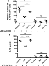
CD4+CD25 T cells from mice gestated in HTX show a decreased differentiation capacity toward CD4+CD25+FOXP3+IL-10+ T cells in vitro assay. (A) The graph shows the absolute number of CD4+CD25+IL-10+FoxP3+ after an in vitro cell culture assay for regulatory T cell (Treg) generation. CD4+CD25 T cells derived from the spleen of offspring gestated under control, Hpx, or Hpx + T4 were cultured in vitro for 5 days under induction media to stimulate the differentiation of Treg in the presence or in the absence of anti-CD3 and anti-CD28 antibodies. Statistical analysis shows a significantly decrease in the absolute number of CD4+CD25+IL-10+FoxP3+ in the in vitro Treg differentiation assay of CD4+CD25 T cells derived from the offspring gestated in Hpx cells. There are not significant differences observed when CD4+CD25 T cells were cultured in the induction medium for Treg but without anti-CD3 and anti-CD28 antibodies n = 3, mean ± SEM p ≤ 0.05 ANOVA and Tukey’s test. (B) The graph shows the content of IL-10 measured by ELISA in the supernatant of CD4+CD25 T cells from spleen of control, Hpx, and Hpx + T4 offspring. CD4+CD25 T cells were in vitro cell cultured for 5 days under induction medium for Treg and stimulated or not with anti-CD3 and anti-CD28 antibodies (see Materials and Methods). Statistical analysis shows a significantly reduced amount of IL-10 of cultured CD4+CD25 T cells of Hpx mice compared to control and Hpx + T4 offspring. There are no significant differences observed when CD4+CD25 T cells were cultured in the induction media for Treg without anti-CD3 and anti-CD28 monoclonal antibodies. n = 3, mean ± SEM ***p < 0.001 and ****p < 0.0001, ANOVA, and Tukey’s test.
Figure 7
Figure 7
Adoptive transfer of CD4+CD25+ T cells derived from the offspring gestated in hypothyroxinemia (Hpx) increases the severity and onset of experimental autoimmune encephalomyelitis (EAE). (A) Representative dot-plot of the percentage of CD4+FOXP3+ T cells after CD4+CD25+ T cells were cultured in vitro for 7 days in the presence of IL-2 and anti-CD3 and anti CD8 antibodies. (B) The graph shows a pathological score of the recipient offspring gestated in Hpx after EAE induction (day 1) that received 1 × 106 CD4+CD25+ T cells from mice gestated in euthyroidism (control) (inverted gray triangle) or Hpx (closed circle) that also suffer EAE. As control it shows also the pathological score of control offspring that were induced with EAE and did not receive CD4+CD25+ T cells (open diamond). N = 3 independent experiments. (C) The graph shows the quantification by flow cytometry of CD4+CD25+IL-10+ FOXP3+ T cells from splenocytes of naïve mice that previously were cultured in vitro for 48 or 72 h. These naïve mice were adoptively transfered with 1 × 106 CD4+CD25+ T cells of the offspring gestated in Hpx (closed circles) or control (inverted gray triangles) that suffer EAE. The CD4+CD25+ T cells that were adoptively transfered are derived from spleen and were purified at day 21 of EAE induction in these mice and cultured in vitro for 7 days in the presence of IL-2 and anti-CD3 and anti-CD28 antibodies. Values represent the mean ± SEM. Statistical analysis was performed using one-way ANOVA and Tukey’s post-test. Statistical significance is indicated as *p < 0.05.

Similar articles

Cited by

References

    1. Brent GA. Mechanisms of thyroid hormone action. J Clin Invest (2012) 122(9):3035–43.10.1172/JCI60047 - DOI - PMC - PubMed
    1. Mondal S, Raja K, Schweizer U, Mugesh G. Chemistry and biology in the biosynthesis and action of thyroid hormones. Angew Chem Int Ed Engl (2016) 55:7606–30.10.1002/anie.201601116 - DOI - PubMed
    1. Opazo MC, Gianini A, Pancetti F, Azkcona G, Alarcon L, Lizana R, et al. Maternal hypothyroxinemia impairs spatial learning and synaptic nature and function in the offspring. Endocrinology (2008) 149:5097–106.10.1210/en.2008-0560 - DOI - PMC - PubMed
    1. Gyllenberg D, Sourander A, Surcel HM, Hinkka-Yli-Salomaki S, Mckeague IW, Brown AS. Hypothyroxinemia during gestation and offspring schizophrenia in a national birth cohort. Biol Psychiatry (2015) 79(12):962–70.10.1016/j.biopsych.2015.06.014 - DOI - PMC - PubMed
    1. Wei W, Wang Y, Dong J, Wang Y, Min H, Song B, et al. Hypothyroxinemia induced by maternal mild iodine deficiency impairs hippocampal myelinated growth in lactational rats. Environ Toxicol (2015) 30:1264–74.10.1002/tox.21997 - DOI - PubMed

Publication types

MeSH terms