Review
. 2018 Jun;15(6):8901-8914.
doi: 10.3892/ol.2018.8487. Epub 2018 Apr 13.

Primary tumours of the calcaneus

Affiliations
Review

Primary tumours of the calcaneus

Litao Yan et al. Oncol Lett. 2018 Jun.

Abstract

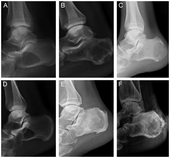
The calcaneus is a rare location for the occurrence and development of primary tumour types. Clinicians are unfamiliar with calcaneal tumour types, which may result in a delay in diagnosis or a missed diagnosis, resulting in unnecessary morbidity and amputation. Heel pain and localized swelling of the ankle are the most common symptoms. X-ray is the first choice for a tentative diagnosis of a calcaneal tumour. The final diagnosis depends on a histological examination. The treatment of calcaneal tumour types varies depending on the Enneking system. The majority of patients with benign tumours heal, except for a few with a palindromia. For malignant tumours, the prognosis is comparatively poor, resulting in disability and a high rate of metastasis. This review describes the spectrum of calcaneal tumour types and specifically illustrates the epidemiology, symptomatology, imagology, histopathology and treatment options that may facilitate diagnosis and improve prognosis.

Keywords: calcaneus; diagnosis; primary tumour; surgery; symptom.


Figures

Figure 1.
Figure 1.
Lateral x-ray of a number of different calcaneal tumour types. (A) Simple bone cysts of the calcaneus. A solitary, sharply demarcated radiolucent osteolysis may be observed in the calcaneus, without a pathologic fracture. (B) Aneurysmal bone cysts of the calcaneus. The lesion is generally identified as a lytic, expansile lesion with an eccentric shape and septations. (C) Chondroblastoma of the calcaneus. The lesion is located in the posterior calcaneus, which is a lytic expansile mass with a sclerotic margin. (D) Intraosseous lipomas of the calcaneus. A well-circumscribed lytic lesion is located at the base of the calcaneus neck in the region of the Ward's triangle. (E) Chondromyxoid fibroma of the calcaneus. The lesion demonstrates a large lytic lesion with sclerotic margins and ‘soap bubble’ appearance. (F) Chondrosarcoma of the calcaneus. The lesion shows a destructive, expansive and pathologic fracture with calcification of the soft tissues.

Similar articles

Cited by

References

    1. Kilgore WB, Parrish WM. Calcaneal tumors and tumor-like conditions. Foot Ankle Clin. 2005;10:541–565. doi: 10.1016/j.fcl.2005.05.002. - DOI - PubMed
    1. Young PS, Bell SW, MacDuff EM, Mahendra A. Primary osseous tumors of the hindfoot: Why the delay in diagnosis and should we be concerned? Clin Orthop Relat Res. 2013;471:871–877. doi: 10.1007/s11999-012-2570-6. - DOI - PMC - PubMed
    1. Hanna SJ, Dasic D, Floyd A. Simple bone cysts of the calcaneus: A report of five cases and a review of the literature. Foot Ankle Int. 2004;25:680–684. doi: 10.1177/107110070402500914. - DOI - PubMed
    1. Polat O, Sağlik Y, Adiguzel HE, Arikan M, Yildiz HY. Our clinical experience on calcaneal bone cysts: 36 cysts in 33 patients. Arch Orthop Trauma Surg. 2009;129:1489–1494. doi: 10.1007/s00402-008-0779-3. - DOI - PubMed
    1. Takada J, Hoshi M, Oebisu N, Ieguchi M, Kakehashi A, Wanibuchi H, Nakamura H. A comparative study of clinicopathological features between simple bone cysts of the calcaneus and the long bone. Foot Ankle Int. 2014;35:374–382. doi: 10.1177/1071100713519600. - DOI - PubMed