. 2018 Jun;15(6):9663-9672.
doi: 10.3892/ol.2018.8584. Epub 2018 Apr 26.

Quercetin induced apoptosis of human oral cancer SAS cells through mitochondria and endoplasmic reticulum mediated signaling pathways

Affiliations

Quercetin induced apoptosis of human oral cancer SAS cells through mitochondria and endoplasmic reticulum mediated signaling pathways

Yi-Shih Ma et al. Oncol Lett. 2018 Jun.

Abstract

Oral cancer is a cause of cancer-associated mortality worldwide and the treatment of oral cancer includes radiation, surgery and chemotherapy. Quercetin is a component from natural plant products and it has been demonstrated that quercetin is able to induce cytotoxic effects through induction of cell apoptosis in a number of human cancer cell lines. However, there is no available information to demonstrate that quercetin is able to induce apoptosis in human oral cancer cells. In the present study, the effect of quercetin on the cell death via the induction of apoptosis in human oral cancer SAS cells was investigated using flow cytometry, Annexin V/propidium iodide (PI) double staining, western blotting and confocal laser microscopy examination, to test for cytotoxic effects at 6-48 h after treatment with quercetin. The rate of cell death increased with the duration of quercetin treatment based on the results of a cell viability assay, increased Annexin V/PI staining, increased reactive oxygen species and Ca2+ production, decreased the levels of mitochondrial membrane potential (ΔΨm), increased proportion of apoptotic cells and altered levels of apoptosis-associated protein expression in SAS cells. The results from western blotting revealed that quercetin increased Fas, Fas-Ligand, fas-associated protein with death domain and caspase-8, all of which associated with cell surface death receptor. Furthermore, quercetin increased the levels of activating transcription factor (ATF)-6α, ATF-6β and gastrin-releasing peptide-78 which indicated an increase in endoplasm reticulum stress, increased levels of the pro-apoptotic protein BH3 interacting-domain death antagonist, and decreased levels of anti-apoptotic proteins B-cell lymphoma (Bcl) 2 and Bcl-extra large which may have led to the decreases of ΔΨm. Additionally, confocal microscopy suggested that quercetin was able to increase the expression levels of cytochrome c, apoptosis-inducing factor and endonuclease G, which are associated with apoptotic pathways. Therefore, it is hypothesized that quercetin may potentially be used as a novel anti-cancer agent for the treatment of oral cancer in future.

Keywords: apoptosis; endoplasmic reticulum mediated signaling pathway; human oral cancer SAS cells; mitochondria signaling pathway; quercetin.


Figures
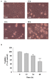
Figure 1.
Figure 1.
Quercetin induced cell morphological changes and decreased the percentage of viable cells in human oral cancer SAS cells. Cells (1×10⁵ cells/well) were placed in a 12-well plate for 24 h and 40 µM quercetin was added to well for 0, 12, 24 and 48 h. (A) Cells were examined and images were captured using a contrast phase microscope. (B) Assays were performed to assess the percentages of viable cells. Each point is mean ± SD of three experiments. **P<0.01 and ***P<0.001 vs. vehicle control (1% DMSO).
Figure 2.
Figure 2.
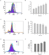
Quercetin induced apoptosis in human oral cancer SAS cells. SAS cells (1×10⁵ cells/ml) in 12-well culture plates were treated with 40 µM quercetin for 0, 24 and 48 h. Results of (A) flow cytometry and (B) assay for percentage of apoptotic cells. *P<0.05 vs. vehicle control (0 h).
Figure 3.
Figure 3.
Quercetin affected the production of ROS and Ca2+ and the levels of MMP in human oral SAS cells. Cells (1×10⁵ cells/well) were treated with 40 µM of quercetin for various time periods prior to being (A) stained by 2,7-dichlorodihydrofluorescein diacetate and (B) ROS levels quantified, (C) stained by DiOC6 and (D) the MMP levels quantified, and (E) stained by Fluo-3/AM and (F) the Ca2+ levels quantified. Data represents mean ± SD of three experiments. *P<0.05, **P<0.01 and ***P<0.001 vs. vehicle control (0 h). ROS, reactive oxygen species; ΔΨm, mitochondrial membrane potential; Fluo-3/AM, Fluo-3 acetoxymethyl ester.
Figure 4.
Figure 4.
Quercetin stimulated the activities of caspase-3, caspase-9 and caspase-8 in SAS cells. Cells were treated with quercetin, and the activities of caspase-3, presented as (A) histogram of FACS and (B) Bar chart; caspase-9, presented as (C) histogram of FACS and (D) Bar chart; and caspase-8, presented as (E) histogram of FACS and (F) Bar chart, were determined by flow cytometric assay. **P<0.01 and ***P<0.001 vs. vehicle control (0 h).
Figure 5.
Figure 5.
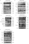
Quercetin affected the levels of apoptosis-associated proteins of SAS cells. Cells were treated with quercetin and the total protein levels were determined and used for SDS page gel electrophoresis. The levels of (A) caspase-2, Bak, Bid, Bad, Bcl-2 and Bcl-x; (B) cytochrome c, Apaf-1, Endo G, AIF and PARP; (C) caspase-9, pro-caspase-3, active-caspase-3, caspase-6 and caspase-7; (D) TRAIL, Fas-L, Fas, FADD and caspase-8; (E) ATF-6α, ATF-6β, XBP-1, IRE-1α and GRP-78 were examined. Bak, Bcl-2 homologous antagonist killer; Bid, BH3 interacting-domain death antagonist; Bad, Bcl-2-associated death promoter, Bcl-2, B cell lymphoma 2; Bcl-xl, Bcl-extra large; Apaf-1, apoptotic protease activating factor; Endo G, endonuclease G; AIF, apoptosis-inducing factor; PARP, poly(ADP-ribose) polymerase; TRAIL, TNF-related apoptosis-inducing ligand; Fas-L, Fas ligand; FADD, fas-associated protein with death domain; ATF-6β, activating transcription factor-6β; XBP-1, X-box binding protein 1; IRE-1α, iron responsive element-1α; GRP-78, gastrin releasing peptide-78.
Figure 6.
Figure 6.
Quercetin promoted the levels of Cyto c, AIF and Endo G in SAS cells. Cells were incubated quercetin, fixed and stained with (A) anti-Cyto c, (B) anti-AIF and (C) anti-Endo G which were then stained by FITC-labeled secondary antibodies (green fluorescence) and the nuclei were stained by PI (red fluorescence). All stained proteins were examined and images were captured using a confocal laser microscopic system. Scale bar, 20 µm. Cyto C, cytochrome c; AIF, apoptosis-inducing factor; Endo G, endonuclease G; FITC, fluorescein isothiocyanate; PI, propidium iodide.
Figure 7.
Figure 7.
Proposed signaling pathways for quercetin induced apoptosis in SAS cells. FADD, fas-associated protein with death domain; ER, endoplasmic reticulum; Cyto c, cytochrome c; AIF, apoptosis-inducing factor; Endo G, endonuclease G; Bcl-2, B cell lymphoma 2; Bax, Bcl-associated X protein; Bid, BH3 interacting-domain death antagonist; GRP-78, gastrin-releasing peptide-78; ATF-6α/β, activating transcription factor-6α/β.

Similar articles

Cited by

References

    1. Maleki D, Ghojazadeh M, Mahmoudi SS, Mahmoudi SM, Pournaghi-Azar F, Torab A, Piri R, Azami-Aghdash S, Naghavi-Behzad M. Epidemiology of oral cancer in Iran: A systematic review. Asian Pac J Cancer Prev. 2015;16:5427–5432. doi: 10.7314/APJCP.2015.16.13.5427. - DOI - PubMed
    1. Krishna A, Singh S, Kumar V, Pal US. Molecular concept in human oral cancer. Natl J Maxillofac Surg. 2015;6:9–15. doi: 10.4103/0975-5950.168235. - DOI - PMC - PubMed
    1. Halboub ES, Al-Anazi YM, Al-Mohaya MA. Characterization of Yemeni patients treated for oral and pharyngeal cancers in Saudi Arabia. Saudi Med J. 2011;32:1177–1182. - PubMed
    1. Chung TT, Pan MS, Kuo CL, Wong RH, Lin CW, Chen MK, Yang SF. Impact of RECK gene polymorphisms and environmental factors on oral cancer susceptibility and clinicopathologic characteristics in Taiwan. Carcinogenesis. 2011;32:1063–1068. doi: 10.1093/carcin/bgr083. - DOI - PubMed
    1. Galbiatti AL, Padovani-Junior JA, Maníglia JV, Rodrigues CD, Pavarino ÉC, Goloni-Bertollo EM. Head and neck cancer: Causes, prevention and treatment. Braz J Otorhinolaryngol. 2013;79:239–247. doi: 10.5935/1808-8694.20130041. (In English, Portuguese) - DOI - PMC - PubMed