Overexpression of hsa-miR-125a-5p enhances proliferation, migration and invasion of head and neck squamous cell carcinoma cell lines by upregulating C-C chemokine receptor type 7
- PMID: 29928346
- PMCID: PMC6004657
- DOI: 10.3892/ol.2018.8564
Overexpression of hsa-miR-125a-5p enhances proliferation, migration and invasion of head and neck squamous cell carcinoma cell lines by upregulating C-C chemokine receptor type 7
Abstract
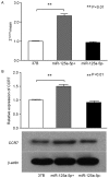
Head and neck squamous cell carcinoma (HNSCC) is usually diagnosed accompanied by lymph node metastasis. C-C chemokine receptor type 7 (CCR7) is associated with the invasion and metastasis of tumors in HNSCC through various signaling pathways. The role of hsa-miR-125a-5p in HNSCC remains unclear. The present study was performed to investigate the association between hsa-miR-125a-5p and CCR7 in HNSCC. Reverse transcription-quantitative polymerase chain reaction was applied to analyze the expression of hsa-miR-125a-5p in clinical samples. Cell Counting Kit-8, Transwell and wound healing assays were used to detect cell proliferation, invasion, and metastasis, respectively, following overexpression of hsa-miR-125a-5p. Changes in protein expression of CCR7 were observed using western blotting. In the survival analysis, Student's t-tests and log rank tests were performed to analyze the association between the expression of hsa-miR-125a-5p, and HNSCC according to the Cancer Genome Atlas database. The expression of hsa-miR-125a-5p was identified to be significantly lower in cancer tissue compared with the corresponding adjacent normal tissues in clinical samples (P=0.038). The results of western blotting indicated that there was a positive regulatory association between hsa-miR-125a-5p and CCR7. Furthermore, overexpression of hsa-miR-125a-5p significantly enhanced the ability of cell proliferation, migration and invasion in HNSCC, with upregulation of CCR7. The results of survival analysis revealed that patients in the low expression group of hsa-miR-125a-5p tended to have longer survival times compared with the high expression group (P=0.045). Altogether, the data raised the possibility that hsa-miR-125a-5p has a significant role in promoting cancer in HNSCC, which may provide a basis for the treatment of HNSCC in molecular targeted therapy. Further studies are required to ascertain the role of hsa-miR-125a-5p in other HNSCC cell lines and in vivo.
Keywords: chemokine receptor type 7; head and neck squamous cell carcinoma; invasion; migration; proliferation.
Figures


Similar articles
-
MiR-125 family improves the radiosensitivity of head and neck squamous cell carcinoma.Mol Biol Rep. 2023 Jun;50(6):5307-5317. doi: 10.1007/s11033-023-08364-x. Epub 2023 May 8. Mol Biol Rep. 2023. Retraction in: Mol Biol Rep. 2024 Jan 30;51(1):239. doi: 10.1007/s11033-024-09283-1. PMID: 37155009 Free PMC article. Retracted.
-
Hsa-miR-125a-3p and hsa-miR-125a-5p are downregulated in non-small cell lung cancer and have inverse effects on invasion and migration of lung cancer cells.BMC Cancer. 2010 Jun 22;10:318. doi: 10.1186/1471-2407-10-318. BMC Cancer. 2010. PMID: 20569443 Free PMC article.
-
Hsa-let-7e-5p Inhibits the Proliferation and Metastasis of Head and Neck Squamous Cell Carcinoma Cells by Targeting Chemokine Receptor 7.J Cancer. 2019 Apr 25;10(8):1941-1948. doi: 10.7150/jca.29536. eCollection 2019. J Cancer. 2019. PMID: 31205553 Free PMC article.
-
miR-125a-5p Functions as Tumor Suppressor microRNA And Is a Marker of Locoregional Recurrence And Poor prognosis in Head And Neck Cancer.Neoplasia. 2019 Sep;21(9):849-862. doi: 10.1016/j.neo.2019.06.004. Epub 2019 Jul 18. Neoplasia. 2019. PMID: 31325708 Free PMC article.
-
Is the Non-Coding RNA miR-195 a Biodynamic Marker in the Pathogenesis of Head and Neck Squamous Cell Carcinoma? A Prognostic Meta-Analysis.J Pers Med. 2023 Jan 31;13(2):275. doi: 10.3390/jpm13020275. J Pers Med. 2023. PMID: 36836509 Free PMC article. Review.
Cited by
-
MIR600HG sponges miR-125a-5p to regulate glycometabolism and cisplatin resistance of oral squamous cell carcinoma cells via mediating RNF44.Cell Death Discov. 2022 Apr 20;8(1):216. doi: 10.1038/s41420-022-01000-w. Cell Death Discov. 2022. Retraction in: Cell Death Discov. 2023 Nov 22;9(1):422. doi: 10.1038/s41420-023-01711-8. PMID: 35443748 Free PMC article. Retracted.
-
MiR-125a-5p regulates the radiosensitivity of laryngeal squamous cell carcinoma via HK2 targeting through the DDR pathway.Front Oncol. 2024 Aug 19;14:1438722. doi: 10.3389/fonc.2024.1438722. eCollection 2024. Front Oncol. 2024. PMID: 39224810 Free PMC article.
-
miR-18a-5p Facilitates Malignant Progression of Head and Neck Squamous Cell Carcinoma Cells via Modulating SORBS2.Comput Math Methods Med. 2021 Oct 18;2021:5953881. doi: 10.1155/2021/5953881. eCollection 2021. Comput Math Methods Med. 2021. Retraction in: Comput Math Methods Med. 2023 Sep 27;2023:9762607. doi: 10.1155/2023/9762607. PMID: 34707683 Free PMC article. Retracted.
-
C-C Chemokine Receptor 7 in Cancer.Cells. 2022 Feb 14;11(4):656. doi: 10.3390/cells11040656. Cells. 2022. PMID: 35203305 Free PMC article. Review.
-
Identification of microRNA Signature and Key Genes Between Adenoma and Adenocarcinomas Using Bioinformatics Analysis.Onco Targets Ther. 2021 Sep 4;14:4707-4720. doi: 10.2147/OTT.S320469. eCollection 2021. Onco Targets Ther. 2021. PMID: 34511938 Free PMC article.
References
-
- Bachelerie F, Ben-Baruch A, Burkhardt AM, Combadiere C, Farber JM, Graham GJ, Horuk R, Sparre-Ulrich AH, Locati M, Luster AD, et al. International union of basic and clinical pharmacology. [corrected]. LXXXIX. Update on the extended family of chemokine receptors and introducing a new nomenclature for atypical chemokine receptors. Pharmacol Rev. 2013;66:1–79. doi: 10.1124/pr.113.007724. - DOI - PMC - PubMed
LinkOut - more resources
Full Text Sources
Other Literature Sources
