. 2018 Jul;16(1):199-210.
doi: 10.3892/ol.2018.8678. Epub 2018 May 9.

Overexpression of α3, β3 and γ2 chains of laminin-332 is associated with poor prognosis in pancreatic ductal adenocarcinoma

Affiliations

Overexpression of α3, β3 and γ2 chains of laminin-332 is associated with poor prognosis in pancreatic ductal adenocarcinoma

Jun Chen et al. Oncol Lett. 2018 Jul.

Abstract

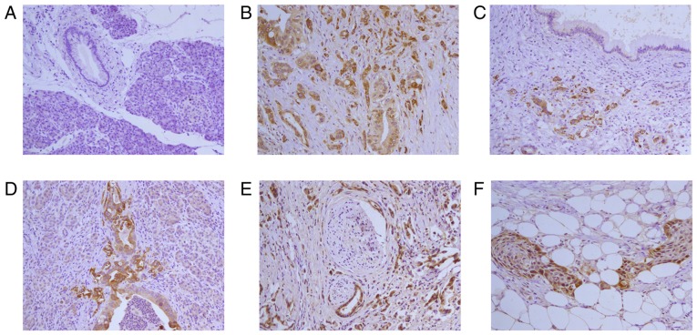
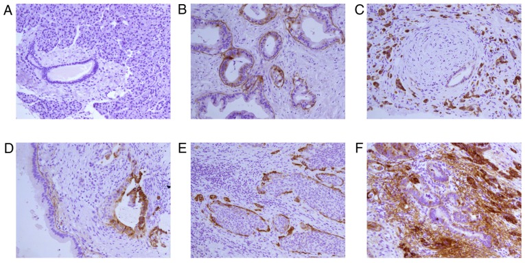
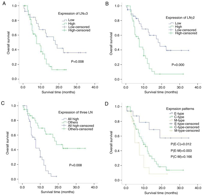
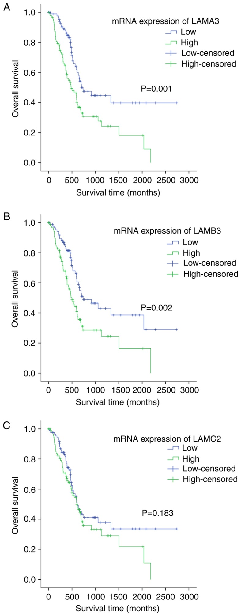
Pancreatic ductal adenocarcinoma (PDA) is a worldwide health problem. Early diagnosis and assessment may enhance the quality of life and survival of patients. The present study investigated the potential correlations between the gene and protein expression of laminin-332 (LM-332 or laminin-5) and clinicopathological factors as well as evaluating its influence on the survival of patients with PDA. The expression of LM-332 subunit mRNAs in pancreatic carcinoma specimens from 37 patients was investigated by reverse transcription-quantitative polymerase chain reaction (RT-qPCR) analysis. Using immunohistochemical methods, the protein expressions of the three chains of LM-322 (LNα3, LNβ3 and LNγ2) were determined in 96 pancreatic carcinoma specimens, for association analysis with clinicopathological characteristics from patient data. The results of the prognosis analysis of three mRNAs expression datasets were validated in The Cancer Genome Atlas datasets. RT-qPCR results indicated that the overall relative values of LNα3 and LNγ2 mRNAs were increased in pancreatic carcinoma compared with the control. In immunostaining analyses LNα3 and LNγ2 expression was observed in all tumor tissues from the 96 patient samples. The expression levels of LNα3, LNβ3 and LNγ2 were associated with each other. LNα3 and LNγ2 positivity was significantly associated with differentiation, depth of invasion and advanced stage (P<0.05). The samples were classified into three groups: Basement membrane (B) type, cytoplasmic (C) type and mixed (M) type, according to their LNγ2 immunohistochemical expression patterns. The B type correlated significantly with differentiation (P=0.010) and the M type was significantly associated with hepatic metastasis (P=0.031). Patients with B-type LNγ2 demonstrated significantly better outcomes than patients with the C or M type (P=0.012 and P=0.003, respectively). Overexpression of the α3, β3 and γ2 chains of LM-332 may serve an important role in the progression and prognosis of PDA.

Keywords: laminin α3; laminin β3; laminin γ2; laminin-332; pancreatic ductal adenocarcinoma.


Figures

Figure 1.
Figure 1.
Immunohistochemistry for LNα3 in pancreatic ductal adenocarcinoma tissues. (A) Pancreatic tissue is negative for staining. (B) Poorly differentiated adenocarcinoma is positive for staining. Intensity of staining for LNα3 in Poorly differentiated domain is more strongly than moderately differentiated domain. (C) Poorly differentiated adenocarcinoma is strong positive for staining. Proliferative duct is negative for staining. (D) Poorly differentiated pancreatic ductal adenocarcinoma positive for staining. Tumor budding was seen in the invasive fronts. Expression of LNα3 is strong. (E) Perineural invasion is observed in poorly differentiated adenocarcinoma. Strong (high expression) stains for LNα3 is shown in tumor cells. (F) Peripancreatic adipo tissue invasion is observed in adenocarcinoma with squamous metaplasia. Strong (high expression) stains for LNα3 is predominantly expressed in cancer cells contacting the stroma at the edge of cancer nests and weakly stains was detected in the center of cancer nests. Magnification, ×200. LNα3, laminin α3.
Figure 2.
Figure 2.
Immunohistochemistry for LNγ2 in pancreatic ductal adenocarcinoma tissues. (A) Peritumoral pancreatic ductal and acinar is negative for staining. (B) Well differentiated adenocarcinoma is positive for staining mainly in the basement membrane. Please note that most of the basement membrane around the duct stained LNγ2 is continuous linear structure. (C) Perineural invasion is observed in poorly differentiated adenocarcinoma. Strong (high expression) stains for LNγ2 is shown in cytoplasm of cancer cells. Please note that the basement membrane around a well-differentiated tubular is positive for staining. (D) The basement membrane in well differentiated adenocarcinoma stained LNγ2 is continuous linear structure. Please note that cytoplasm of cancer cells in tumor budding is stained strongly and the structure of basement membrane is absence. (E) Adenocarcinoma is accompanied with squamous metaplasia. Strong (high expression) stains for LNγ2 is predominantly expressed in the cytoplasm of cancer cells contacting the stroma at the edge of cancer nests and negative stains was detected in the center of cancer nests. (F) The cytoplasm of well differentiated glandular in the center of adenocarcinoma stained LNγ2 is very weakly and the basement membrane is continuous linear structure. The cytoplasm of moderately-poorly differentiated glandular at the edge of adenocarcinoma stained LNγ2 is strongly, the structure of basement membrane is absence and a lot of linear and flocculent basement membrane-like material is observed in extracellular matrix of adenocarcinoma. Magnification, ×200. LNγ2, laminin γ2.
Figure 3.
Figure 3.
Correlation between LAMA3, LAMC2, three LN and three patterns of LAMC2 immunohistochemical expression in pancreatic cancer patients. (A) Kaplan-Meier plots for overall survival for a discriminatory median LAMA3 immunohistochemical expression, (B) LAMC2 (C) three LN and (D) three patterns of LAMC2. P-values were calculated using the log-rank test. LNα3 (laminin α3) and γ2 (laminin γ2) chains are encoded by the LAMA3 and LAMC2 genes, respectively.
Figure 4.
Figure 4.
Correlation between LAMA3, LAMB3 and LAMC2 mRNA expression and prognosis in pancreatic cancer patients. (A) Kaplan-Meier plots for overall survival for a discriminatory median LAMA3 mRNA expression, from TCGA sequencing data to assess prognostic accuracy, (B) LAMB3 and (C) LAMC2. P-values were calculated using the log-rank test. LNα3 (laminin α3), β3 (laminin β3) and γ2 (laminin γ2) chains are encoded by the LAMA3, LAMB3, and LAMC2 genes, respectively.

Similar articles

Cited by

References

    1. Miner JH, Yurchenco PD. Laminin functions in tissue morphogenesis. Annu Rev Cell Dev Biol. 2004;20:255–284. doi: 10.1146/annurev.cellbio.20.010403.094555. - DOI - PubMed
    1. Marinkovich MP. Laminin 332 in squamous-cell carcinoma. Nature Rev Cancer. 2007;7:370–380. doi: 10.1038/nrc2089. - DOI - PubMed
    1. Ii M, Yamamoto H, Taniguchi H, Adachi Y, Nakazawa M, Ohashi H, Tanuma T, Sukawa Y, Suzuki H, Sasaki S, et al. Co-expression of laminin β3 and γ2 chains and epigenetic inactivation of laminin α3 chain in gastric cancer. Int J Oncol. 2011;39:593–599. - PubMed
    1. Guess CM, Quaranta V. Defining the role of laminin-332 in carcinoma. Matrix Biol. 2009;28:445–455. doi: 10.1016/j.matbio.2009.07.008. - DOI - PMC - PubMed
    1. Yamashita H1, Tripathi M, Harris MP, Liu S, Weidow B, Zent R, Quaranta V. The role of a recombinant fragment of laminin-332 in integrin α3β1-dependent cell binding, spreading and migration. Biomaterials. 2010;31:5110–5121. doi: 10.1016/j.biomaterials.2010.03.003. - DOI - PMC - PubMed