

Advanced choroidal melanoma with a desirable aesthetic outcome after enucleation: A case report
- PMID: 29928440
- PMCID: PMC6006346
- DOI: 10.3892/ol.2018.8661
Advanced choroidal melanoma with a desirable aesthetic outcome after enucleation: A case report
Abstract
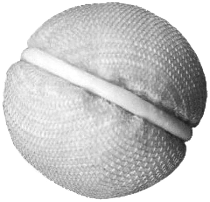
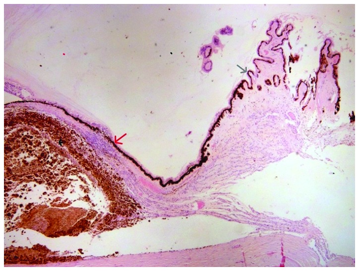
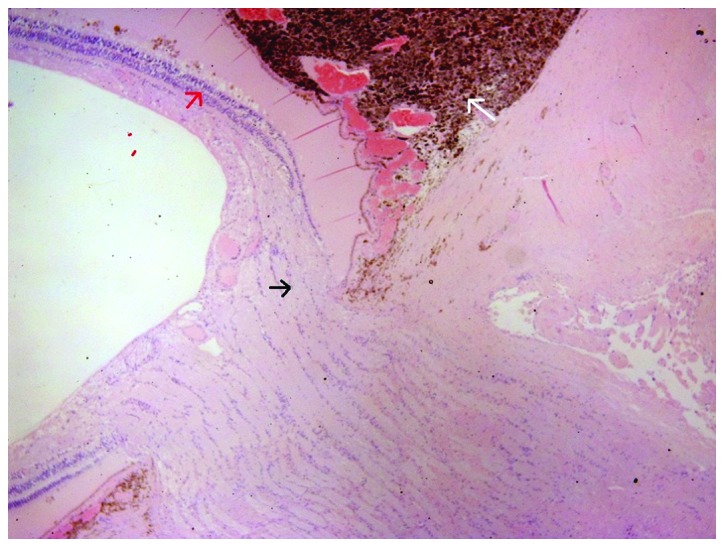
Choroidal melanoma is a rare ocular tumor. The present study reports the case of a 66-year-old male who presented with chronic headache and progressive visual loss. Physical eye examination and combined A- and B-mode ultrasonography detected choroidal melanoma. Due to tumor characteristics the eye was enucleated restoring the orbital volume with a 22 mm intraorbital bioceramic sphere implant. The eye was subjected to histopathological examination that confirmed the choroidal melanoma, 2 cm in diameter and 0.8 cm in elevation, occupying almost half of the globe. Microscopically, the neoplasm comprises mostly of epithelioid cells and fewer Type B spindle cells, with intense pigmentation. AJCC staging for the melanoma was T4b. The patient was fitted with an artificial eye after enucleation. Thirteen months after initial diagnosis, liver metastases were confirmed during his scheduled follow-up.
Keywords: choroidal; enucleation; eye; melanoma; ocular.
Figures
References
-
- Kanski J, Bowling B. Clinical Ophthalmology: A Systematic Approach (7th ed) Elsevier/Saunders; New York: 2011. pp. 501–504.
LinkOut - more resources
Full Text Sources
Other Literature Sources