Primary cauda equina lymphoma diagnosed by nerve biopsy: A case report and literature review
- PMID: 29928449
- PMCID: PMC6006481
- DOI: 10.3892/ol.2018.8629
Primary cauda equina lymphoma diagnosed by nerve biopsy: A case report and literature review
Abstract
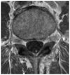
Primary cauda equina lymphoma (CEL) is a rare malignant tumor among various neoplasms that affects the cauda equina nerve roots. The present case report described the case of a 65-year-old man who presented with cauda equina syndrome with progressive motor palsy in the legs and gait disturbance over the last 5 months. Magnetic resonance (MR) images showed enlargement of the cauda equina occupying the dural sac from the L1-S1 level with isointensity to the spinal cord signal on both T1- and T2-weighted imaging. Enhancement of the cauda equina was seen on contrast MR images. On F-18 2-fluoro-2-deoxy-glucose positron emission tomography examination, diffuse accumulation of 2-fluoro-2-deoxy-glucose was observed in the cauda equina with a maximum standardized uptake value of 4.9. Based on elevation of soluble interleukin 2 receptor in cerebrospinal fluid and a biopsy of the enlarging cauda equina, a diagnosis of CEL of the diffuse large B-cell type was made. The present case report provided a detailed case discussion and a review of the available literature on this rare entity, focusing on clinical characteristics and imaging of primary CEL.
Keywords: FDG-PET/CT; MR imaging; cauda equina lymphoma; nerve biopsy; sIL-2R.
Figures


Similar articles
-
Primary Cauda Equina Lymphoma Mimicking Meningioma.J Clin Med. 2024 Aug 22;13(16):4959. doi: 10.3390/jcm13164959. J Clin Med. 2024. PMID: 39201102 Free PMC article. Review.
-
Primary Malignant Lymphoma of the Cauda Equina Diagnosed after Decompression for Lumbar Spinal Stenosis: A Case Report.Tohoku J Exp Med. 2023 Aug 23;260(4):341-346. doi: 10.1620/tjem.2023.J047. Epub 2023 Jun 8. Tohoku J Exp Med. 2023. PMID: 37286520
-
Primary cauda equina lymphoma: case report and literature review.Nagoya J Med Sci. 2014 Aug;76(3-4):349-54. Nagoya J Med Sci. 2014. PMID: 25741044 Free PMC article.
-
[Cauda equina syndrome due to recurrent malignant lymphoma of the spinal cord. A case report].Rinsho Shinkeigaku. 1999 Oct;39(10):1071-4. Rinsho Shinkeigaku. 1999. PMID: 10655773 Review. Japanese.
-
F-18 Fluorodeoxyglucose Positron Emission Tomography Computed Tomography Findings in an Interesting Case of Primary Cauda Equina Lymphoma with Literature Review.Indian J Nucl Med. 2021 Oct-Dec;36(4):425-428. doi: 10.4103/ijnm.ijnm_75_21. Epub 2021 Dec 15. Indian J Nucl Med. 2021. PMID: 35125761 Free PMC article.
Cited by
-
Multiple secondary cauda equina non-Hodgkin's lymphoma: a case report and literature review.BMC Cancer. 2019 Jun 17;19(1):594. doi: 10.1186/s12885-019-5800-4. BMC Cancer. 2019. PMID: 31208357 Free PMC article.
-
Primary Cauda Equina Lymphoma Treated with CNS-Centric Approach: A Case Report and Literature Review.J Blood Med. 2021 Jul 21;12:645-652. doi: 10.2147/JBM.S325264. eCollection 2021. J Blood Med. 2021. PMID: 34321945 Free PMC article.
-
Primary Cauda Equina Lymphoma Mimicking Meningioma.J Clin Med. 2024 Aug 22;13(16):4959. doi: 10.3390/jcm13164959. J Clin Med. 2024. PMID: 39201102 Free PMC article. Review.
-
Primary peripheral nerve lymphoma: a case report and literature review.Neurol Sci. 2024 Apr;45(4):1447-1454. doi: 10.1007/s10072-023-07192-y. Epub 2023 Nov 22. Neurol Sci. 2024. PMID: 37991640 Review.
-
Non-Hodgkin Lymphoma of Cauda Equina: A Diagnostic Conundrum: Case Report.Adv Biomed Res. 2023 Apr 25;12:95. doi: 10.4103/abr.abr_36_21. eCollection 2023. Adv Biomed Res. 2023. PMID: 37288018 Free PMC article.
References
-
- Deckert M, Paulus W, Kluin PM, Ferry JA. Diffuse large B-cell lymphoma of the CNS. In: Louis DN, Ohgaki H, Wiestler OD, Cavenee WK, editors. WHO Classification of Tumours of the Central Nervous System. 4th edition. Vol. 1. IAR Press; Lyon: 2016. pp. 272–275. WHO/IARC Classification of Tumours, Revised.
LinkOut - more resources
Full Text Sources
Other Literature Sources
