. 2018 Jun 1;9(42):26834-26851.
doi: 10.18632/oncotarget.25515.

Mechanisms for mTORC1 activation and synergistic induction of apoptosis by ruxolitinib and BH3 mimetics or autophagy inhibitors in JAK2-V617F-expressing leukemic cells including newly established PVTL-2

Affiliations

Mechanisms for mTORC1 activation and synergistic induction of apoptosis by ruxolitinib and BH3 mimetics or autophagy inhibitors in JAK2-V617F-expressing leukemic cells including newly established PVTL-2

Shinya Ishida et al. Oncotarget. .

Abstract

The activated JAK2-V617F mutant is very frequently found in myeloproliferative neoplasms (MPNs), and its inhibitor ruxolitinib has been in clinical use, albeit with limited efficacies. Here, we examine the signaling mechanisms from JAK2-V617F and responses to ruxolitinib in JAK2-V617F-positive leukemic cell lines, including PVTL-2, newly established from a patient with post-MPN secondary acute myeloid leukemia, and the widely used model cell line HEL. We have found that ruxolitinib downregulated the mTORC1/S6K/4EBP1 pathway at least partly through inhibition of the STAT5/Pim-2 pathway with concomitant downregulation of c-Myc, MCL-1, and BCL-xL as well as induction of autophagy in these cells. Ruxolitinib very efficiently inhibited proliferation but only modestly induced apoptosis. However, inhibition of BCL-xL/BCL-2 by the BH3 mimetics ABT-737 and navitoclax or BCL-xL by A-1331852 induced caspase-dependent apoptosis involving activation of Bak and Bax synergistically with ruxolitinib in HEL cells. On the other hand, the putative pan-BH3 mimetic obatoclax as well as chloroquine and bafilomycin A1 inhibited autophagy at its late stage and induced apoptosis in PVTL-2 cells synergistically with ruxolitinib. The present study suggests that autophagy as well as the anti-apoptotic BCL-2 family members, regulated at least partly by the mTORC1 pathway downstream of STAT5/Pim-2, protects JAK2-V617F-positive leukemic cells from ruxolitinib-induced apoptosis depending on cell types and may contribute to development of new strategies against JAK2-V617F-positive neoplasms.

Keywords: BH3 mimetic; JAK2-V617F; MPN; apoptosis; mTOR.


Conflict of interest statement

CONFLICTS OF INTEREST The authors declare no conflicts of interest.

Figures

Figure 1
Figure 1. Morphological, cytogenetic, and genetic analyses of PVTL-2 cells
(A) A cytospin preparation of PVTL-2 cells. May-Grunwald-Giemsa staining. (B) Giemsa-banded cytogenetic analysis showing a near-tetraploid consensus karyotype, 83,XX,-X,-X,-2,-5,der(5;7)(p10;q10),-7,-8,del(12)(q?)x2,-13,-14,-15,-16,-16,-18,-20,-20,-22,add(22)(q11.2),+7mar. (C) Direct sequence analysis of the JAK2 gene obtained by PCR from PVTL-2 cells. Nucleotide sequences around the codon coding for V617 in normal JAK2 or F617 in the JAK2 mutant are shown with the mutated nucleotide and amino acid sequences indicated in red.
Figure 2
Figure 2. Down stream signaling mechanisms from JAK2-V617F in PVTL-2 and HEL cells
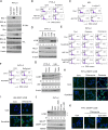
(A) PVTL-1, PVTL-2, and HEL cells were treated for 3 h with 1 μM ruxolitinib or left untreated as control, as indicated, and subjected to immunoblot analysis using antibodies against indicated proteins. Abbreviations: JAK-PY, phospho-Y1007/1008-JAK2; STAT5-PY, phospho-Y694-STAT5. (B) The JAK family members indicated were immunoprecipitated from lysates of PVTL-2 and analyzed by immunoblot analysis using antibodies against indicated proteins. JAK2 was immunoprecipitated with the monoclonal antibody (CS-3230) and was detected using the polyclonal antibody (SC-294), which cross-reacted with JAK3. Positions of JAK3 are indicated by asterisks. PY: anti-phosphotyrosine. (C) PVTL-2 or HEL cells were treated with indicated concentrations of ruxolitinib for 6 h and analyzed. Abbreviations: STAT3-PY, phospho-Y705-STAT3; MEK-P, phospho-S217/221-MEK; Erk-PY, phospho-T202/Y204-Erk; GSK3-P, phospho-S9-GSK3ß; PRAS40-P, phospho-T246-PRAS40; S6K-PT, phospho-T389-p70S6 kinase; 4EBP1-nonP, non-phospho-T46-4EBP1. (D, E) PVTL-2 or HEL cells were treated for 6 h with 1 μM JAKI-1 or 1 μM ruxolitinib, as indicated, and subjected to the cap-binding assay to analyze the eIF4E-eIF4G complex formation. Proteins affinity purified with m7-GTP-sepharose (m7-GTP) as well as total cell lysates (TCL) were subjected to Western blot analysis. (F, G) PVTL-2 or HEL cells transduced with STAT5A1*6 or vector control cells, as indicated, were treated with indicated concentrations of ruxolitinib for 24 h and subjected to immunoblot analysis. Abbreviations: S6RP-P, phospho-S240/244-S6RP; 4EBP1-P, phospho-T37/46-4EBP1. (H) PVTL-2 or HEL cells, as indicated, were treated for 6 h in ASF104 medium with 0.5 μM PP242 or 1 μM of indicated inhibitors and analyzed. Abbreviations: MK, MK-2206; AZD, AZD1208; 4EBP1-S65P, phospho-S65-4EBP1.
Figure 3
Figure 3. Ruxolitinib effectively inhibits proliferation of PVTL-2 and HEL cells without distinctively inducing cell death
(A, B) PVTL-2 or HEL cells, as indicated, were cultured with indicated concentrations of ruxolitinib for 48 h. Viable cell numbers were measured by the Cell counting Kit-8. Relative cell numbers expressed as percentages of cell numbers without ruxolitinib from triplicate samples are plotted with four-parameter logistic curves obtained by using ImageJ software with calculated IC50 (μM) indicated. (C, D) PVTL-2 or HEL cells, as indicated, were cultured with indicated concentrations of ruxolitinib for indicated days. Viable cell numbers and viability were counted and plotted. Each data point represents the mean of triplicate determinations, with error bars indicating standard errors.
Figure 4
Figure 4. Ruxolitinib induces apoptosis synergistically with obatoclax in PVTL-2 cells and with ABT-737 or obatoclax in HEL cells through caspase-dependent mechanisms involving activation of Bax and Bak
(A) PVTL-2 or HEL cells, as indicated, were cultured with 1 μM ABT-737 or 0.5 μM obatoclax in the presence or absence of 1 μM ruxolitinib, as indicated, for 48 h and analyzed for the cellular DNA content by flow cytometry. Percentages of apoptotic cells with sub-G1 DNA content are indicated. (B) HEL cells were cultured with indicated concentrations of A-1331852 (A-13) in the presence or absence of 1 μM ruxolitinib, as indicated, for 48 h and analyzed. (C) HEL cells were cultured with 1 μM ABT-737 and 100 μM Boc-d-FMK in the presence or absence of 1 μM ruxolitinib, as indicated, for 24 h and analyzed for activation of Caspase-3 by flow cytometry. Percentages of cells with cleaved Caspase-3 are indicated. (D) HEL cells were cultured with 1 μM ruxolitinib and 1 μM ABT-737 in the presence or absence of 100 μM Boc-d-FMK, as indicated, for 48 h and analyzed for the cellular DNA content. (E) HEL cells were cultured with 1 μM ABT-737, 100 μM Boc-d-FMK, and 1 μM ruxolitinib, as indicated, for 16 h and analyzed for activation of Bak by flow cytometry. Percentages of cells with activated Bak are indicated. (F) HEL cells transduced with the wild-type BCL-xL (WT) or its mutants (Mut1, Mut8) and vector control cells (Cont.) were cultured for 24 h with or without 1 μM ruxolitinib, as indicated, and subjected to immunoblot analysis using antibodies against indicated proteins. (G) HEL cells transduced with the wild-type BCL-xL or its mutants (Mut1, Mut8) and vector control cells, as indicated, were treated with the combination of 1 μM ruxolitinib and 1 μM ABT-737 for 48 h or 16 h to be subjected to flow cytometric analyses for the cellular DNA content or activation of Bax and Bak, respectively. Percentages of apoptotic cells with sub-G1 DNA content and activated Bak (Act. Bak) or Bax (Act. Bax) are indicated.
Figure 5
Figure 5. Inhibition of autophagy induced by ruxolitinib leads to induction of apoptosis in PVTL-2 cells
(A) PVTL-2 cells were cultured with 1 μM ruxolitinib, 1 μM ABT-737, 10 μM A-1210477, or 0.5 μM obatoclax, as indicated, for 8 h and lysed. Immunoprecipitates (IP) obtained with anti-Bim and total cell lysates (TCL) were subjected to immunoblot analysis using antibodies against indicated proteins. (B) PVTL-2 cells were cultured with 1 μM ABT-737, 10 μM A-1210477, and 1 μM ruxolitinib, as indicated, for 48 h and analyzed for the cellular DNA content by flow cytometry. Percentages of apoptotic cells with sub-G1 DNA content are indicated. (C) HEL cells were cultured with 1 μM ABT-737 and 10 μM A-1210477, as indicated, for 48 h and analyzed. (D) PVTL-2 cells were treated with 1 μM ruxolitinib (Ruxo), 1 μM ABT-737 (ABT), 10 μM A-1210477 (A-12), 0.5 μM obatoclax (Obat), or 40 μM chloroquine (CQ) for 8 h and subjected to immunoblot analysis using antibodies against indicated proteins. Positions of LC3B-I and LC3B-II as well as the relative ratio of LC3B-II/LC3B-I (II/I) determined by densitometric analysis are indicated. (E) PVTL-2 cells were cultured with 1 μM ruxolitinib, 1 μM ABT-737, 0.5 μM obatoclax, or 40 μM chloroquine, as indicated, for 36 h and analyzed for the cellular DNA content by flow cytometry. Percentages of apoptotic cells with sub-G1 DNA content are indicated. (F) PVTL-2 cells were cultured with 1 μM ruxolitinib and 100 nM bafilomycin A1, as indicated, for 48 h and analyzed. (G) PVTL-2 or HEL cells were treated with 1.5 μM ruxolitinib (Ruxo) or 1.5 μM fedratinib (Fedra), as indicated, for 16 h and subjected to immunoblot analysis using antibodies against indicated proteins. Relative expression levels of p62 were determined by densitometric analysis and are shown below the panel. (H) PVTL-2 cells transduced with EGFP-LC3B were treated with or without 1 μM ruxolitinib for 6 h with or without 40 μM chloroquine or 0.5 μM obatoclax added during the last 2 h. Cells were stained with DAPI for nuclear staining and analyzed by confocal immunofluorescence microscopy. Representative merged images of EGFP-LC3B (green) and DAPI (blue) are shown. (I) HEL cells transduced with EGFP-LC3B were treated with or without 1 μM ruxolitinib for 4 h in the presence or absence of 50 μM chloroquine and analyzed by confocal immunofluorescence microscopy. (J) PVTL-2 cells were treated for 6 h with 1 μM of ruxolitinib (Ruxo), GDC-0941 (GDC), PP242, MK-2206 (MK), or AZD1208 (AZD), as indicated, and analyzed. Abbreviations: S6K-PT, phospho-T389-p70S6 kinase; 4EBP1-nonP, non-phospho-T46-4EBP1. (K) PVTL-2 cells transduced with EGFP-LC3B were left untreated for control or treated with or without 1 μM GDC-0941 for 6 h with 40 μM chloroquine added during the last 2 h and analyzed by confocal immunofluorescence microscopy.
Figure 6
Figure 6. A schematic model of intracellular signaling mechanisms by which JAK2-V617F regulates proliferation, autophagy, and apoptosis in leukemic cells and their inhibition by various small molecule inhibitors
Abbreviations: CQ, chloroquine; Bafilo., bafilomycin A1.

Similar articles

Cited by

References

    1. Springuel L, Renauld JC, Knoops L. JAK kinase targeting in hematologic malignancies: a sinuous pathway from identification of genetic alterations towards clinical indications. Haematologica. 2015;100:1240–1253. https://doi.org/10.3324/haematol.2015.132142. - DOI - PMC - PubMed
    1. Ihle JN, Gilliland DG. Jak2: normal function and role in hematopoietic disorders. Curr Opin Genet Dev. 2007;17:8–14. https://doi.org/10.1016/j.gde.2006.12.009. - DOI - PubMed
    1. Spivak JL. Myeloproliferative Neoplasms. N Engl J Med. 2017;376:2168–2181. https://doi.org/10.1056/NEJMra1406186. - DOI - PubMed
    1. Bose P, Verstovsek S. JAK2 inhibitors for myeloproliferative neoplasms: what is next? Blood. 2017;130:115–125. https://doi.org/10.1182/blood-2017-04-742288. - DOI - PMC - PubMed
    1. Quentmeier H, MacLeod RA, Zaborski M, Drexler HG. JAK2 V617F tyrosine kinase mutation in cell lines derived from myeloproliferative disorders. Leukemia. 2006;20:471–476. https://doi.org/10.1038/sj.leu.2404081. - DOI - PubMed