. 2018;8(1):412.
Epub 2018 Jan 9.

Towards a circuit-level understanding of hippocampal CA1 dysfunction in Alzheimer's disease across anatomical axes

Affiliations

Towards a circuit-level understanding of hippocampal CA1 dysfunction in Alzheimer's disease across anatomical axes

Arjun V Masurkar. J Alzheimers Dis Parkinsonism. 2018.

Abstract

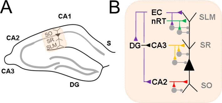
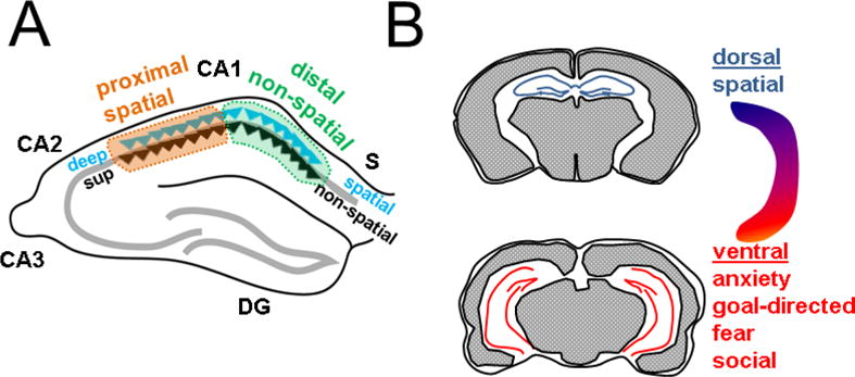
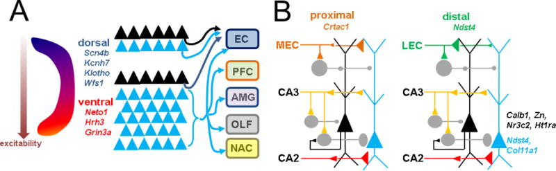
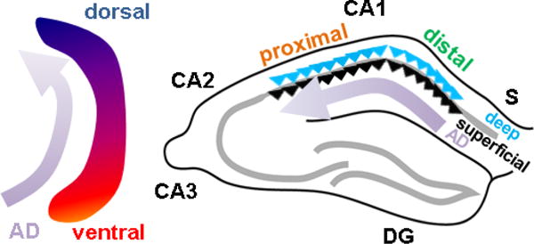
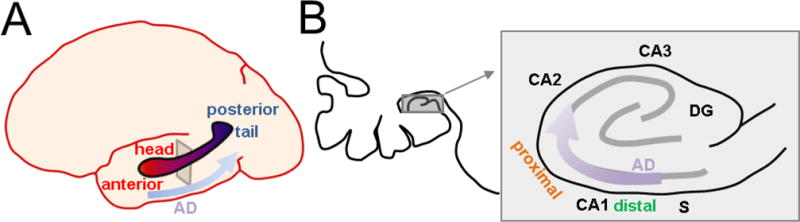
The hippocampus has been a primary region of study with regards to synaptic and functional changes in Alzheimer’s disease (AD) due to its involvement in early stages, specifically area CA1. However, most work in this area has treated CA1 as a homogeneous structure comprised of uniform neural circuits. Yet, there is a plethora of evidence that CA1 varies in its structure and function across anatomical axes. Here I review the heterogeneity of the functional and circuit architecture of hippocampal area CA1 across three primary anatomical axes. I also summarize evidence that AD differentially affects these subregions, as well as hypotheses as to why this may occur.

Keywords: Alzheimer’s disease; Hippocampus; CA1; entorhinal cortex; Pyramidal neuron.


Figures

Figure 1
Figure 1. Overview of CA1 circuitry
A Hippocampal CA1 in relation to other subregions: subiculum (S), CA2, CA3, dentate gyrus (DG). CA1 pyramidal neuron is indicated with labeled strata stratum oriens (SO), stratum radiatum (SR) and stratuma lacunosum moleculare (SLM). B. CA1 pyramidal neuron excitatory (triangle) and inhibitory (circle) inputs to different parts of its apical dendrite in SR, SLM and basal dendrite in SO. Distal apical dendrite receives direct input from entorhinal cortex (EC) and nucleus reuniens of thalamus (nRT). Indirect EC input arrives at the proximal apical dendrite, via DG and CA3, or at the basal dendrite, from CA2. Each pathway can elicit inhibition in feedforward fashion.
Figure 2
Figure 2. Functional heterogeneity of CA1 across anatomical axes
A Across transverse axis, spatial encoding is more robust towards CA2 (proximal); non-spatial encoding is more robust towards S (distal). Across the radial axis, deep neurons (blue triangle) show more spatial tuning and superficial neurons (black triangle) appear to be specialized for non-spatial processing. B. Across the longitudinal axis, dorsal CA1 (top) shows more robust spatial responses whereas ventral CA1 (below) is specialized for various affective and motivational behaviors.
Figure 3
Figure 3. Intrinsic and circuit heterogeneity of CA1 across anatomical axes
A Longitudinal axis intrinsic differences include differential expression of physiologically relevant genes (italics) and increased electrical excitability in ventral CA1. Circuit differences derive from increased diversity of axonal projections in ventral CA1 (below), mainly from deep (blue triangle) rather than superficial neurons (black triangle). Targets include EC, prefrontal cortex (PFC), amygdala (AMG), olfactory areas (OLF), and nucleus accumbens (NAC). Dorsal CA1 neurons (top) project back to EC. B. Transverse and radial axis molecular differences are few (italics). Circuit differences derive from preferential targeting of proximal CA1 by medial entorhinal cortex (MEC) and distal CA1 by lateral entorhinal cortex (LEC). Moreover, MEC favors deep and LEC favors superficial neurons. Deep neurons also receive more proximal inhibition, partially via superficial neurons. CA2 preferentially excites deep neurons. Variability of inhibition to the basal dendrite is not known (not shown).
Figure 4
Figure 4. Spatiotemporal evolution of Alzheimer disease across rodent CA1 axes
In mouse models, Alzheimer disease (AD) pathology begins in ventral hippocampus and progresses dorsally (left) and begins in distal CA1 at the CA1-S border, and progresses proximally to CA2 (right). Distribution across the radial axis is unknown.
Figure 5
Figure 5. Spatiotemporal evolution of Alzheimer disease across human CA1 axes
A Biomarkers of Alzheimer disease (AD) begin anteriorly in the hippocampal head and progress posteriorly towards the hippocampal tail. B. Left, expansion of the coronal cross-section indicated in A. showing position of the hippocampus in the temporal lobe (gray square). Right, detailed view of human hippocampus indicating that AD pathology begins in distal CA1 at the CA1-S border, and progresses proximally to CA2. Distribution across the radial axis is unknown.

Similar articles

Cited by

References

    1. Ahn JH, So SP, Kim NY, Kim HJ, Yoon SY, Kim DH. c-Jun N-terminal Kinase (JNK) induces phosphorylation of amyloid precursor protein (APP) at Thr668, in okadaic acid-induced neurodegeneration. BMB Rep. 2016;49:376–381. - PMC - PubMed
    1. Amaral DG, Dolorfo C, Alvarez-Royo P. Organization of CA1 projections to the subiculum: a PHA-L analysis in the rat. Hippocampus. 1991;1:415–435. - PubMed
    1. Arszovszki A, Borhegyi Z, Klausberger T. Three axonal projection routes of individual pyramidal cells in the ventral CA1 hippocampus. Front Neuroanat. 2014;8:53. - PMC - PubMed
    1. Baimbridge KG, Miller JJ. Immunohistochemical localization of calcium-binding protein in the cerebellum, hippocampal formation and olfactory bulb of the rat. Brain Res. 1982;245:223–229. - PubMed
    1. Basu J, Siegelbaum SA. The Corticohippocampal Circuit, Synaptic Plasticity, and Memory. Cold Spring Harb Perspect Biol. 2015;7 - PMC - PubMed

LinkOut - more resources