

Case Reports
doi: 10.1016/j.eucr.2018.06.004.
eCollection 2018 Sep.
Pedunculated and obstructive Wilms' tumor: A rare presentation in a 2 year-old male
Affiliations
- PMID: 29928591
- PMCID: PMC6008277
- DOI: 10.1016/j.eucr.2018.06.004
Item in Clipboard
Case Reports
Pedunculated and obstructive Wilms' tumor: A rare presentation in a 2 year-old male
Urol Case Rep.
.
Abstract
Wilms' tumor manifesting as an obstructing ureteral mass is extremely rare. Herein, we report an unusual case in which a child presented with a clinical picture concerning for and suggestive of ureteropelvic junction obstruction (UPJO), but was instead found to have an intrapelvic pedunculated Wilms' tumor with extension into the proximal ureter. We discuss the patient's diagnostic workup, radiographic, operative and pathologic findings, as well as important lessons learned from this unusual case.
Keywords: Cancer; Nephrectomy; Pediatric urology; Wilms' tumor.
Figures

Renal US demonstrating a 2.3 cm hemorrhagic debris-filled focus within the left UPJ and proximal ureter, obstructing the renal pelvis.
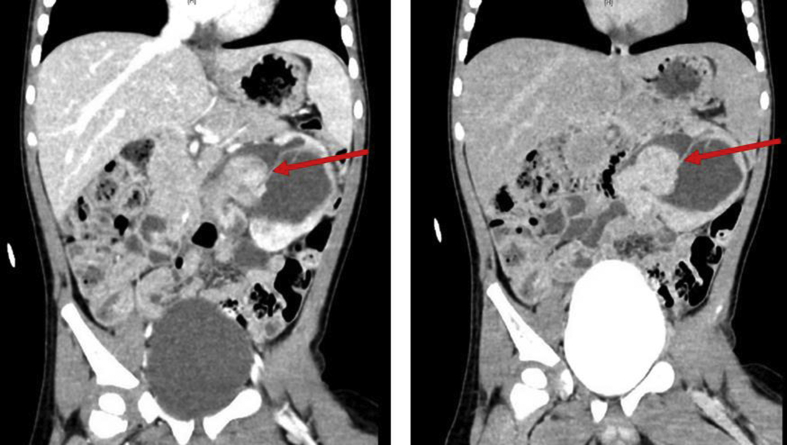
CT with contrast arterial phase (left) and delayed phase (right) demonstrated an enhancing, rounded mass at the level of the left UPJ with extension into the proximal ureter along with several other smaller enhancing mass-like lesions within the lower pole collecting system.
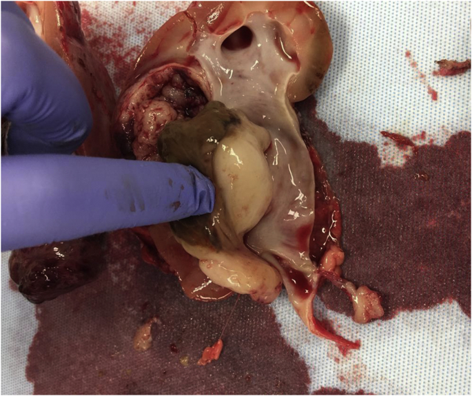
Specimen from left nephrectomy. The mass has a friable pink peripheral rim with both solid and cystic components. (For interpretation of the references to colour in this figure legend, the reader is referred to the Web version of this article.)

Microscopic image at 200×. Triphasic pattern with blastemal, epithelial and stromal components.
References
-
- Breslow N., Olshan A., Beckwith J.B., Green D.M. Epidemiology of Wilms tumor. Med Pediatr Oncol. 1993;21(3):172–181. - PubMed
-
- Coppes M.J., Haber D.A., Grundy P.E. Genetic events in the development of Wilms' tumor. N Engl J Med. 1994;331(9):586–590. - PubMed
-
- Ritchey ML, Shamberger RC. Pediatric Urologic Oncology: Renal and Adrenal. Campbell-Walsh Urology, 11e: Elsevier, Inc: p3559–3581.
-
- Konety B.R., Vaena D.A., Williams R.D. Chapter 22. Renal parenchymal neoplasms. In: McAninch J.W., Lue T.F., editors. Smith & Tanagho's General Urology, 18e. The McGraw-Hill Companies; New York, NY: 2013.
Publication types
LinkOut - more resources
Full Text Sources
Other Literature Sources
