. 2018 Jun 13:5:737-743.
doi: 10.1016/j.toxrep.2018.06.007. eCollection 2018.

Complex toxicity as disruption of adipocyte or osteoblast differentiation in human mesenchymal stem cells under the mixed condition of TBBPA and TCDD

Affiliations

Complex toxicity as disruption of adipocyte or osteoblast differentiation in human mesenchymal stem cells under the mixed condition of TBBPA and TCDD

Hideki Kakutani et al. Toxicol Rep. .

Erratum in

Abstract

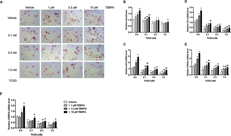
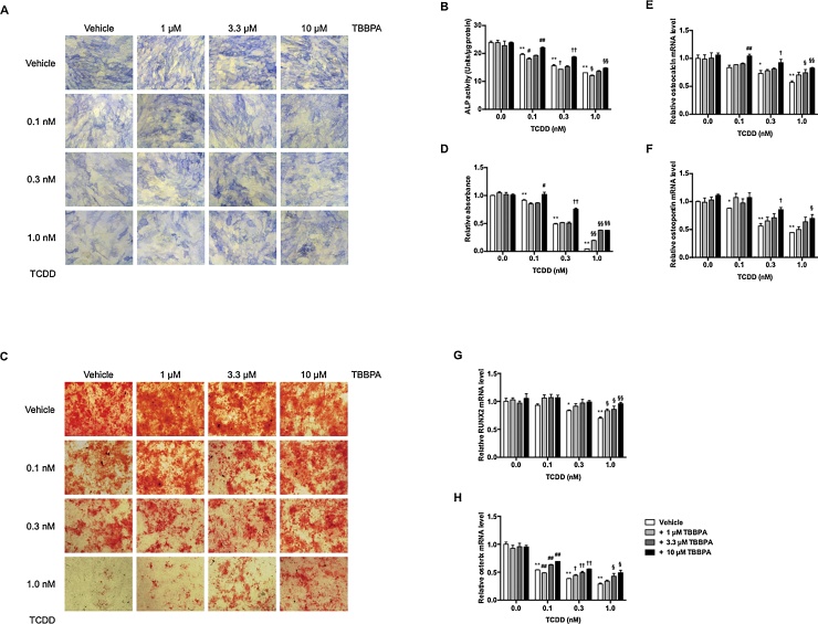
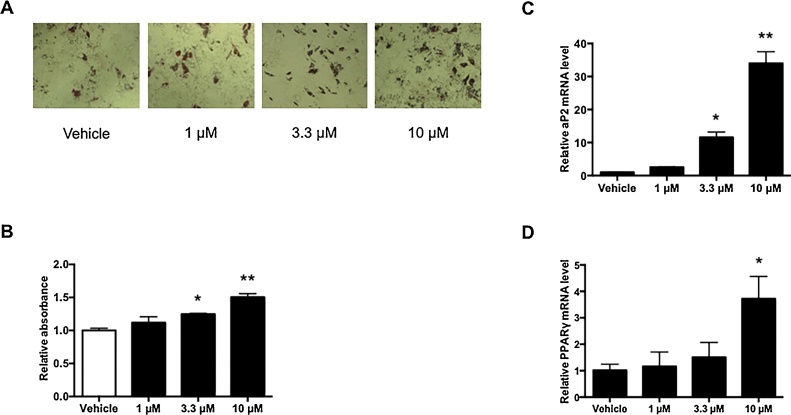
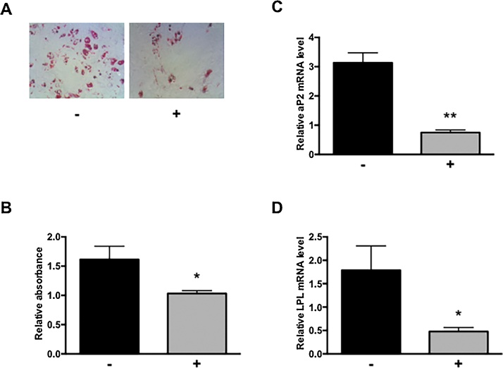
People are frequently and unintentionally exposed to many chemical compounds, such as environmental pollutants and endocrine-disrupting chemicals (EDCs), in food and from the atmosphere. In particular, endocrine-disrupting TBBPA and dioxins are found in human breast milk and in the body. Conventional studies evaluate toxicity by administering a single substance to cells or animals, but evaluation of the toxicity of mixtures of these ingested compounds is essential for "true" toxicological assessment. We evaluated toxic effects in vitro using human mesenchymal stem cells (hMSCs). TBBPA increased the number of lipid droplets, and upregulated the expression of adipocyte-related mRNA, aP2 and LPL, through a PPARγ-dependent mechanism. TCDD suppressed lipid droplets and adipocyte-related mRNA levels. Adipocyte differentiation was stimulated by TBBPA and inhibited by TCDD in a dose-dependent manner. TBBPA did not influence osteoblast differentiation, but TCDD suppressed ALP staining and activity, calcium deposition, and osteoblast-related mRNA levels. In a mixture of TBBPA and TCDD, TBBPA inhibited TCDD suppression of adipocyte and osteoblast differentiation in a dose-dependent manner. Interestingly, we observed lipid droplets in TBBPA-treated cells differentiated into osteoblasts. These results suggest that TBBPA and TCDD disrupted differentiation into adipocytes and osteoblasts and contributes to a more complete toxicological understanding of exposure to these chemical substances.

Keywords: 2,3,7,8-tetrachlorodibenzo-p-dioxin; ALP, alkaline phosphatase; Adipocyte differentiation; BFRs, brominated flame retardants; C/EBPα, CCAAT-enhancer-binding protein alpha; DOHaD, developmental origins of health and disease; EDCs, endocrine-disrupting chemicals; Human mesenchymal stem cell; LPL, lipoprotein lipase; MSC, mesenchymal stem cell; Osteoblast differentiation; PCDDs/DFs, polychlorinated dibenzo-p-dioxins and dibenzofurans; PPARγ, peroxisome proliferator activated receptor gamma; RUNX2, runt-related transcription factor 2; TBBPA, tetrabromobisphenol A; TCDD, 2,3,7,8-tetrachlorodibenzo-p-dioxin; Tetrabromobisphenol A; aP2, adipocyte-specific protein 2.


Figures

Fig. 1
Fig. 1
Effects of TBBPA and TCDD on adipocyte differentiation in hMSCs. hMSCs were incubated with 1, 3.3, and 10 μM TBBPA and/or 0.0, 0.1, 0.3, and 1 nM TCDD for 21 days. (A) Lipid droplets were stained with oil red O. Morphological changes were observed under a microscope at 40× magnification. (B) Quantitative measurement of lipid droplets stained with oil red O. Oil red O in lipid droplets was eluted with isopropanol. The relative absorbance at 550 nm was expressed as the fold induction as compared with the vehicle. After incubation, aP2 (C), LPL (D), PPARγ (E), and C/EBPα (F) mRNA levels were measured by quantitative real-time PCR. The relative mRNA level was expressed as the fold induction as compared with the vehicle. The data are presented as means ± SD (n = 5). *P < 0.05, **P < 0.01, compared with vehicle-treated cells in 0.0 nM TCDD. #P < 0.05, ##P < 0.01, compared with vehicle-treated cells in 0.1 nM TCDD. †P < 0.05, ††P < 0.01, compared with vehicle-treated cells in 0.3 nM TCDD. §P < 0.05, §§P < 0.01, compared with vehicle-treated cells in 1.0 nM TCDD.
Fig. 2
Fig. 2
Effects of TBBPA and TCDD on osteoblast differentiation in hMSCs. hMSCs were incubated with 1, 3.3, and 10 μM TBBPA and/or 0.0, 0.1, 0.3, and 1 nM TCDD for 14 or 21 days. At Day 14, ALP staining (A) and activity (B) were measured. At Day 21, calcium deposits were stained with alizarin red S (C). Morphological changes were observed under a microscope at 40× magnification. (D) Quantitative measurement of calcium deposites stained with alizarin red S. Alizarin red S in calcium deposites was eluted with 10% acetic acid. The relative absorbance at 450 nm was expressed as the fold induction as compared with the vehicle. At Day 21, osteocalcin (E), osteopontin (F), RUNX2 (G), and osterix (H) mRNA levels were measured by quantitative real-time PCR. The relative mRNA level was expressed as the fold induction as compared with the vehicle. The data are presented as means ± SD (n = 5). *P < 0.05, **P < 0.01, compared with vehicle-treated cells in 0.0 nM TCDD. #P < 0.05, ##P < 0.01, compared with vehicle-treated cells in 0.1 nM TCDD. †P < 0.05, ††P < 0.01, compared with vehicle-treated cells in 0.3 nM TCDD. §P < 0.05, §§P < 0.01, compared with vehicle-treated cells in 1.0 nM TCDD.
Fig. 3
Fig. 3
TBBPA-induced lipogenesis in hMSC osteoblast differentiation. hMSCs were incubated with 1, 3.3, and 10 μM TBBPA in osteoblast differentiation medium for 21 days. (A) Lipid droplets were stained with oil red O. Morphological changes were observed under a microscope at 40× magnification. (B) Quantitative measurement of lipid droplets stained with oil red O. Oil red O in lipid droplets was eluted with isopropanol. The relative absorbance at 550 nm was expressed as the fold induction as compared with the vehicle. aP2 (C) and PPARγ (D) mRNA levels were measured by quantitative real-time PCR. The relative mRNA level was expressed as the fold induction as compared with the vehicle. The data are presented as means ± SD (n = 5). *P < 0.05, **P < 0.01, significantly different from that in vehicle-treated cells.
Fig. 4
Fig. 4
Involvement of PPARγ in TBBPA-induced lipogenesis. hMSCs differentiated into osteoblast with 10 μM TBBPA in the absence (-) or presence (+) of 1 μM GW9662 for 21 days. (A) Lipid droplets were stained with oil red O. Morphological changes were observed under a microscope at 40× magnification. (B) Quantitative measurement of lipid droplets stained with oil red O. Oil red O in lipid droplets was eluted with isopropanol. The relative absorbance at 550 nm was expressed as the fold induction as compared with the vehicle. aP2 (C), LPL (D), and PPARγ (E) mRNA levels were measured by quantitative real-time PCR. The relative mRNA level was expressed as the fold induction as compared with the vehicle. The data are presented as means ± SD (n = 5). *P < 0.05, **P < 0.01, significantly different from that in the absence of GW9662.

References

    1. Buchanan D.L., Sato T., Peterson R.E., Cooke P.S. Antiestrogenic effects of 2,3,7,8-tetrachlorodibenzo-p-dioxin in mouse uterus: critical role of the aryl hydrocarbon receptor in stromal tissue. Toxicol. Sci. 2000;57:302–311. - PubMed
    1. Meerts I.A., Letcher R.J., Hoving S., Marsh G., Bergman A., Lemmen J.G. In vitro estrogenicity of polybrominated diphenyl ethers, hydroxylated PDBEs, and polybrominated bisphenol A compounds. Environ. Health Perspect. 2001;109:399–407. - PMC - PubMed
    1. Chamorro-Garcia R., Sahu M., Abbey R.J., Laude J., Pham N., Blumberg B. Transgenerational inheritance of increased fat depot size, stem cell reprogramming, and hepatic steatosis elicited by prenatal exposure to the obesogen tributyltin in mice. Environ. Health Perspect. 2013;121:359–366. - PMC - PubMed
    1. Manikkam M., Tracey R., Guerrero-Bosagna C., Skinner M.K. Plastics derived endocrine disruptors (BPA, DEHP and DBP) induce epigenetic transgenerational inheritance of obesity, reproductive disease and sperm epimutations. PloS One. 2013;8 - PMC - PubMed
    1. Newbold R.R., Padilla-Banks E., Jefferson W.N. Environmental estrogens and obesity. Mol. Cell. Endocrinol. 2009;304:84–89. - PMC - PubMed