. 2018 Apr 14;5(6):687-696.
doi: 10.1002/acn3.563. eCollection 2018 Jun.

Cardiac responses to viewing facial emotion differentiate frontotemporal dementias

Affiliations

Cardiac responses to viewing facial emotion differentiate frontotemporal dementias

Charles R Marshall et al. Ann Clin Transl Neurol. .

Abstract

Objective: To establish proof-of-principle for the use of heart rate responses as objective measures of degraded emotional reactivity across the frontotemporal dementia spectrum, and to demonstrate specific relationships between cardiac autonomic responses and anatomical patterns of neurodegeneration.

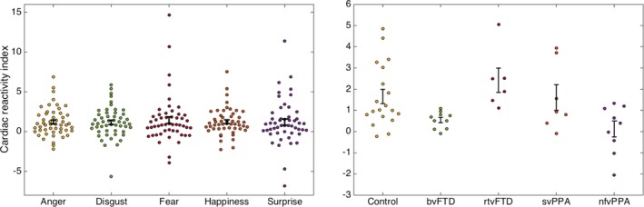
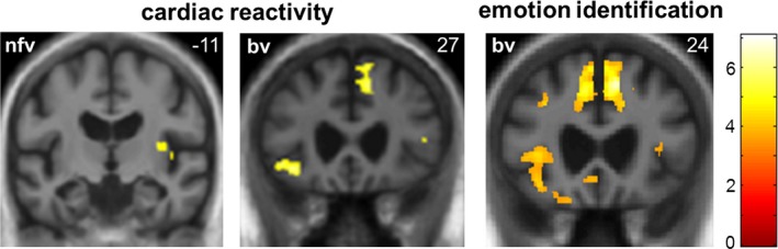
Methods: Thirty-two patients representing all major frontotemporal dementia syndromes and 19 healthy older controls performed an emotion recognition task, viewing dynamic, naturalistic videos of facial emotions while ECG was recorded. Cardiac reactivity was indexed as the increase in interbeat interval at the onset of facial emotions. Gray matter associations of emotional reactivity were assessed using voxel-based morphometry of patients' brain MR images.

Results: Relative to healthy controls, all patient groups had impaired emotion identification, whereas cardiac reactivity was attenuated in those groups with predominant fronto-insular atrophy (behavioral variant frontotemporal dementia and nonfluent primary progressive aphasia), but preserved in syndromes focused on the anterior temporal lobes (right temporal variant frontotemporal dementia and semantic variant primary progressive aphasia). Impaired cardiac reactivity correlated with gray matter atrophy in a fronto-cingulo-insular network that overlapped correlates of cognitive emotion processing.

Interpretation: Autonomic indices of emotional reactivity dissociate from emotion categorization ability, stratifying frontotemporal dementia syndromes and showing promise as novel biomarkers. Attenuated cardiac responses to the emotions of others suggest a core pathophysiological mechanism for emotional blunting and degraded interpersonal reactivity in these diseases.


Figures

Figure 1
Figure 1
Cardiac reactivity indices by emotion and participant group. Plots show individual participants’ mean cardiac reactivity index (mean percentage change in RR interval, see text) to viewing each of the assessed universal facial emotions (left) and mean overall cardiac reactivity index across viewed emotions, separately for each participant group (right; note change of scale on y‐axis). Error bars represent standard error of the mean. bvFTD, patients with behavioral variant frontotemporal dementia; Control, healthy control group; nfvPPA, patients with nonfluent variant primary progressive aphasia; rtvFTD, patients with right temporal variant frontotemporal dementia; svPPA, patients with semantic variant primary progressive aphasia.
Figure 2
Figure 2
Neuroanatomical correlates of heart rate response to viewing facial emotion and emotion identification in patients. Statistical parametric maps of regional gray matter volume associated with change in RR interval and performance on a facial emotion identification task (derived from a voxel‐based morphometric analysis) are shown for patients with behavioral variant frontotemporal dementia (bv) and nonfluent variant primary progressive aphasia (nfv; these syndromic groups showed an attenuated heart rate response relative to healthy controls). Maps have been overlaid on representative coronal sections of the normalized study‐specific T1‐weighted group mean brain MR image, thresholded at P < 0.001 uncorrected over the whole brain for the purpose of display; regional local maxima (see text) were significant at P < 0.05FWE corrected for multiple comparisons within prespecified anatomical regions of interest. The MNI coordinate (mm) of the plane of each section is indicated (the right hemisphere is on the right in each case) and the color bar codes T values.

References

    1. Warren JD, Rohrer JD, Rossor MN. Clinical review. Frontotemporal dementia. BMJ 2013;347:f4827. - PMC - PubMed
    1. Ranasinghe KG, Rankin KP, Pressman PS, et al. Distinct subtypes of behavioral variant frontotemporal dementia based on patterns of network degeneration. JAMA Neurol 2016;73:1078–1088. - PMC - PubMed
    1. Chan D, Anderson V, Pijnenburg Y, et al. The clinical profile of right temporal lobe atrophy. Brain 2009;132(Pt 5):1287–1298. - PubMed
    1. Couto B, Manes F, Montañés P, et al. Structural neuroimaging of social cognition in progressive non‐fluent aphasia and behavioral variant of frontotemporal dementia. Front Hum Neurosci 2013;7:467. - PMC - PubMed
    1. Rosen HJ, Pace‐Savitsky K, Perry RJ, et al. Recognition of emotion in the frontal and temporal variants of frontotemporal dementia. Dement Geriatr Cogn Disord 2004;17:277–281. - PubMed

LinkOut - more resources