. 2018 Jun 22;16(1):66.
doi: 10.1186/s12915-018-0536-1.

Determinants of the cytosolic turnover of mitochondrial intermembrane space proteins

Affiliations

Determinants of the cytosolic turnover of mitochondrial intermembrane space proteins

Lukasz Kowalski et al. BMC Biol. .

Abstract

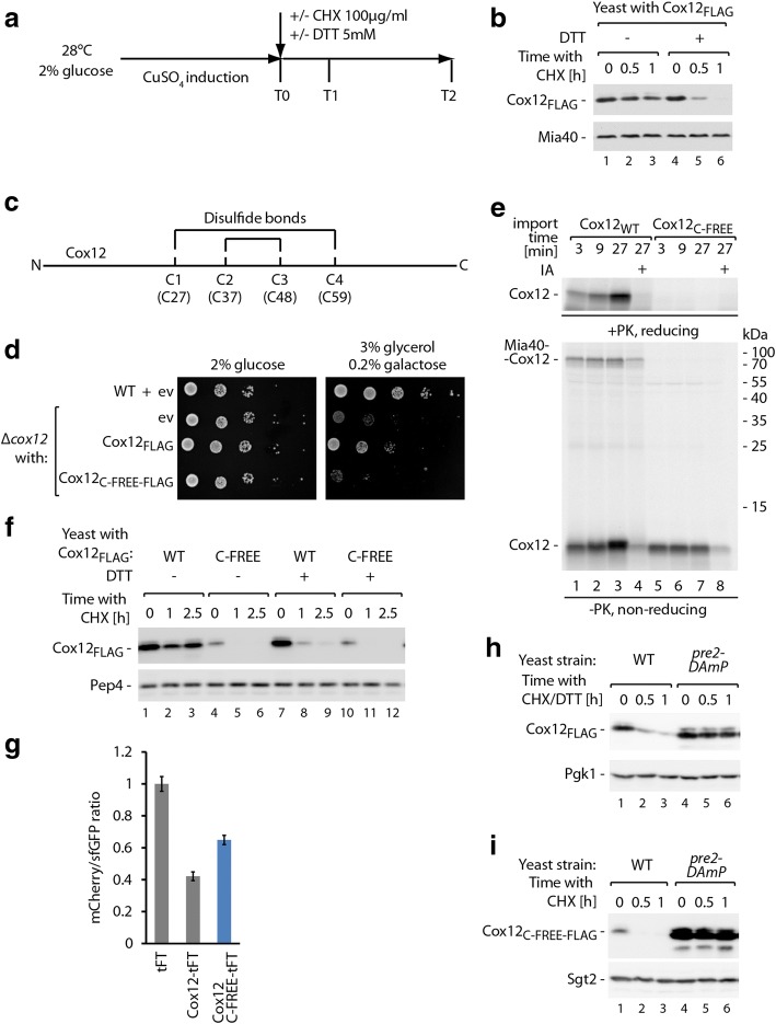
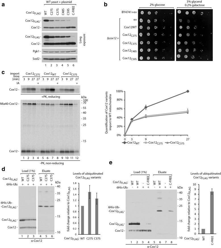
Background: The proteome of mitochondria comprises mostly proteins that originate as precursors in the cytosol. Before import into the organelle, such proteins are exposed to cytosolic quality control mechanisms. Multiple lines of evidence indicate a significant contribution of the major cytosolic protein degradation machinery, the ubiquitin-proteasome system, to the quality control of mitochondrial proteins. Proteins that are directed to the mitochondrial intermembrane space (IMS) exemplify an entire class of mitochondrial proteins regulated by proteasomal degradation. However, little is known about how these proteins are selected for degradation.

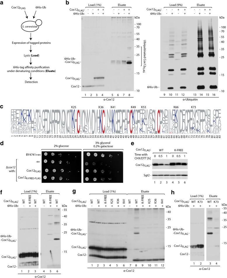
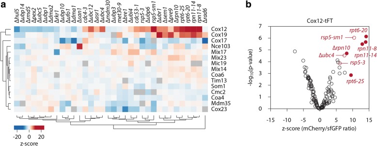
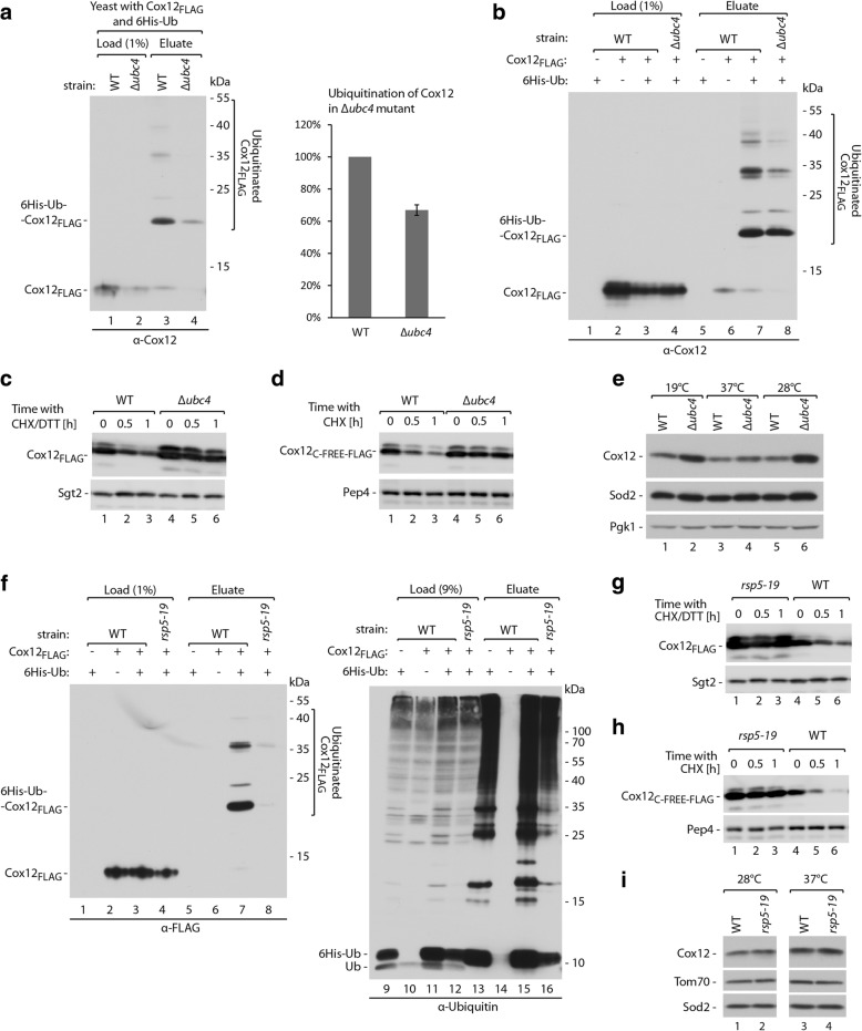
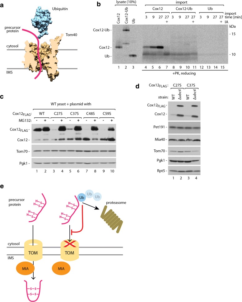
Results: The present study revealed the heterogeneous cytosolic stability of IMS proteins. Using a screening approach, we found that different cytosolic factors are responsible for the degradation of specific IMS proteins, with no single common factor involved in the degradation of all IMS proteins. We found that the Cox12 protein is rapidly degraded when localized to the cytosol, thus providing a sensitive experimental model. Using Cox12, we found that lysine residues but not conserved cysteine residues are among the degron features important for protein ubiquitination. We observed the redundancy of ubiquitination components, with significant roles of Ubc4 E2 ubiquitin-conjugating enzyme and Rsp5 E3 ubiquitin ligase. The amount of ubiquitinated Cox12 was inversely related to mitochondrial import efficiency. Importantly, we found that precursor protein ubiquitination blocks its import into mitochondria.

Conclusions: The present study confirms the involvement of ubiquitin-proteasome system in the quality control of mitochondrial IMS proteins in the cytosol. Notably, ubiquitination of IMS proteins prohibits their import into mitochondria. Therefore, ubiquitination directly affects the availability of precursor proteins for organelle biogenesis. Importantly, despite their structural similarities, IMS proteins are not selected for degradation in a uniform way. Instead, specific IMS proteins rely on discrete components of the ubiquitination machinery to mediate their clearance by the proteasome.

Keywords: Cox12; Intermembrane space; Mitochondria; Proteasome; Protein degradation; Protein import; Ubiquitination.


Conflict of interest statement

Ethics approval and consent to participate

Not applicable.

Competing interests

The authors declare that they have no competing interests.

Publisher’s Note

Springer Nature remains neutral with regard to jurisdictional claims in published maps and institutional affiliations.

Figures

Fig. 1
Fig. 1
Diverse cytosolic stability of IMS proteins revealed by the tandem fluorescent protein timer approach. a Schematic illustration of a tandem fluorescent protein timer (tFT) fusion. The protein of interest is tagged with two fluorescent proteins with different maturation kinetics. The fluorescent signal ratio of slowly maturing mCherry protein and the rapidly maturing sfGFP protein reflects the half-life of the entire protein fusion. b Tenfold dilutions of WT cells that expressed the indicated plasmid-borne tFT fusions or an empty vector control were spotted on selective medium agar plates with either glucose or glycerol as the main carbon source. c Live confocal imaging of WT cells that expressed the indicated plasmid-borne tFT fusions and an empty vector control. Yeast were grown in minimal selective media with 3% glycerol at 24 °C. Prior to imaging, cells were stained with MitoTracker Deep Red FM and calcofluor white to label mitochondria and the cell wall, respectively. d Ratio of mCherry and sfGFP fluorescent signals measured in WT cells that expressed the indicated plasmid-borne tFT fusions. Cells were cultured in liquid selective media with 2% glucose at 28 °C. The ratio for the tFT alone was set to 1. The data are expressed as mean ± SEM. n = 6. ev, empty vector; N-deg, N-terminal degron; tFT, tandem fluorescent protein timer; WT, wild-type
Fig. 2
Fig. 2
Disturbance of Cox12 protein oxidative folding triggers its rapid proteasome-mediated degradation. a Schematic illustration of the cycloheximide (CHX) chase experiments. b Degradation of Cox12FLAG with or without 5 mM DTT treatment, tested using CHX chase. DTT was added in parallel to CHX as indicated. c Schematic representation of Cox12 protein with indicated positions of cysteine residues and disulfide bonds. d Growth test of WT and Δcox12 yeast transformed with a plasmid that carried Cox12FLAG or Cox12C-FREE-FLAG under control of the galactose-inducible promoter or an empty vector control. Tenfold dilutions were spotted on selective minimal medium plates with a carbon source as indicated and grown at 28 °C. e Import of radiolabeled Cox12 or Cox12C-FREE into mitochondria isolated from WT cells. The incubation times are indicated. Pretreatment with 50 mM IA was used as a negative control. f Degradation of Cox12FLAG and Cox12C-FREE-FLAG, tested using CHX chase with or without DTT. g mCherry/sfGFP fluorescent signal ratio measured in WT cells that expressed plasmid-borne Cox12-tFT, Cox12C-FREE-tFT, and empty tFT. Cells were cultured in selective medium with glucose at 28 °C. The fluorescence ratio for the empty tFT was set to 1. Data are expressed as mean ± SEM, n = 6. Cox12-tFT and empty tFT data are the same as in Fig. 1d. h, i The degradation of Cox12FLAG (h) or Cox12C-FREE-FLAG (i) in WT or pre2-DAmP yeast, tested using CHX chase. In parallel to CHX, 5 mM DTT was added (h). b, f, h, i The plasmid-borne Cox12FLAG was expressed using the copper-inducible promoter; yeast were cultured in selective medium with 2% glucose at 28 °C. Proteins were analyzed by SDS-PAGE and immunodetection (b, f, h, i) or autoradiography (e). CHX, cycloheximide; DTT, dithiothreitol; ev, empty vector; IA, iodoacetamide; PK, proteinase K; WT, wild-type
Fig. 3
Fig. 3
Lysine residues are required for Cox12 ubiquitination and proteasomal degradation. a Experimental scheme of 6His-tagged ubiquitin affinity purification. b Affinity purification of ubiquitinated Cox12FLAG via 6His-ubiquitin. c Amino acid residue frequencies in Cox12 proteins among the selected fungal species. Amino acid numbering is according to S. cerevisiae protein. The data are presented in WebLogo format; cysteine and lysine residues are marked with red and blue, respectively. Positions of lysine residues that are present in S. cerevisiae Cox12 are indicated. d Growth test of WT and Δcox12 yeast transformed with a plasmid that carried Cox12FLAG or its mutant with all seven lysine residues mutated into arginine residues (Cox12K-FREE-FLAG) under the control of the galactose-inducible promoter and an empty vector control. Tenfold dilutions were spotted on selective minimal medium with a carbon source as indicated and grown at 28 °C. e Degradation of Cox12FLAG and Cox12K-FREE-FLAG, tested using CHX chase experiments. The plasmid-borne Cox12FLAG or Cox12K-FREE-FLAG were expressed in WT cells using the copper-inducible promoter. Yeast were cultured in selective medium with 2% glucose at 28 °C. Samples were collected at the indicated time points, starting from the addition of CHX and DTT. f Affinity purification of ubiquitinated Cox12FLAG and Cox12K-FREE-FLAG via 6His-tagged ubiquitin. g Affinity purification of ubiquitinated Cox12FLAG, Cox12K-FREE-FLAG, and Cox12K-FREE-FLAG variants with single lysine residues reintroduced at positions 25, 36, or 41. h Affinity purification of ubiquitinated Cox12FLAG or its variant that possessed only a single lysine residue at position 73. b, f, g, h The plasmid-borne Cox12FLAG variants were expressed in WT cells under control of the galactose-inducible promoter. Yeast were cultured in modified selective medium with 3% glycerol at 28 °C. b, e, f, g, h Proteins were analyzed by SDS-PAGE and immunodetection. 6His-Ub, 6His-tagged ubiquitin; CHX, cycloheximide; DTT, dithiothreitol; ev, empty vector; WT, wild-type
Fig. 4
Fig. 4
Screens for machinery involved in the degradation of IMS proteins. a Summary heat map of the screens for components of the ubiquitin-proteasome system that are involved in the degradation of the indicated tFT-tagged IMS proteins. Changes in protein stability (z-scores) are color-coded from blue (decrease) to red (increase). Only mutants that significantly affected the stability of at least one tFT-tagged protein (5% false discovery rate and |z-score| > 4) are shown. b Volcano plot of the screen results with Cox12-tFT, with z-scores for changes in protein stability on the x-axis and the negative logarithm of p values adjusted for multiple testing on the y-axis. 19S proteasome mutants are represented by filled circles
Fig. 5
Fig. 5
Involvement of the E2 ubiquitin-conjugating enzyme Ubc4 and the E3 ubiquitin ligase Rsp5 in Cox12 ubiquitination and degradation. a, b Affinity purification of ubiquitinated Cox12FLAG via 6His-Ub from WT or Δubc4 yeast cells. The plasmid-borne Cox12FLAG was expressed under the control of a galactose-inducible promoter (a) or a copper-inducible promoter (b). Yeast were cultured at 28 °C in modified selective medium with 3% glycerol (a) or 2% glucose (b). Monoubiquitinated Cox12FLAG levels were quantified and normalized to ubiquitin-free Cox12FLAG levels from the load fractions (a). The level of normalized monoubiquitinated Cox12FLAG from WT yeast was set to 100%. Data are expressed as mean ± SEM. n = 3. c, d Degradation of Cox12FLAG (c) or Cox12C-FREE-FLAG (d) in WT and Δubc4 yeast, tested using CHX chase. The plasmid-borne Cox12FLAG and Cox12C-FREE-FLAG were expressed using the copper-inducible promoter. Yeast were cultured in selective medium with 2% glucose at 28 °C. e Cellular protein levels in WT and Δubc4 yeast grown in minimal medium with 3% glycerol at indicated temperatures. f Affinity purification of ubiquitinated Cox12FLAG via 6His-Ub in WT and rsp5-19 yeast. The plasmid-borne Cox12FLAG was expressed under the control of the copper-inducible promoter. Yeast were cultured in modified selective medium with 2% glucose at 37 °C. Degradation of Cox12FLAG (g) or Cox12C-FREE-FLAG (h) in WT and rsp5-19 yeast. The plasmid-borne Cox12FLAG or Cox12C-FREE-FLAG were expressed under the control of the copper-inducible promoter. Yeast were cultured in selective medium with 2% glucose at 28 °C. c, d, g, h Samples were collected at the indicated time points, starting from the addition of CHX. In c, g, 5 mM DTT was added in parallel to CHX. i Cellular protein levels in WT and rsp5-19 yeast grown in minimal medium with 3% glycerol at indicated temperatures. Proteins were analyzed by SDS-PAGE and immunodetection (all panels). 6His-Ub, 6His-tagged ubiquitin; CHX, cycloheximide; DTT, dithiothreitol; WT, wild-type
Fig. 6
Fig. 6
Less-efficient import to mitochondria results in increased protein ubiquitination and decreased cellular accumulation. a Cellular accumulation of Cox12FLAG, Cox12C27S-FLAG, Cox12C37S-FLAG, Cox12C48S-FLAG, Cox12C59S-FLAG, and Cox12C-FREE-FLAG proteins in WT yeast. Yeast were cultured on selective minimal medium supplemented with glycerol as a carbon source at 28 °C. The expression of plasmid-borne Cox12FLAG variants was driven by the galactose-inducible promoter. To induce expression, the medium was supplemented with 0.5% galactose for 6 h. b Growth test of WT and Δcox12 yeast transformed with plasmids that carried Cox12FLAG or one of its single cysteine residue mutants (C27S, C37S, C48S, C59S) and an empty vector control. Tenfold dilutions were spotted on selective minimal medium with a carbon source as indicated and grown at 28 °C. c Import of radiolabeled Cox12, Cox12C27S-FLAG, or Cox12C37S-FLAG into mitochondria isolated from WT cells. Incubation times are indicated. Pretreatment with 50 mM IA was used as a negative control. The results were quantified, and the amount of WT Cox12 that was imported during 27 min of incubation was set to 100%. The data are expressed as mean ± SEM, n = 3. d, e Affinity purification of ubiquitinated Cox12FLAG and Cox12C27S-FLAG, Cox12C37S-FLAG (d), or Cox12C-FREE-FLAG (e) from WT cells via 6His-Ub. Levels of monoubiquitinated Cox12FLAG variants were quantified and normalized to ubiquitin-free Cox12FLAG variants from the load fractions. The ubiquitinated Cox12FLAG level was set to 100%. The data are expressed as mean ± SEM. n = 3. Yeast were cultured in liquid modified selective medium with 3% glycerol at 28 °C, and plasmid-borne Cox12FLAG variants were expressed under the control of the galactose-inducible promoter. Proteins were analyzed by SDS-PAGE and immunodetection (a, d, e) or autoradiography (c). 6His-Ub, 6His-tagged ubiquitin; ev, empty vector; IA, iodoacetamide; PK, proteinase K; WT, wild-type
Fig. 7
Fig. 7
Cox12 protein ubiquitination prevents its import into mitochondria. a Juxtaposition of the three-dimensional density map of ubiquitin [PDB:1UBQ structure] and cross-section of the three-dimensional density map of the Tom40 protein-conducting channel based on homology modeling. b Import of radiolabeled Cox12, Cox12 head-to-tail fusion with ubiquitin (Cox12-Ub), and ubiquitin (Ub) into the mitochondria isolated from WT cells. The incubation times are indicated. Pretreatment with 50 mM IA was used as a negative control for MIA-dependent import. c Cellular accumulation of Cox12FLAG, Cox12C27S-FLAG, Cox12C37S-FLAG, Cox12C48S-FLAG, and Cox12C59S-FLAG proteins in WT yeast with or without MG132 proteasome inhibitor treatment. Yeast were cultured in liquid modified selective medium with 3% glycerol at 28 °C; plasmid-borne Cox12FLAG variants were expressed under the control of the galactose-inducible promoter. d Cellular accumulation of Cox12C27S-FLAG or Cox12C37S-FLAG proteins in WT and Δubc4 yeast. Yeast were cultured in liquid selective medium with 3% glycerol at 28 °C; plasmid-borne Cox12FLAG variants were expressed under the control of the galactose-inducible promoter. e Mitochondrial import of precursors of MIA substrate proteins is sensitive to ubiquitination. Ubiquitinated precursor proteins are rerouted from the mitochondrial import pathway to proteasomal degradation. Proteins were analyzed by SDS-PAGE and autoradiography (b) or immunodetection (c, d). IA, iodoacetamide; PK, proteinase K; Ub, ubiquitin; WT, wild-type

Comment in

Similar articles

Cited by

References

    1. Ciechanover A. Intracellular protein degradation: from a vague idea thru the lysosome and the ubiquitin-proteasome system and onto human diseases and drug targeting. Biochim Biophys Acta. 2012;1824(1):3–13. doi: 10.1016/j.bbapap.2011.03.007. - DOI - PubMed
    1. Walter P, Ron D. The unfolded protein response: from stress pathway to homeostatic regulation. Science. 2011;334(6059):1081–1086. doi: 10.1126/science.1209038. - DOI - PubMed
    1. Topf U, Wrobel L, Chacinska A. Chatty mitochondria: keeping balance in cellular protein homeostasis. Trends Cell Biol. 2016;26(8):577–586. doi: 10.1016/j.tcb.2016.03.002. - DOI - PubMed
    1. Newmeyer DD, Ferguson-Miller S. Mitochondria: releasing power for life and unleashing the machineries of death. Cell. 2003;112(4):481–490. doi: 10.1016/S0092-8674(03)00116-8. - DOI - PubMed
    1. Rizzuto R, De Stefani D, Raffaello A, Mammucari C. Mitochondria as sensors and regulators of calcium signalling. Nat Rev Mol Cell Biol. 2012;13(9):566–578. doi: 10.1038/nrm3412. - DOI - PubMed

Publication types

MeSH terms

Substances

LinkOut - more resources