Review
. 2018 Jun 22;123(1):107-128.
doi: 10.1161/CIRCRESAHA.118.312017.

Metabolic Coordination of Physiological and Pathological Cardiac Remodeling

Affiliations
Review

Metabolic Coordination of Physiological and Pathological Cardiac Remodeling

Andrew A Gibb et al. Circ Res. .

Abstract

Metabolic pathways integrate to support tissue homeostasis and to prompt changes in cell phenotype. In particular, the heart consumes relatively large amounts of substrate not only to regenerate ATP for contraction but also to sustain biosynthetic reactions for replacement of cellular building blocks. Metabolic pathways also control intracellular redox state, and metabolic intermediates and end products provide signals that prompt changes in enzymatic activity and gene expression. Mounting evidence suggests that the changes in cardiac metabolism that occur during development, exercise, and pregnancy as well as with pathological stress (eg, myocardial infarction, pressure overload) are causative in cardiac remodeling. Metabolism-mediated changes in gene expression, metabolite signaling, and the channeling of glucose-derived carbon toward anabolic pathways seem critical for physiological growth of the heart, and metabolic inefficiency and loss of coordinated anabolic activity are emerging as proximal causes of pathological remodeling. This review integrates knowledge of different forms of cardiac remodeling to develop general models of how relationships between catabolic and anabolic glucose metabolism may fortify cardiac health or promote (mal)adaptive myocardial remodeling. Adoption of conceptual frameworks based in relational biology may enable further understanding of how metabolism regulates cardiac structure and function.

Keywords: exercise; glucose; heart failure; hypertrophy; mitochondria; pregnancy; systems biology.


Figures

Figure 1.
Figure 1.
Primary functions of metabolism. Schematic summarizing the importance of metabolism for useable energy (ATP production), the synthesis of cellular building blocks (Biosynthesis), control of NAD(P)+/NAD(P)H pools (Control of redox state), and regulation of signals in the cell, which includes allosterism, metabolite signaling, and post-translational protein modifications (Signaling).
Figure 2.
Figure 2.
Overview of metabolic substrates used for cardiac ATP production. Schematic representing the physiological range of circulating substrates (dark blue) and the utilization of those substrates by the heart for ATP production (light blue). The percentages in bold represent the expected cardiac reliance on each substrate by the normal heart under physiological conditions. The range of physiological circulating levels were primarily from the HMDB database (www.hmdb.ca). *Lactate and ketone levels, when elevated to high levels (eg, during exercise or dietary interventions), may influence cardiac metabolism more than that estimated here (eg, for lactate; for ketone bodies). BCAA indicates branched-chain amino acid.
Figure 3.
Figure 3.
Schematic of glycolysis and ancillary glucose metabolism pathways. Once glucose enters the cell, it can enter the polyol pathway or it can be phosphorylated to form glucose-6-phosphate (G6P), which isomerizes to fructose-6-phosphate (F6P). The F6P is then phosphorylated by phosphofructokinase (PFK)—the major rate-limiting and committed step in glycolysis—to fructose-1,6-bisphosphate (F-1,6-BP). These reactions comprise the preparatory phase of glycolysis and use ATP. In addition, glycolytic metabolites in this phase of glycolysis serve as precursors to the pentose phosphate pathway (PPP), the hexosamine biosynthetic pathway (HBP), and the glycogen synthetic pathway. The payoff phase commences with the splitting of F-1,6-BP into dihydroxyacetone phosphate (DHAP) and glyceraldehyde-3-phosphate (GAP). DHAP is a precursor for the glycerolipid pathway (GLP), important for the synthesis of glycerophospholipids. GAP is oxidized and phosphorylated to 1,3-bisphosphoglycerate. The formation of 3-phosphoglycerate (3PG) then provides precursors for the serine biosynthesis pathway (SBP), which yields serine and glycine and intersects with one-carbon metabolism to regulate the abundance of epigenetic modifiers such as methyl donors and α-ketoglutarate. HK indicates hexokinase; OAA, oxaloacetate; PC, pyruvate kinase; PEP, phosphoenolpyruvate; PFK, phosphofructokinase; and PKM, pyruvate kinase M.
Figure 4.
Figure 4.
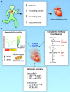
Metabolic mechanisms of exercise-induced cardiac growth. A, During exercise, changes in cardiac workload, circulating substrates, and hormones influence cardiac metabolism. The catabolic pathways invoked and the degree by which cardiac metabolism changes are dependent on the type and duration of exercise. B, Metabolic mechanisms that integrate to promote cardiac growth: exercise may diminish the activity phosphofructokinase (PFK), which leads to diminished glucose catabolism (ie, decreased glycolysis and glucose oxidation). In the exercise-adapted, resting state, glucose catabolism appears elevated above the untrained state. Such periodicity in glucose metabolism seems to regulate the exercise gene program, that is, by decreasing Cebpb expression and augmenting levels of Cited4. In addition, the metabolic periodicity caused by exercise may influence mitochondrial dynamics and help maintain healthy pools of mitochondria. The PFK step of glycolysis is important for coordinating the activity of ancillary biosynthetic pathways, which not only provide material causes for cardiac growth but may also influence the epigenetic landscape. Changes in the intracellular abundance of metabolites such as AMP and glucose-6-phosphate (G6P) may directly activate key kinases involved in cardiac adaptation. Also, circulating metabolites [eg, palmitoleate (C16:1n7)] may activate signaling pathways that promote cardiac growth. 3PG indicates 3-phosphoglycerate; F-1,6-BP, fructose-1,6-bisphosphate; F6P, fructose-6-phosphate; FFA, free fatty acids; GAP, glyceraldehyde-3-phosphate; GLP, glycero(phospho)lipid synthesis pathway; HBP, hexosamine biosynthetic pathway; and SBP, serine biosynthesis pathway. Illustration credit: Ben Smith.
Figure 5.
Figure 5.
Metabolic changes induced by pregnancy. Heart catabolism in the mother and fetus are driven not only by intrinsic metabolic programs in the heart (eg, progesterone-dependent PDK4 [pyruvate dehydrogenase kinase 4] upregulation in the maternal heart) but also by changes in circulating substrate levels. Shown are the directional changes compared with a relevant context (denoted by the *). FGF21 indicates fibroblast growth factor 21. Illustration credit: Ben Smith.
Figure 6.
Figure 6.
Changes in substrate catabolism associated with pathological cardiac remodeling. A, Metabolic changes in the hypertrophied and failing heart: glycolysis and ketone oxidation are higher and fatty acid oxidation and branched-chain amino acid (BCAA) oxidation are lower in the hypertrophied and failing heart. This appears to be caused by decreases in mitochondrial oxidative capacity, BCAA catabolic gene expression, and insulin sensitivity, and increases in phosphofructokinase (PFK) activity, glucose transporter 1 (GLUT1) expression, β-hydroxybutyrate dehydrogenase (BDH) and succinyl CoA:3-oxoacid CoA-transferase (SCOT) abundance. Moreover, higher circulating levels of ketones may also contribute to the diverse metabolic changes occurring during hypertrophy and heart failure. B, Metabolic changes in the diabetic heart: fatty acid and ketone oxidation are higher, and glucose/lactate oxidation and glycolysis are lower in the diabetic heart. This appears to be caused by decreased insulin sensitivity, elevated circulating levels of fatty acids and ketone bodies, alterations in substrate transport, higher pyruvate dehydrogenase kinase 4 (PDK4) abundance, lower PFK activity, lower acetyl CoA carboxylase (ACC) activity, and higher malonyl-CoA decarboxylase (MCD) activity. Illustration credit: Ben Smith.
Figure 7.
Figure 7.
Generalized model of how metabolism regulates cardiac health and remodeling. Stimuli that promote physiological cardiac growth (eg, exercise, pregnancy) augment fatty acid, ketone, and lactate catabolism, which collectively supplant glucose as a metabolic fuel. In contrast, conditions that instigate pathological remodeling are characterized by metabolic changes that dysregulate glucose uptake and increase reliance of the heart on glucose for fuel. The metabolic changes may cause either adaptive or maladaptive remodeling by modulating the activity of ancillary biosynthetic pathways of glucose metabolism. In the context of diabetes mellitus, excess substrate may cause ancillary biosynthetic pathway dysfunction and redox stress, which could contribute to the development of cardiomyopathy. Illustration credit: Ben Smith.

References

    1. Taegtmeyer H, Lam T, Davogustto G. Cardiac metabolism in perspective. Compr Physiol. 2016;6:1675–1699. doi: 10.1002/cphy.c150056. - PubMed
    1. Doenst T, Nguyen TD, Abel ED. Cardiac metabolism in heart failure: implications beyond ATP production. Circ Res. 2013;113:709–724. - PMC - PubMed
    1. Opie LH. Metabolism of the heart in health and disease. II. Am Heart J. 1969;77:100–122 contd. - PubMed
    1. Opie LH. Heart Physiology: From Cell to Circulation. Philadelphia, PA: Lippincott Williams & Wilkins; 2004. pp. 308–354.
    1. Schönekess BO. Competition between lactate and fatty acids as sources of ATP in the isolated working rat heart. J Mol Cell Cardiol. 1997;29:2725–2733. doi: 10.1006/jmcc.1997.0504. - PubMed

Publication types