. 2018 Jul 2;215(7):1853-1868.
doi: 10.1084/jem.20170779. Epub 2018 Jun 21.

Trim33 mediates the proinflammatory function of Th17 cells

Affiliations

Trim33 mediates the proinflammatory function of Th17 cells

Shinya Tanaka et al. J Exp Med. .

Abstract

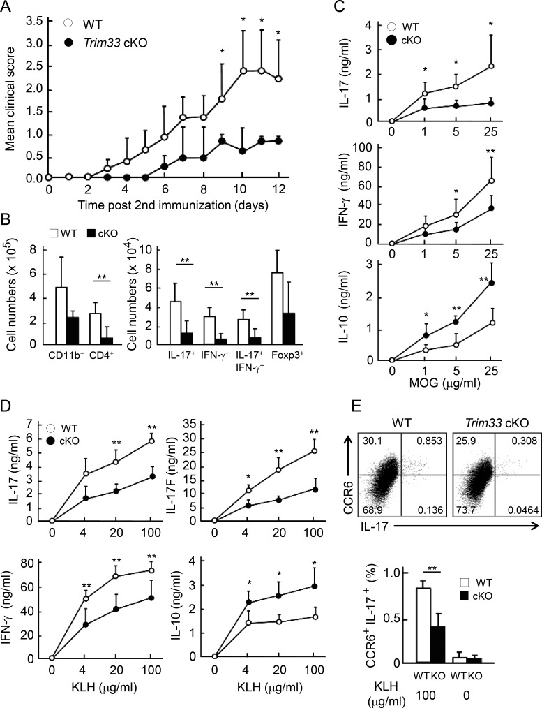
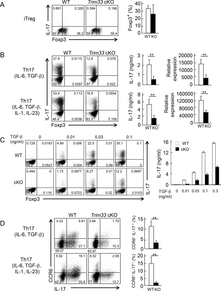
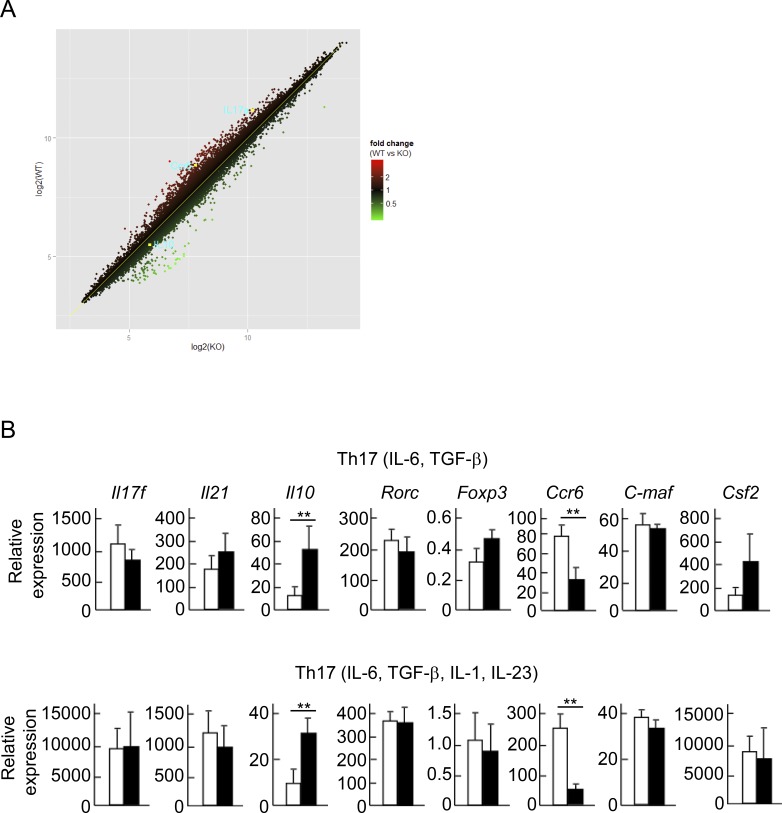
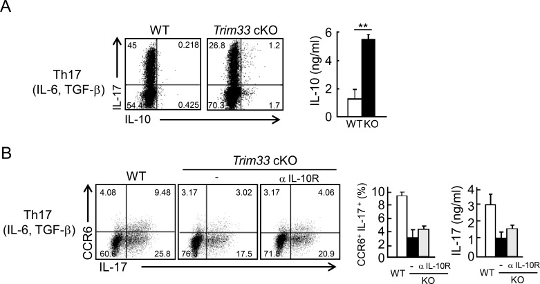
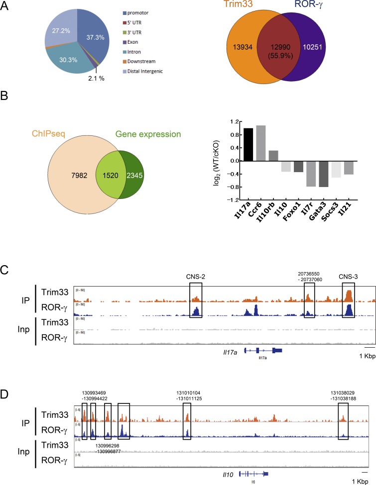
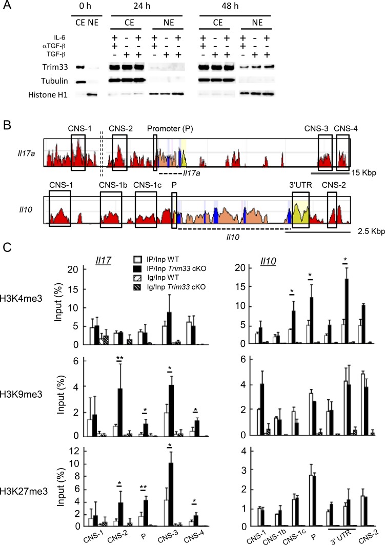
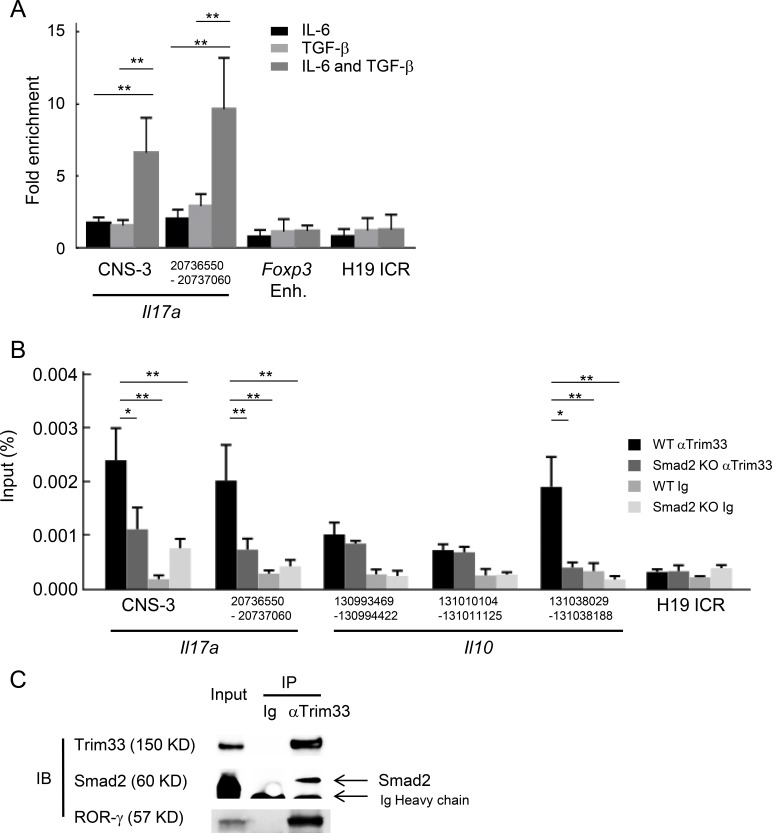
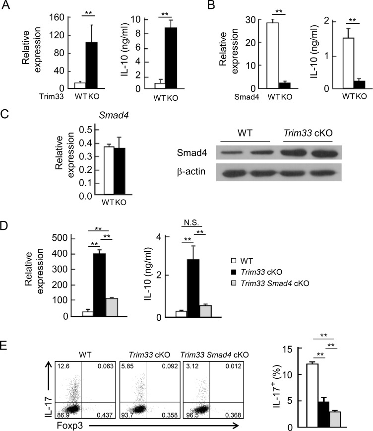
Transforming growth factor-β (TGF-β) regulates reciprocal regulatory T cell (T reg) and T helper 17 (Th17) differentiation, the underlying mechanism of which is still not understood. Here, we report that tripartite motif-containing 33 (Trim33), a modulator of TGF-β signaling that associates with Smad2, regulates the proinflammatory function of Th17 cells. Trim33 deficiency in T cells ameliorated an autoimmune disease in vivo. Trim33 was required for induction in vitro of Th17, but not T reg cells. Moreover, Smad4 and Trim33 play contrasting roles in the regulation of IL-10 expression; loss of Trim33 enhanced IL-10 production. Furthermore, Trim33 was recruited to the Il17a and Il10 gene loci, dependent on Smad2, and mediated their chromatin remodeling during Th17 differentiation. Trim33 thus promotes the proinflammatory function of Th17 cells by inducing IL-17 and suppressing IL-10 expression.


Figures

Figure 1.
Figure 1.
Trim33 is indispensable for Th17 response in vivo. (A) Trim33 f/f (WT n = 5; open circle) and Trim33 f/f CD4-Cre (Trim33 cKO n = 5; closed circle) mice were immunized with MOG peptides in CFA to induce EAE. Disease scores are shown. (B) Absolute numbers of central nervous system–infiltrating cells were analyzed. CD11b+or CD4+ cells are presented in left panel. IL-17+, IFN-γ+, IL-17+IFN-γ+, and Foxp3+ cells in CD4+ cells are shown in right panel. (C) Splenocytes of immunized mice shown in A were restimulated with MOG peptide for 72 h. IL-17, IL-10, and IFN-γ were measured by ELISA. (D) WT (n = 4; open circle) and Trim33 cKO (n = 4; closed circle) mice were immunized with KLH in CFA. After 7 d, dLN cells of immunized mice were restimulated with KLH for 72 h, and cytokine expression was assessed by ELISA. (E) dLN cells were restimulated for 24 h, as performed in D, followed by costaining of CCR6 and IL-17. Frequency of CCR6+ and IL-17+ double-positive cells in WT and the KO mice is shown. Data are representative of three independent experiments. Error bars show means ± SD. *, P < 0.05; **, P < 0.01.
Figure 2.
Figure 2.
Trim33 is required for Th17 but not iT reg cell differentiation in vitro. (A) Naive CD4+ T cells (CD25CD44loCD62Lhi) isolated from WT (white) and Trim33 cKO (black) mice were stimulated by anti-CD3 and anti-CD28 mAbs under Th17 and iT reg skewing conditions. After 4 d, cultured cells were restimulated. Foxp3 and IL-17 expression in iT reg cell condition are shown (left). Frequency of Foxp3+ cells is presented (right). (B) Foxp3 and IL-17 expression in Th17 conditions are shown (left). IL-17 expression was measured by ELISA (middle) and quantitative PCR (right). Data were normalized to b-actin. (C) Naive CD4+ T cells isolated from WT and Trim33 cKO mice were cultured under Th17 skewing conditions with indicated concentrations of TGF-β. IL-17 expression was detected upon restimulation by intracellular cytokine staining (left) and ELISA (right). (D) Costaining of CCR6 and IL-17 in Th17 conditions is presented (left). Frequency of CCR6+ IL-17+ cells is shown (right). Data are representative of three independent experiments. Error bars show means ± SD. **, P < 0.01.
Figure 3.
Figure 3.
Identification of Trim33-target genes during Th17 differentiation. (A) Naive CD4+ T cells isolated from WT and Trim33 cKO mice were cultured in Th17 skewing conditions (IL-6, TGF-β, anti–IL-4 antibody, and anti–IFN-γ antibody). The cells were restimulated with anti-CD3 antibody and subjected to microarray analysis. (B) Expression of Th17-related genes in WT and Trim33 KO T cells cultured under Th17 conditions, as cultured in Fig. 2, was assessed by quantitative PCR. Data shown combine results from three independent experiments. Error bars show means ± SD. **, P < 0.01.
Figure 4.
Figure 4.
Trim33 deficiency results in IL-10 up-regulation in Th17 cells. (A) Naive CD4+ T cells isolated from WT (white) and Trim33 cKO (black) mice were stimulated by anti-CD3 and anti-CD28 antibodies under Th17 skewing conditions. After restimulation, costaining of IL-17 and IL-10 was performed by intracellular staining (left), and IL-10 production was measured by ELISA (right). (B) Naive CD4+ T cells isolated from WT and Trim33 cKO mice were stimulated by anti-CD3 and anti-CD28 antibodies under Th17 skewing conditions with or without anti–IL-10R antibody (WT, white; KO, black; and KO+anti–IL-10R, gray). Costaining of CCR6 and IL-17 was performed after restimulation (left). The statistical analysis of CCR6+IL-17+ cells is shown (middle). IL-17 expression was measured by ELISA (right). Data are representative of three independent experiments. Error bars show means ± SD. **, P < 0.01.
Figure 5.
Figure 5.
Trim33 ChIP-seq analysis. (A) Distribution of Trim33 ChIP-seq peaks in Th17 cells (left). Occupancy of ChIP-seq binding peaks of Trim33 and ROR-γ (right). (B) Integration of Trim33 binding peak data with gene expression analysis (left). Among 1,520 genes, genes related to the function and differentiation of Th cells are shown with their expression changes of WT versus KO (right). (C and D) ChIP-seq peaks of Trim33 and ROR-γ in Il17a (C) and Il10 (D) gene loci were shown. Data are representative of two independent experiments.
Figure 6.
Figure 6.
Trim33 regulates histone modifications at the Il17a and Il10 gene loci. (A) Extracts from both nuclei and cytosol were isolated from CD4+ T cells before stimulation (0 h) and stimulated with indicated conditions after 24 and 48 h. Trim33, tubulin (cytosolic protein) and histone H1 (nucleic protein) were detected by immunoblot. Data are representative of three independent experiments. (B) Schematic diagrams indicate comparison of genomic sequence of murine Il17 and Il10 gene loci with human. (C) Histone modifications were assessed by ChIP analysis. Naive CD4+ T cells isolated from WT and Trim33 cKO mice were stimulated under Th17 skewing conditions. Chromatin isolated from these cells was immunoprecipitated with antibodies against H3K4me3, H3K9me3, H3K27me3, and control Ig. Genomic regions modified with histone marks were detected by PCR with primer sets of each CNS. Data are representative of two independent experiments. Error bars show means ± SD. *, P < 0.05; **, P < 0.01.
Figure 7.
Figure 7.
Cooperative regulation of IL-17 and IL-10 by Trim33, Smad2, and ROR-γ. (A) Trim33 binding was assessed by ChIP-PCR analysis. PCR amplification was conducted with input DNA and I.P. DNA prepared from chromatin precipitated with anti-Trim33 antibodies, with C57BL/6 CD4+ T cells stimulated by anti-CD3 and anti-CD28 antibodies under indicated conditions. Primer for the H19 ICR locus was used as a negative control. Data are representative of three independent experiments. (B) Trim33 ChIP was performed with WT and Smad2 KO CD4+ T cells cultured under Th17 skewing conditions. (C) Physical association of Trim33 with Smad2 and ROR-γ in Th17 cells. Lysates of Th17 cells were subjected to immunoprecipitation with anti-Trim33 antibody or isotype Ig, followed by immunoblot analysis with anti-Trim33, Smad2, and ROR-γ antibodies. Data are representative of four independent experiments. Error bars show means ± SD. *, P < 0.05; **, P < 0.01.
Figure 8.
Figure 8.
Reciprocal IL-10 regulation by Trim33 and Smad4. (A) Naive CD4+ T cells isolated from WT and Trim33 cKO mice were stimulated by anti-CD3 and anti-CD28 antibodies under Th17 skewing conditions. On day 3, the cultured cells and supernatant were harvested, followed by measurement of IL-10 at the mRNA (left) and protein (right) levels. (B) IL-10 production was measured with WT and Smad4 KO Th17 cells, as done in A. (C) Smad4 expression was detected at the mRNA (left) and protein (right) levels in WT and Trim33 KO Th17 cells. β-Actin was used as loading control. Data are representative of three independent experiments. (D) IL-10 production was measured in WT, Trim33 KO, and Trim33 × Smad4 KO Th17 cells, as described in A. (E) IL-17 production by WT, Trim33 KO, and Trim33 × Smad4 KO Th17 cells, after restimulation. Data are representative of two independent experiments. Error bars show means ± SD. **, P < 0.01.

Comment in

  • TRIMming TGF-β signals in Th17 cells.
    Rapaport AS, Ouyang W. Rapaport AS, et al. J Exp Med. 2018 Jul 2;215(7):1775-1776. doi: 10.1084/jem.20180986. Epub 2018 Jun 26. J Exp Med. 2018. PMID: 29945999 Free PMC article.

References

    1. Agricola E., Randall R.A., Gaarenstroom T., Dupont S., and Hill C.S.. 2011. Recruitment of TIF1γ to chromatin via its PHD finger-bromodomain activates its ubiquitin ligase and transcriptional repressor activities. Mol. Cell. 43:85–96. 10.1016/j.molcel.2011.05.020 - DOI - PubMed
    1. Akimzhanov A.M., Yang X.O., and Dong C.. 2007. Chromatin remodeling of interleukin-17 (IL-17)-IL-17F cytokine gene locus during inflammatory helper T cell differentiation. J. Biol. Chem. 282:5969–5972. 10.1074/jbc.C600322200 - DOI - PubMed
    1. Bettelli E., Das M.P., Howard E.D., Weiner H.L., Sobel R.A., and Kuchroo V.K.. 1998. IL-10 is critical in the regulation of autoimmune encephalomyelitis as demonstrated by studies of IL-10- and IL-4-deficient and transgenic mice. J. Immunol. 161:3299–3306. - PubMed
    1. Bettelli E., Carrier Y., Gao W., Korn T., Strom T.B., Oukka M., Weiner H.L., and Kuchroo V.K.. 2006. Reciprocal developmental pathways for the generation of pathogenic effector TH17 and regulatory T cells. Nature. 441:235–238. 10.1038/nature04753 - DOI - PubMed
    1. Chu G.C., Dunn N.R., Anderson D.C., Oxburgh L., and Robertson E.J.. 2004. Differential requirements for Smad4 in TGFbeta-dependent patterning of the early mouse embryo. Development. 131:3501–3512. 10.1242/dev.01248 - DOI - PubMed

Publication types

MeSH terms