. 2018 Aug;177(4):1439-1452.
doi: 10.1104/pp.18.00320. Epub 2018 Jun 21.

An Assembly Factor Promotes Assembly of Flavinated SDH1 into the Succinate Dehydrogenase Complex

Affiliations

An Assembly Factor Promotes Assembly of Flavinated SDH1 into the Succinate Dehydrogenase Complex

Katharina Belt et al. Plant Physiol. 2018 Aug.

Abstract

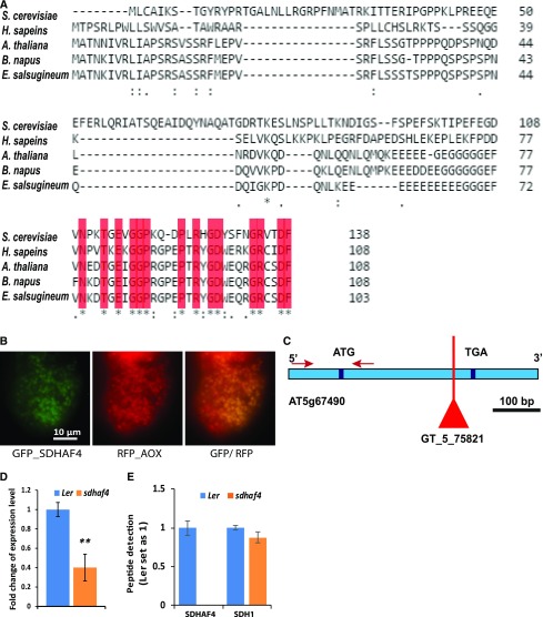
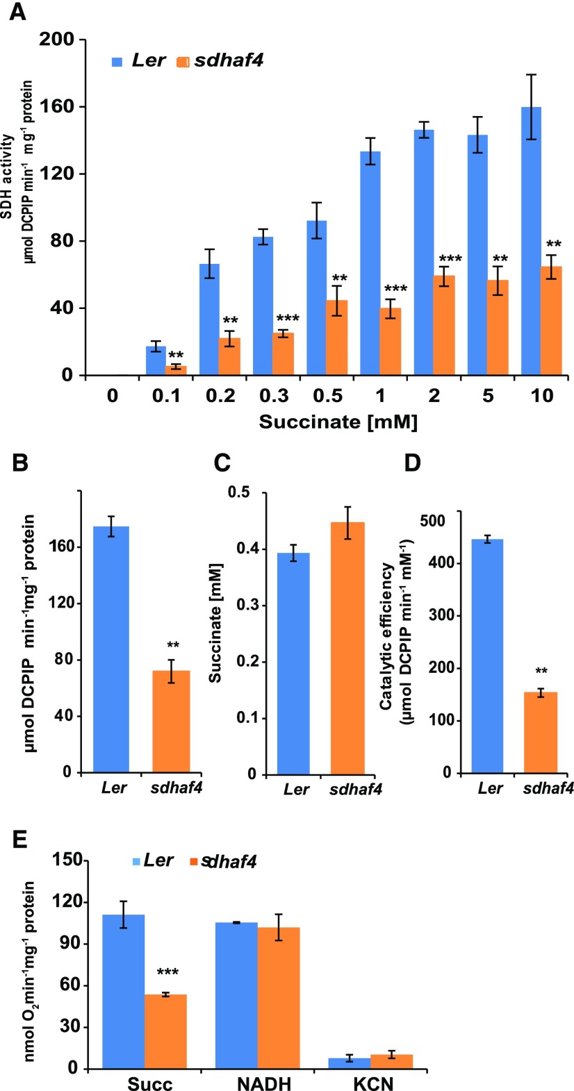
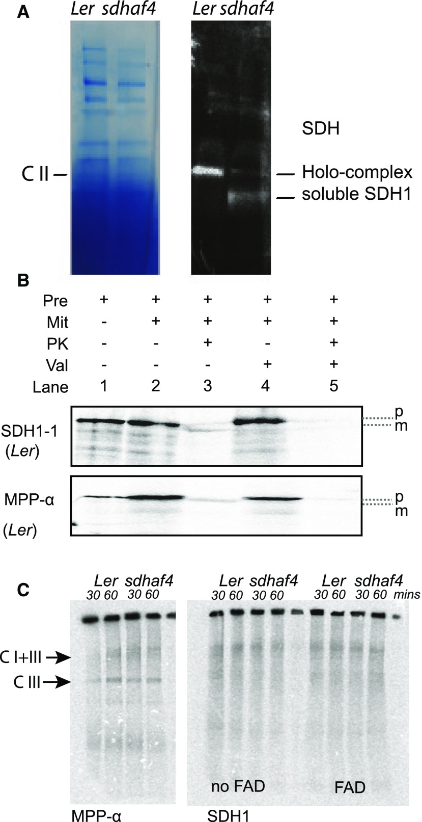
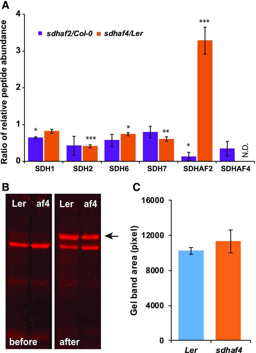
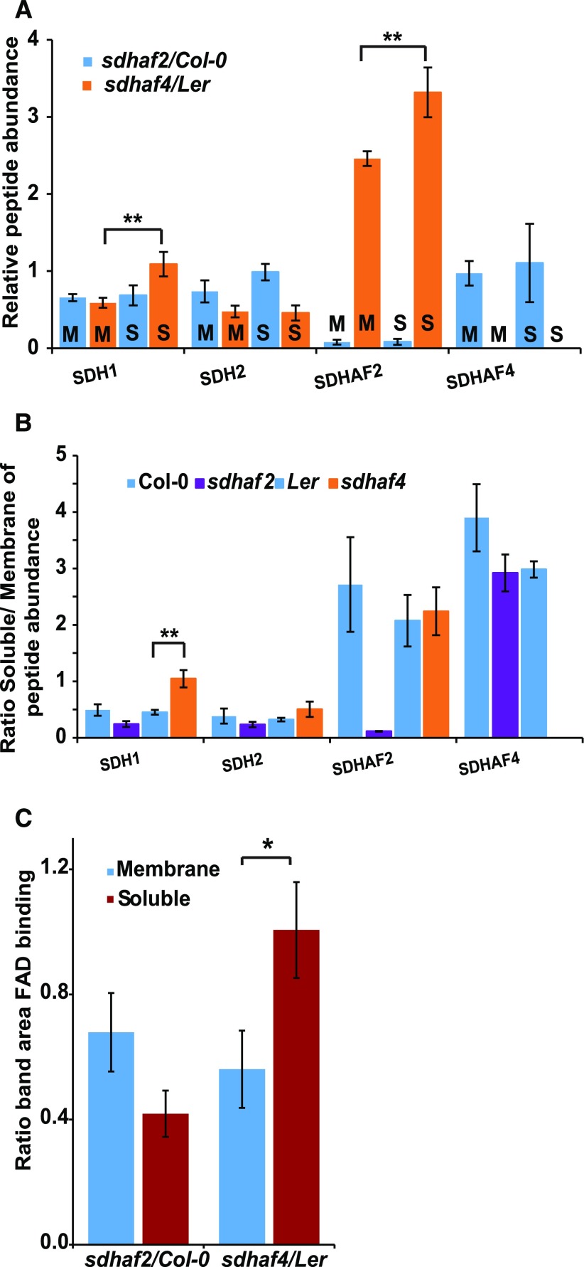
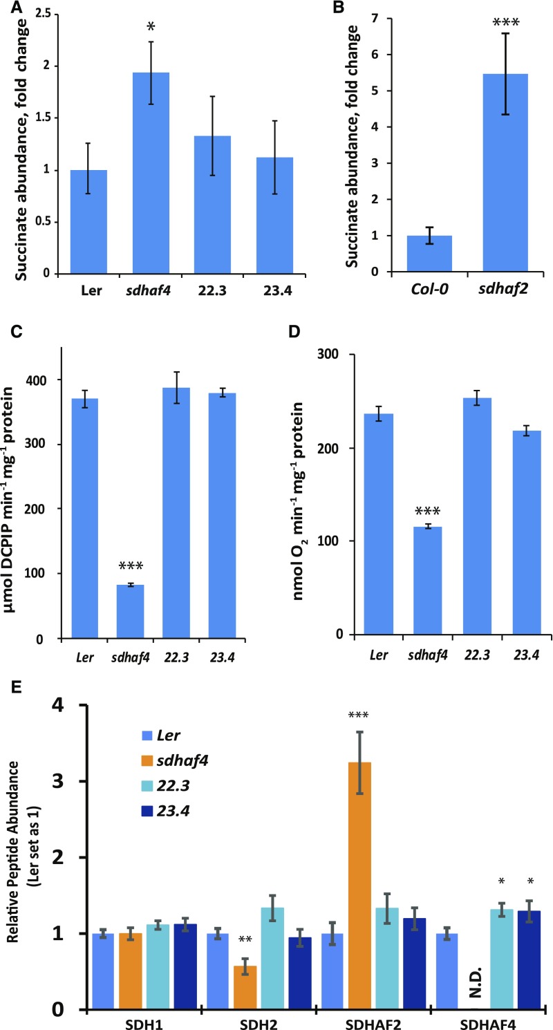
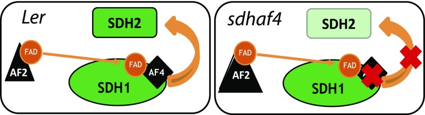
Succinate dehydrogenase (Complex II; SDH) plays an important role in mitochondrial respiratory metabolism. The SDH complex consists of four core subunits and multiple cofactors, which must be assembled correctly to ensure enzyme function. To date, only an assembly factor (SDHAF2) required for FAD insertion into subunit SDH1 has been identified in plants. Here, we report the identification of Arabidopsis (Arabidopsis thaliana) At5g67490 as a second SDH assembly factor. Knockout of At5g67490 (sdhaf4) did not cause any phenotypic variation in seedlings but resulted in a decrease in both SDH activity and the succinate-dependent respiration rate as well as increased accumulation of succinate. Mass spectrometry analyses revealed stable levels of FAD-SDH1 in sdhaf4, together with increased levels of the FAD-SDH1 assembly factor, SDHAF2, and reduced levels of SDH2 compared with the wild type. Loss of SDHAF4 in sdhaf4 inhibited the formation of the SDH1/SDH2 intermediate, leading to the accumulation of soluble SDH1 in the mitochondrial matrix and reduced levels of SDH1 in the membrane. The increased levels of SDHAF2 suggest that the stabilization of soluble FAD-SDH1 depends on SDHAF2 availability. We conclude that SDHAF4 acts on FAD-SDH1 and promotes its assembly with SDH2, thereby stabilizing SDH2 and enabling its full assembly with SDH3/SDH4 to form the SDH complex.


Figures

Figure 1.
Figure 1.
SDHAF4 sequences contain a conserved region at the C terminus among different species, the Arabidopsis protein is located within mitochondria, and sdhaf4 is an effective knockout Arabidopsis line at the protein level for SDHAF4. A, Sequence alignment of SDHAF4 protein between S. cerevisiae (YBR269C), human (NM_145267), Arabidopsis (At5g67490), B. napus (XM_013885343), and E. salsugenium (XM_006393894; Clustal Omega). Conserved regions are highlighted in red. Asterisks indicate identical residues, colons indicates conserved substitution, and periods indicate semiconserved substitution. B, GFP_SDHAF4 and RFP_AOX constructs were expressed transiently in Arabidopsis cell culture. GFP localization of SDHAF4 in mitochondria was performed by microscopy imaging. C, Location of the T-DNA insertion in At5g67490 (SDHAF4) within the exon region. Red arrows indicate the binding sites of the primers used for RT-qPCR. D, SDHAF4 gene expression in Ler and sdhaf4. RT-qPCR using primers outside the T-DNA region (red arrows in C) was performed to determine SDHAF4 expression. Expression levels were normalized to actin, and the expression of SDHAF4 in Ler was set as 1. E, SDH1 and SDHAF4 peptides were detected and quantified using mass spectrometry. Mitochondria isolated from Ler and sdhaf4 were used, and protein-specific peptides were used for identification. Samples were normalized to ATP synthase. Shown is the ratio of sdhaf4 to Ler. Error bars indicate se (n = 4). **, P ≤ 0.01 (Student’s t test).
Figure 2.
Figure 2.
sdhaf4 shows lower SDH activity and succinate-dependent oxygen consumption compared with Ler. A, SDH activity at different succinate concentrations in the range of 0.1 to 10 mm. B, Kinetic analysis using the Michaelis-Menten formula was performed to determine maximum SDH velocity. C, Km value of SDH for succinate. D, Catalytic efficiency in sdhaf4 and Ler. E, Oxygen consumption in the presence of 5 mm succinate or 1 mm NADH. Error bars indicate se. Student’s t test was performed to determine significant differences between genotypes (n = 4): **, P < 0.01 and ***, P < 0.001.
Figure 3.
Figure 3.
Less SDH1 is incorporated into the SDH holo-complex, and it accumulates as a soluble protein in sdhaf4. A, SDH1 antibody was used to detect SDH1 abundance in whole mitochondria samples from Ler and sdhaf4 loaded on a BN gel and blotted on a PVDF membrane. Two bands were detected, potentially representing SDH holo-complex and soluble SDH1 protein, as indicated. B, Import of [35S]Met-labeled SDH1 and MPP-α into Ler mitochondria analyzed by SDS-PAGE. Pre, Precursor; Mit, mitochondria; PK, proteinase K; Val, valinomycin (inhibitor for import across the IMM); p, precursor protein; m, mature protein. C, [35S]Met-labeled SDH1 and MPP-α imported into Ler and sdhaf4 mitochondria for 30 and 60 min and separated by BN-PAGE. C I+III, ComplexI+III; C I, Complex I.
Figure 4.
Figure 4.
Abundances of SDH2 and SDHAF2, but not FAD-bound SDH1, are altered in sdhaf4. A, MRM was used to detect peptides from SDH subunits and assembly factors. Shown are the ratios of peptides (mutant to wild type) based on whole mitochondria protein samples (50 µg; n = 3). N.D. indicates not detected in sdhaf4. Error bars indicate se. *, P < 0.05; **, P < 0.01; and ***, P < 0.001 (Student’s t test for sdhaf2:Col-0 or sdhaf4:Ler). B, A FAD-bound protein assay was performed to compare FAD binding with SDH1 in sdhaf4 (af4) and Ler. Mitochondrial proteins (10 µg) were separated by SDS-PAGE, following a gel incubation in 10% (v/v) acetic acid for 30 min. FAD fluorescence scans were performed before and after the acetic acid treatment using Typhoon Trio Laser (Amersham Biosciences) and filters Cy5 (670 bp) and Cy3 (580 bp). The FAD-bound SDH1 band became visible after acetic acid incubation (marked with a black arrow). C, Quantification of FAD-bound SDH1 bands on gels, with three replicates.
Figure 5.
Figure 5.
SDH1 accumulates as a soluble FAD-bound protein in sdhaf4. A, Peptide abundance in soluble (S) and membrane (M) mitochondrial protein fractions compared between mutant lines and the wild type (n = 3). B, Ratio of protein abundance in soluble fractions to that in membrane fractions within each genotype (n = 3). C, Comparison of the gel band area of FAD-bound SDH1 between mutant lines and the wild type from soluble and mitochondrial membrane fractions (n = 4). *, P ≤ 0.08 and **, P ≤ 0.05, by Student’s t test. Error bars indicate se.
Figure 6.
Figure 6.
35S-SDHAF4 complements SDH activity in sdhaf4. A, Fold change of succinate abundance in sdhaf4 and two independent complementation lines (22.3 and 23.4) compared with Ler (n = 4). B, Fold change of succinate abundance in sdhaf2 compared with Col-0 (n = 4). C, SDH enzyme activity at 10 mm succinate. D, Oxygen consumption in the presence of 5 mm succinate. E, Protein peptide detection of SDH subunits SDH1 and SDH2 and SDH assembly factors SDHAF2 and SDHAF4 (n = 4). Error bars indicate se; N.D., not detected. Student’s t test was used to determine significant differences between sdhaf4 and Ler complementation lines: *, P < 0.05; **, P < 0.01; and ***, P < 0.001.
Figure 7.
Figure 7.
Schematic diagram of FAD insertion into SDH1 and the assembly of SDH1 with SDH2. Left, As a first step in SDH1 assembly, SDHAF2 (AF2) is required to insert the FAD cofactor into SDH1. SDHAF4 (AF4) likely binds to the FAD site in SDH1 and promotes the assembly of SDH1 with SDH2. Right, Loss of SDHAF4 causes the decreased assembly of SDH1 with SDH2, leading to the degradation of SDH2. Loss of AF4 also induces the accumulation of AF2, presumably improving the stability of SDH1-FAD.

Similar articles

Cited by

References

    1. Araújo WL, Nunes-Nesi A, Osorio S, Usadel B, Fuentes D, Nagy R, Balbo I, Lehmann M, Studart-Witkowski C, Tohge T, et al. (2011) Antisense inhibition of the iron-sulphur subunit of succinate dehydrogenase enhances photosynthesis and growth in tomato via an organic acid-mediated effect on stomatal aperture. Plant Cell 23: 600–627 - PMC - PubMed
    1. Bafunno V, Giancaspero TA, Brizio C, Bufano D, Passarella S, Boles E, Barile M (2004) Riboflavin uptake and FAD synthesis in Saccharomyces cerevisiae mitochondria: involvement of the Flx1p carrier in FAD export. J Biol Chem 279: 95–102 - PubMed
    1. Belt K, Huang S, Thatcher LF, Casarotto H, Singh KB, Van Aken O, Millar AH (2017) Salicylic acid-dependent plant stress signaling via mitochondrial succinate dehydrogenase. Plant Physiol 173: 2029–2040 - PMC - PubMed
    1. Burger G, Lang BF, Reith M, Gray MW (1996) Genes encoding the same three subunits of respiratory complex II are present in the mitochondrial DNA of two phylogenetically distant eukaryotes. Proc Natl Acad Sci USA 93: 2328–2332 - PMC - PubMed
    1. Carrie C, Murcha MW, Kuehn K, Duncan O, Barthet M, Smith PM, Eubel H, Meyer E, Day DA, Millar AH, et al. (2008) Type II NAD(P)H dehydrogenases are targeted to mitochondria and chloroplasts or peroxisomes in Arabidopsis thaliana. FEBS Lett 582: 3073–3079 - PubMed

Publication types

MeSH terms

LinkOut - more resources