. 2018 Sep 3;217(9):3091-3108.
doi: 10.1083/jcb.201712029. Epub 2018 Jun 21.

Cytosolic Hsp70 and Hsp40 chaperones enable the biogenesis of mitochondrial β-barrel proteins

Affiliations

Cytosolic Hsp70 and Hsp40 chaperones enable the biogenesis of mitochondrial β-barrel proteins

Tobias Jores et al. J Cell Biol. .

Abstract

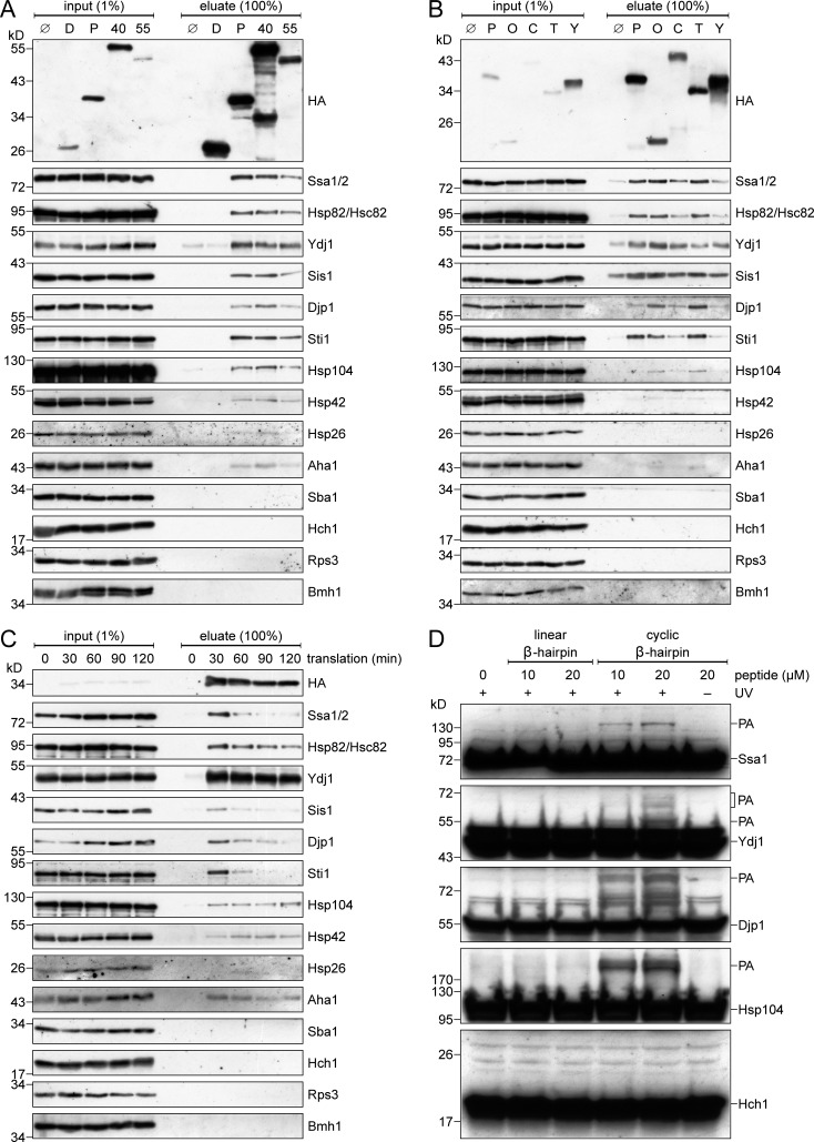
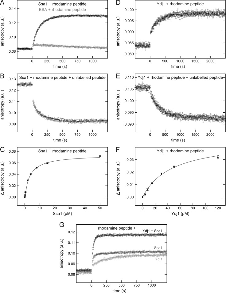
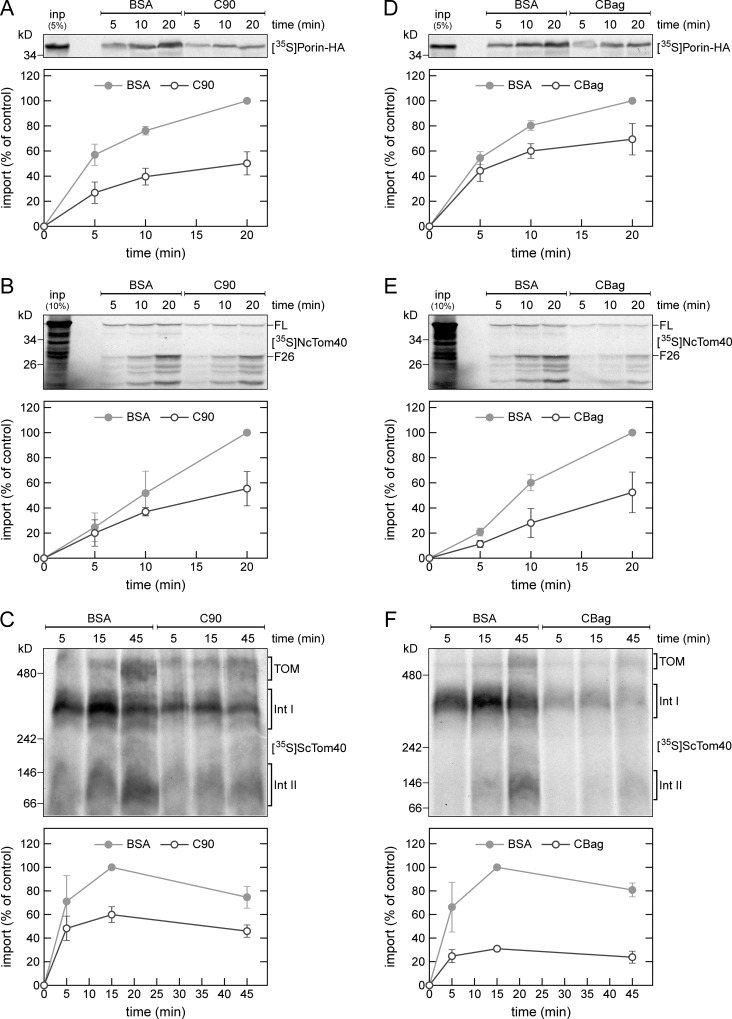
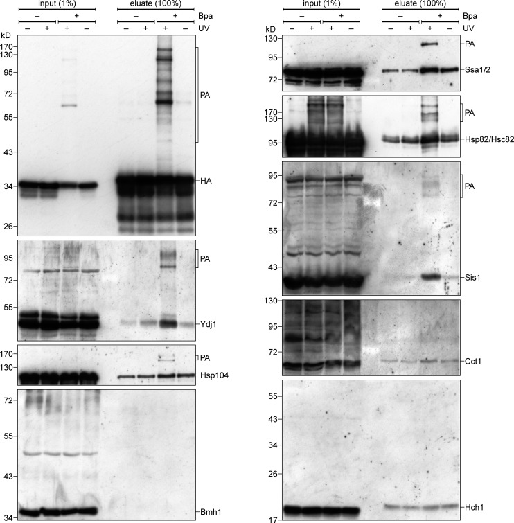
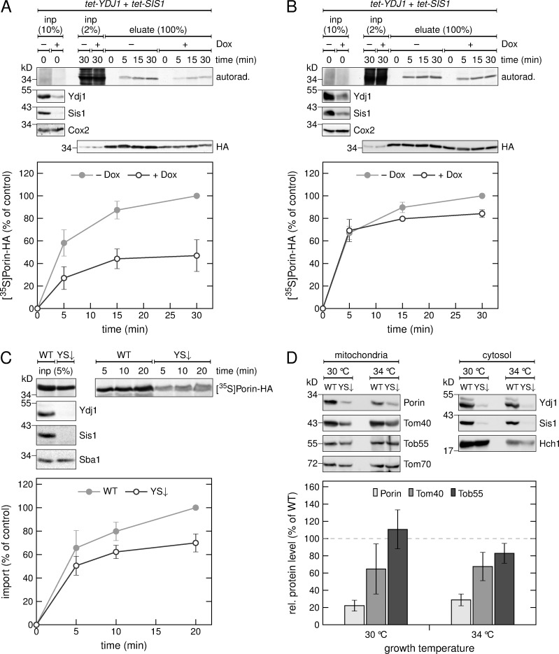
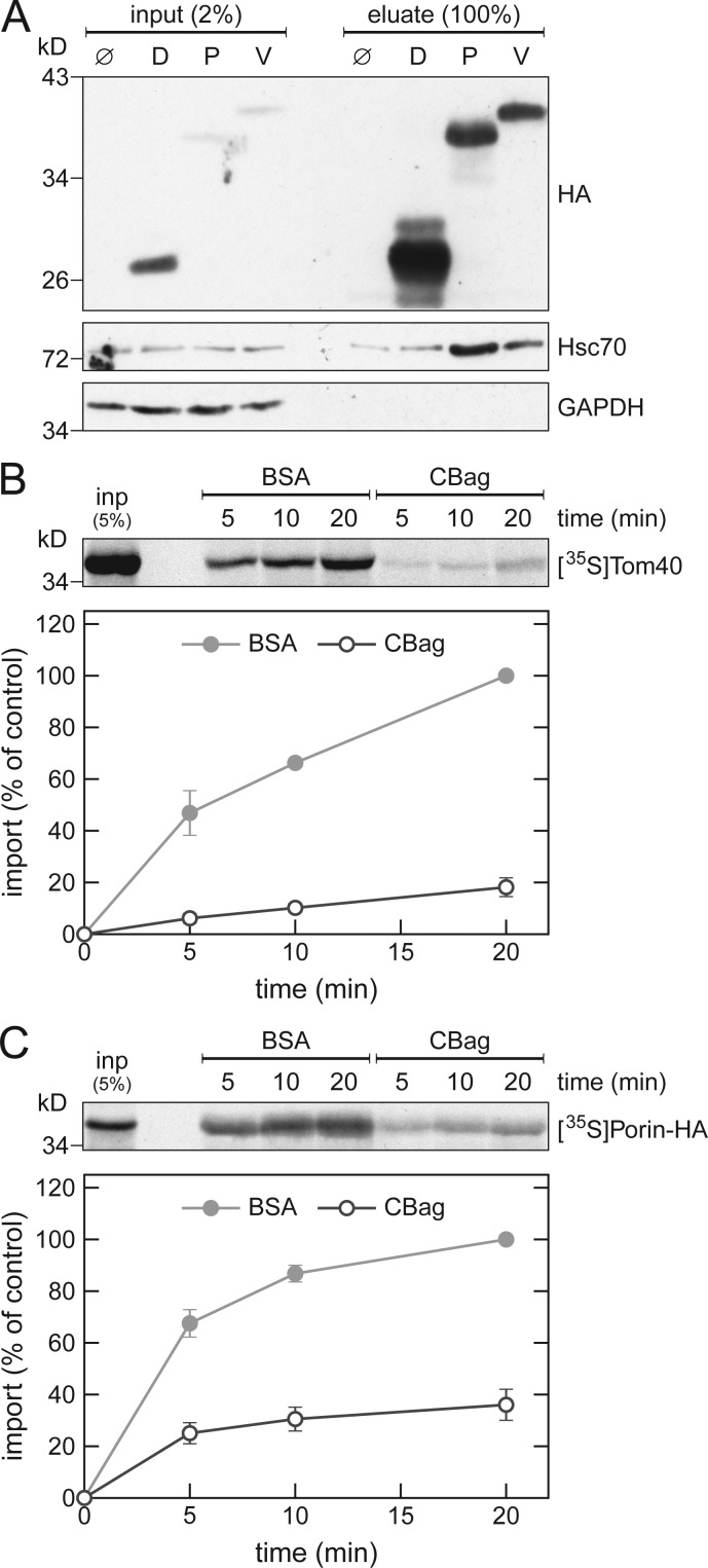
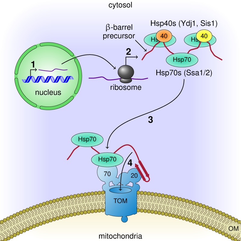
Mitochondrial β-barrel proteins are encoded in the nucleus, translated by cytosolic ribosomes, and then imported into the organelle. Recently, a detailed understanding of the intramitochondrial import pathway of β-barrel proteins was obtained. In contrast, it is still completely unclear how newly synthesized β-barrel proteins reach the mitochondrial surface in an import-competent conformation. In this study, we show that cytosolic Hsp70 chaperones and their Hsp40 cochaperones Ydj1 and Sis1 interact with newly synthesized β-barrel proteins. These interactions are highly relevant for proper biogenesis, as inhibiting the activity of the cytosolic Hsp70, preventing its docking to the mitochondrial receptor Tom70, or depleting both Ydj1 and Sis1 resulted in a significant reduction in the import of such substrates into mitochondria. Further experiments demonstrate that the interactions between β-barrel proteins and Hsp70 chaperones and their importance are conserved also in mammalian cells. Collectively, this study outlines a novel mechanism in the early events of the biogenesis of mitochondrial outer membrane β-barrel proteins.


Figures

Figure 1.
Figure 1.
Cytosolic chaperones interact with newly synthesized β-barrel proteins. (A and B) In vitro translation reactions using yeast extracts without mRNA (Ø) or programmed with mRNA encoding DHFR-HA (D), Porin-HA (P), Tom40-HA (40), Tob55Δ120-HA (55), Om14-HA (O), Cyc3-HA (C), Tim23-HA (T), or Yah1-HA (Y) were subjected to a pull-down with anti-HA beads. Samples from the input and the eluates were analyzed by SDS-PAGE and immunodecoration with the indicated antibodies. (C) Yeast extracts programmed with mRNA encoding for Porin-HA were incubated for the indicated times. Afterward, the reactions were subjected to an anti-HA pull-down and analyzed as in A. (D) Yeast extracts were incubated with the indicated Bpa-containing peptide at the specified concentrations. Some samples were either illuminated with UV light (+UV) to induce cross-linking or were left in darkness (−UV). Then, the samples were analyzed by SDS-PAGE and immunodecoration with the indicated antibodies. PA, specific photoadducts.
Figure 2.
Figure 2.
A cyclic β-hairpin peptide binds directly to Ssa1 or Ydj1. (A–F) The fluorescence anisotropy of a rhodamine-labeled cyclic β-hairpin peptide was measured in the presence of 10 µM Ssa1 (A–C, black circles), 30 µM BSA (A, gray circles), or 30 µM Ydj1 (D–F). After association of the rhodamine-labeled peptide with the indicated proteins (A and D), the samples were supplemented with a 100-fold excess of the unlabeled peptide and dissociation was recorded (B and E). For affinity determinations, the rhodamine-labeled peptide was mixed with the indicated concentrations of either Ssa1 (C) or Ydj1 (F), and the difference in anisotropy (Δ anisotropy) of the bound and free peptide was plotted against the (co)chaperone concentration. The data were fitted using a hyperbolic regression curve (gray). (G) The fluorescence anisotropy of a rhodamine-labeled cyclic β-hairpin peptide was measured in the presence of 5 µM Ssa1 (dark gray circles), 30 µM Ydj1 (light gray circles), or both proteins (black circles).
Figure 3.
Figure 3.
Cytosolic chaperones are required for the in vitro import of β-barrel proteins. (A–F) Top, radiolabeled precursor proteins of Porin-HA (A and D), N. crassa Tom40 (NcTom40; B and E), or yeast Tom40 (ScTom40; C and F) were produced in yeast extract, and subjected to in vitro import reactions using isolated mitochondria. Before the import reaction, the mitochondria were mixed with either C90 or BSA (A–C). Alternatively, the translation reactions were supplemented with either CBag or BSA (D–F). After import for the indicated times, the mitochondria were subjected to carbonate extraction (A and D) or were treated with proteinase K (B and E). The samples were subjected to SDS-PAGE (A, B, D, and E) or BN-PAGE (C and F) and autoradiography. Bottom, intensities of the bands corresponding to Porin-HA, the protease-protected fragment F26 of NcTom40, or assembly intermediate I of Tom40 from three independent experiments were quantified and the mean intensity from the 20-min (A, B, D, and E) or 15-min (C and F) import in the presence of BSA was set to 100%. Error bars represent ± SD. FL, full-length. F26, protease-protected fragment of 26 kD. The migration of the assembled TOM complex and assembly intermediates (Int) I and II of Tom40 are indicated.
Figure 4.
Figure 4.
Cytosolic chaperones interact in vivo with a β-hairpin peptide. Yeast cells expressing either control hp18(VDAC)-DHFR-HA (−Bpa) or a variant with Bpa inserted in the β-hairpin (+Bpa) were subjected to in vivo photo cross-linking (+UV) or kept in the dark (−UV). Next, the cells were lysed and subjected to a pull-down with anti-HA beads. Samples from the input and the eluates were analyzed by SDS-PAGE and immunodecoration with the indicated antibodies.
Figure 5.
Figure 5.
The cochaperones Ydj1 and Sis1 are required for the in vivo biogenesis of Porin. (A and B) HA-tagged Porin was expressed in a strain with tetracycline-repressible promoters controlling the expression of YDJ1 and SIS1 in the absence (−) or presence (+) of doxycycline (Dox) followed by methionine starvation. Synthesis of radiolabeled proteins was initiated by addition of [35S]Met to the medium, and cells were harvested after the indicated time periods. Then, a crude mitochondrial fraction (A) or the whole cell lysate (B) were obtained. The samples were solubilized and subjected to a pull-down with anti-HA beads. Input samples from the whole cell lysate (inp) and the eluates were analyzed by SDS-PAGE, autoradiography (autorad.) and immunodecoration with the indicated antibodies. Bottom, intensities of the bands corresponding to Porin-HA from three independent experiments were quantified, and the mean intensity from the 30-min samples without doxycycline was set to 100%. Error bars represent ± SD. (C) Top, radiolabeled Porin-HA was translated in yeast extract from WT yeast cells or from cells depleted for Ydj1 and Sis1 (YS↓). The translation reactions were subjected to in vitro import reactions using mitochondria isolated from YS↓ cells. After import for the indicated times, the mitochondria were subjected to carbonate extraction. The samples were analyzed by SDS-PAGE, autoradiography ([35S]Porin-HA) and immunodecoration with the indicated antibodies. Bottom, the intensities of the bands corresponding to Porin-HA were quantified and the intensity from import of Porin-HA translated in the WT strain was set to 100%. Error bars represent ± SD. (D) Top, a crude mitochondrial and a cytosolic fraction were isolated from WT or YS↓ yeast cells analyzed by SDS-PAGE and immunodecoration with the indicated antibodies. Bottom, intensities of the bands in the mitochondrial fraction from three independent experiments were quantified and normalized to the level of Tom70. The level of the proteins in the WT strain was set to 100%. Error bars represent ± SD.
Figure 6.
Figure 6.
Mammalian Hsc70 is involved in the biogenesis of β-barrel proteins. (A) In vitro translation reactions using rabbit reticulocyte lysate without mRNA (Ø) or programmed with mRNA encoding DHFR-HA (D), Porin-HA (P), or VDAC1-HA (V) were subjected to a pull-down with anti-HA beads. Samples from the input and the eluates were analyzed by SDS-PAGE and immunodecoration with the indicated antibodies. (B and C) Top, rabbit reticulocyte lysate was used to synthesize radiolabeled human Tom40 (B) or yeast Porin (C). After translation, the lysate was supplemented with either CBag or BSA and subjected to in vitro import reactions using mitochondria isolated from human (B) or yeast (C) cells. After import for the indicated times, the mitochondria were treated with proteinase K (B) or were subjected to carbonate extraction (C), and the samples were analyzed by SDS-PAGE and autoradiography. Bottom, intensities of the bands corresponding to Tom40 or Porin from three independent experiments were quantified and the mean intensity from the 20-min import with BSA was set to 100%. Error bars represent ± SD.
Figure 7.
Figure 7.
Working model for the early events in the biogenesis of β-barrel proteins. Mitochondrial β-barrel proteins are encoded by nuclear genes (1). Upon their synthesis on cytosolic ribosomes (2), they associate with Hsp70 and Hsp40 chaperones that keep them in an import-competent conformation. On the mitochondrial surface, the chaperone–substrate complex can dock to the import receptor Tom70 while the targeting signal in the form of a β-hairpin motif is recognized by the import receptor Tom20 (3). Then, the newly synthesized protein is translocated across the OM through the import pore of the TOM complex (4).

References

    1. Asai T., Takahashi T., Esaki M., Nishikawa S., Ohtsuka K., Nakai M., and Endo T.. 2004. Reinvestigation of the requirement of cytosolic ATP for mitochondrial protein import. J. Biol. Chem. 279:19464–19470. 10.1074/jbc.M401291200 - DOI - PubMed
    1. Bhangoo M.K., Tzankov S., Fan A.C.Y., Dejgaard K., Thomas D.Y., and Young J.C.. 2007. Multiple 40-kDa heat-shock protein chaperones function in Tom70-dependent mitochondrial import. Mol. Biol. Cell. 18:3414–3428. 10.1091/mbc.e07-01-0088 - DOI - PMC - PubMed
    1. Borchert N., Dieterich C., Krug K., Schütz W., Jung S., Nordheim A., Sommer R.J., and Macek B.. 2010. Proteogenomics of Pristionchus pacificus reveals distinct proteome structure of nematode models. Genome Res. 20:837–846. 10.1101/gr.103119.109 - DOI - PMC - PubMed
    1. Bösl B., Grimminger V., and Walter S.. 2006. The molecular chaperone Hsp104--a molecular machine for protein disaggregation. J. Struct. Biol. 156:139–148. 10.1016/j.jsb.2006.02.004 - DOI - PubMed
    1. Caplan A.J., Cyr D.M., and Douglas M.G.. 1992. YDJ1p facilitates polypeptide translocation across different intracellular membranes by a conserved mechanism. Cell. 71:1143–1155. 10.1016/S0092-8674(05)80063-7 - DOI - PubMed

Publication types

MeSH terms

Substances

LinkOut - more resources