. 2018 Sep 3;217(9):3019-3029.
doi: 10.1083/jcb.201707081. Epub 2018 Jun 21.

Cdk1 phosphorylation of the dynein adapter Nde1 controls cargo binding from G2 to anaphase

Affiliations

Cdk1 phosphorylation of the dynein adapter Nde1 controls cargo binding from G2 to anaphase

Caitlin L Wynne et al. J Cell Biol. .

Abstract

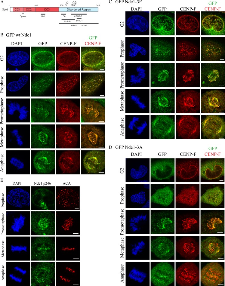
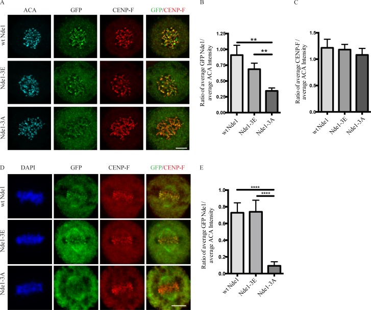
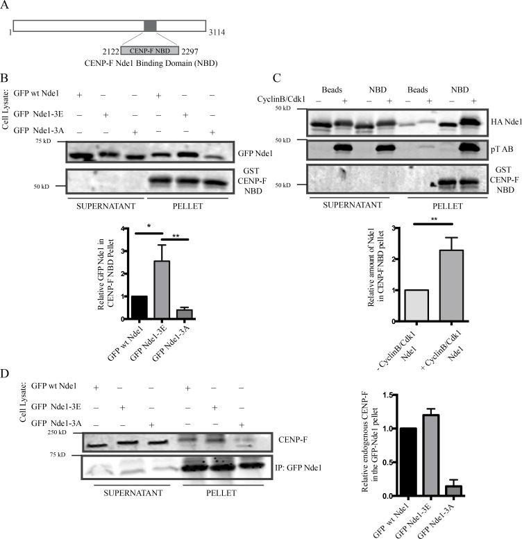
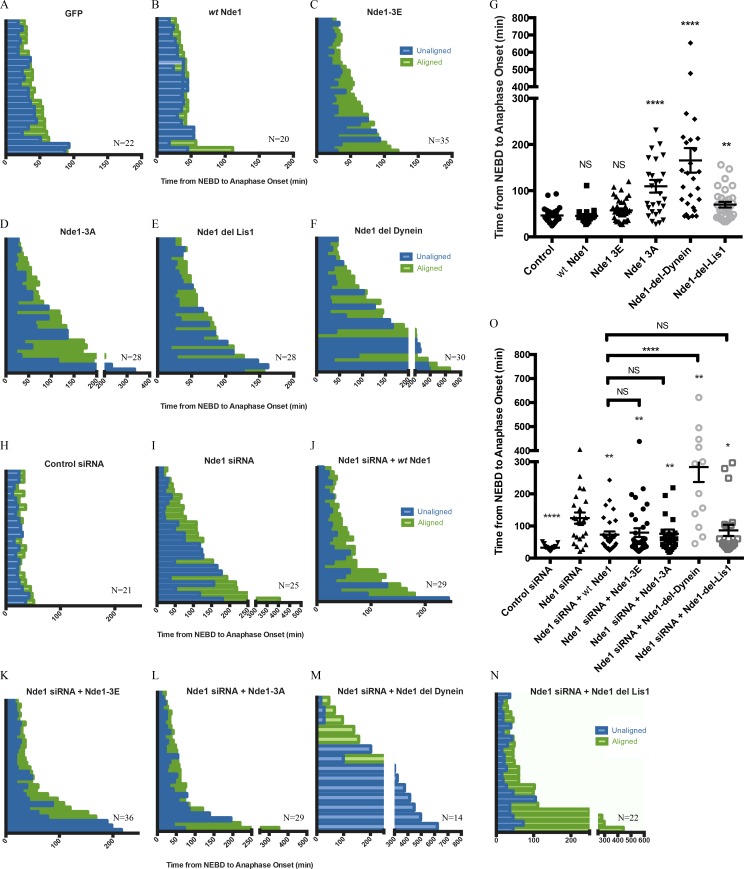
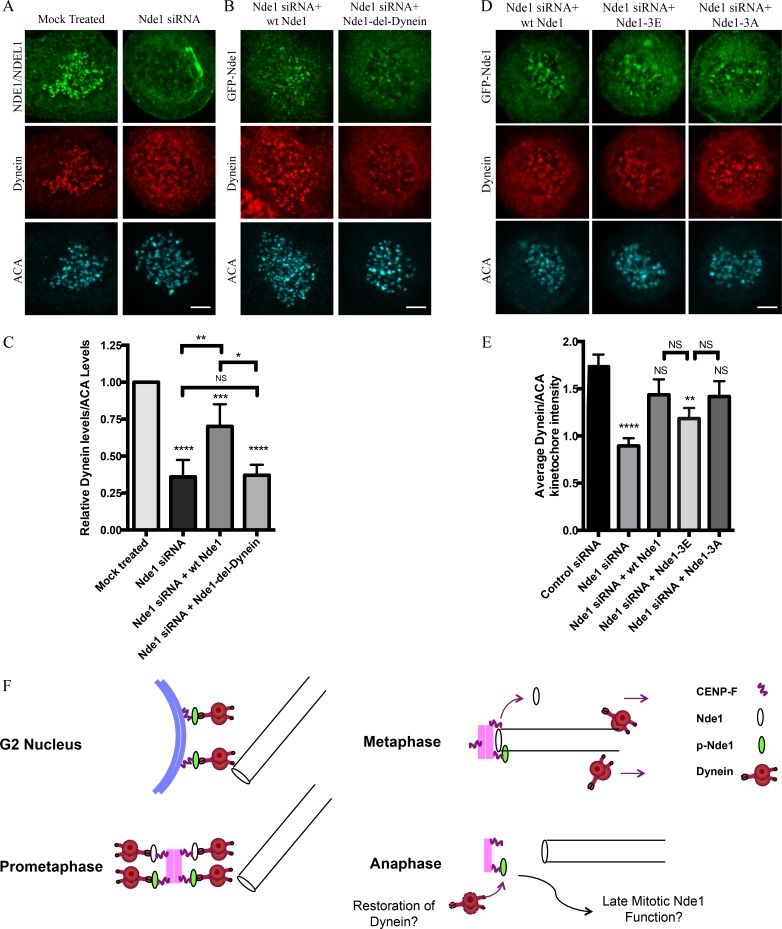
Cytoplasmic dynein is involved in diverse cell cycle-dependent functions regulated by several accessory factors, including Nde1 and Ndel1. Little is known about the role of these proteins in dynein cargo binding, and less is known about their cell cycle--dependent dynein regulation. Using Nde1 RNAi, mutant cDNAs, and a phosphorylation site-specific antibody, we found a specific association of phospho-Nde1 with the late G2-M nuclear envelope and prophase to anaphase kinetochores, comparable to the pattern for the Nde1 interactor CENP-F. Phosphomutant-Nde1 associated only with prometaphase kinetochores and showed weaker CENP-F binding in in vitro assays. Nde1 RNAi caused severe delays in mitotic progression, which were substantially rescued by both phosphomimetic and phosphomutant Nde1. Expression of a dynein-binding-deficient Nde1 mutant reduced kinetochore dynein by half, indicating a major role for Nde1 in kinetochore dynein recruitment. These results establish CENP-F as the first well-characterized Nde1 cargo protein, and reveal phosphorylation control of Nde1 cargo binding throughout a substantial fraction of the cell cycle.


Figures

Figure 1.
Figure 1.
Subcellular localization of Cdk1 phosphomimetic and phosphomutant Nde1 in G2 and mitosis. (A) Diagram of Nde1 showing Cdk1 phosphorylation sites (T215, T243, and T246) and interaction sites. (B–D) HeLa cells were transfected with GFP-tagged WT, phosphomimetic, and phosphomutant Nde1 and examined for localization to the G2 NE and mitotic kinetochores. GFP was detected by immunocytochemistry. WT and phospho-Nde1 colocalize with CENP-F at the NE and prophase–anaphase kinetochores. The phosphomutant Nde1 was weakly detected at these sites and absent from metaphase and anaphase kinetochores. (E) HeLa cells were stained with CDK1-phospho-specific antibody (p246; Alkuraya et al., 2011), which reacted with prometaphase-to-anaphase kinetochores, consistent with the distribution of the expressed phosphomimetic Nde1. Bars, 5 µm.
Figure 2.
Figure 2.
Effects of Nde1 phosphorylation on localization at unattached kinetochores. (A) Distribution of GFP-WT and phosphorylation-state mutant Nde1 in nocodazole-treated HeLa cells showing clear localization of each construct to kinetochores in the absence of MTs. The intensity of phosphomutant Nde1 appeared to be decreased relative to WT and phosphomimetic nde1. Bar, 5 µm. (B) Quantification of (anti-GFP vs. anti-ACA immunofluorescence intensity). Student’s t test showed a significant decrease in phosphomutant Nde1 intensity relative to the other conditions. **, P < 0.005. (C) Quantification of mean CENP-F intensity relative to mean ACA immunofluorescence signal. ANOVA statistical analysis showed no significant difference among the three conditions. Mean ± SEM for three independent experiments is represented. (D) HeLa cells transfected Nde1 phosphorylation-state cDNAs were blocked at metaphase with the proteasome inhibitor MG132, and kinetochores were examined for GFP immunofluorescence intensity. The phosphomutant Nde1-3A was absent from the kinetochores, revealing that Cdk1 phosphorylation of Nde1 blocks its dynein-mediated removal from these sites. Bar, 5 μm. (E) Quantification of GFP Nde1 signal intensity at kinetochores relative to ACA immunofluorescence. Student’s t test identified a significant decrease in levels of GFP Nde1-3A at kinetochores. ****, P < 0.0001.
Figure 3.
Figure 3.
Nde1 Cdk1 phosphorylation of Nde1 enhances CENP-F interaction. (A) Diagram showing CENP-F fragment used for biochemical analysis (NBD: amino acids 2,122–2,297). (B) GST-CENP-F NBD pull-down of GPF-Nde1 constructs from HeLa cells demonstrates increased binding of GFP-phosphomimetic Nde1. (C) GST-CENP-F NBD pull-down of bacterially expressed HA-Nde1 after in vitro phosphorylation with recombinant Cdk1/CyclinB. Levels of Nde1 in the CENP-F NBD pull-downs were quantified and again revealed an increased binding of Cdk1-phosphorylated Nde1 to the GST CENP-F NBD fragment. (D) Anti-GFP immunoprecipitation of endogenous CENP-F with GFP-tagged Nde1 phosphorylation state constructs. Immunoprecipitation of GFP WT Nde1 and GFP-Nde1-3E each specially coprecipitated endogenous CENP-F. Mean ± SEM of three independent experiments is represented. **, P < 0.005; *, P < 0.05.
Figure 4.
Figure 4.
Role of Nde1 phosphorylation in mitotic progression. (A–F) Duration of mitotic events for individual H2B-RFP HeLa cells expressing GFP or GFP-tagged WT Nde1, Nde1-3E, Nde1-3A, Nde1-del-dynein, or Nde1-del-Lis1. (G) Duration of prometaphase (“unaligned”) and metaphase (“aligned”) is shown and replotted as the complete time from NEBD to anaphase onset. Mean ± SEM for each condition is shown. Mann–Whitney statistical tests revealed a significant increase in the time from NEBD to anaphase onset for cells expressing GFP Nde1-3A, GFP Nde1-del-dynein, and GFP Nde1-del-Lis1 relative to GFP alone control. (H–N) Same as above, but in H2B-RFP–expressing HeLa cells treated with control or Nde1 siRNAs for 48 h and then rescued with GFP, WT Nde1, Nde1-3E, Nde1-3A, Nde1-del-dynein, or Nde1-del-Lis1 for 24 h before live-cell imaging. (O) Data replotted as complete time from NEBD to anaphase onset. Mann–Whitney statistical tests were performed to compare Nde1 siRNA to each GFP Nde1 rescue condition. All of the Nde1 constructs to some extent rescued effects of Nde1RNAi, whereas Nde1-del-dynein showed no such effect. Mean ± SEM for each condition is shown. **, P < 0.01; *, P < 0.05; ****, P < 0.0001.
Figure 5.
Figure 5.
Requirement of Nde1 for dynein recruitment to the kinetochore. (A) HeLa cells treated with Nde1 siRNA, exposed to nococazole, and immunostained for endogenous NDE1/NDEL1 or dynein intermediate chain. (B) Nde1 siRNA-treated HeLa cells rescued with expression of GFP WT Nde1 or GFP Nde1-del-dynein and stained for GFP and dynein intermediate chain. (C) Quantification of mean kinetochore dynein levels relative to mean ACA immunofluorescence signal. Dynein/ACA values were plotted ± SEM. Paired Student’s t test was performed to analyze significance between conditions. (D) Nde1 siRNA-treated HeLa cells rescued by expression of WT Nde1, Nde1-3E, or Nde1-3A and stained for GFP and dynein intermediate chain. Bars, 5 µm. (E) Quantification of mean dynein intensity relative to mean ACA immunofluorescence signal. Mean ± SEM of three independent experiments is represented. Student’s t tests were performed to analyze significance between conditions and revealed a significant difference in the levels of kinetochore dynein between Nde1 siRNA and WT Nde1, Nde1-3E, or Nde1-3A. *, P < 0.05; **, P < 0.005; ***, P < 0.001; ****, P < 0.0001. (F) Role of Cdk1 phosphorylation in G2-M Nde1 behavior. Phosphorylated Nde1 is shown associating with the G2 NE and then kinetochores from prophase into anaphase when dephosphorylated Nde1 is lost from these sites. Although dynein is undetectable at normal anaphase kinetochores, we speculate that persistent pNde1 might serve in a late dynein-mediated kinetochore attachment correction mechanism.

Similar articles

Cited by

References

    1. Alkuraya F.S., Cai X., Emery C., Mochida G.H., Al-Dosari M.S., Felie J.M., Hill R.S., Barry B.J., Partlow J.N., Gascon G.G., et al. . 2011. Human mutations in NDE1 cause extreme microcephaly with lissencephaly [corrected]. Am. J. Hum. Genet. 88:536–547. 10.1016/j.ajhg.2011.04.003 - DOI - PMC - PubMed
    1. Baffet A.D., Hu D.J., and Vallee R.B.. 2015. Cdk1 Activates Pre-mitotic Nuclear Envelope Dynein Recruitment and Apical Nuclear Migration in Neural Stem Cells. Dev. Cell. 33:703–716. 10.1016/j.devcel.2015.04.022 - DOI - PMC - PubMed
    1. Barisic M., Sohm B., Mikolcevic P., Wandke C., Rauch V., Ringer T., Hess M., Bonn G., and Geley S.. 2010. Spindly/CCDC99 is required for efficient chromosome congression and mitotic checkpoint regulation. Mol. Biol. Cell. 21:1968–1981. 10.1091/mbc.e09-04-0356 - DOI - PMC - PubMed
    1. Bolhy S., Bouhlel I., Dultz E., Nayak T., Zuccolo M., Gatti X., Vallee R., Ellenberg J., and Doye V.. 2011. A Nup133-dependent NPC-anchored network tethers centrosomes to the nuclear envelope in prophase. J. Cell Biol. 192:855–871. 10.1083/jcb.201007118 - DOI - PMC - PubMed
    1. Chan Y.W., Fava L.L., Uldschmid A., Schmitz M.H., Gerlich D.W., Nigg E.A., and Santamaria A.. 2009. Mitotic control of kinetochore-associated dynein and spindle orientation by human Spindly. J. Cell Biol. 185:859–874. 10.1083/jcb.200812167 - DOI - PMC - PubMed

Publication types

MeSH terms

LinkOut - more resources