. 2018 Jun 20;10(6):1454-1473.
doi: 10.18632/aging.101482.

Naked mole rat cells display more efficient excision repair than mouse cells

Affiliations

Naked mole rat cells display more efficient excision repair than mouse cells

Alexei Evdokimov et al. Aging (Albany NY). .

Abstract

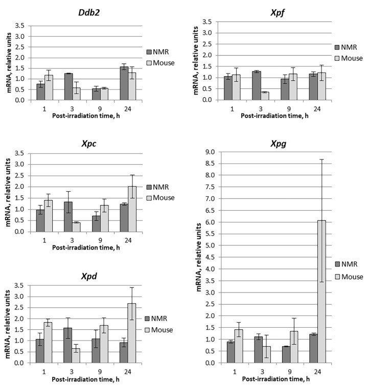
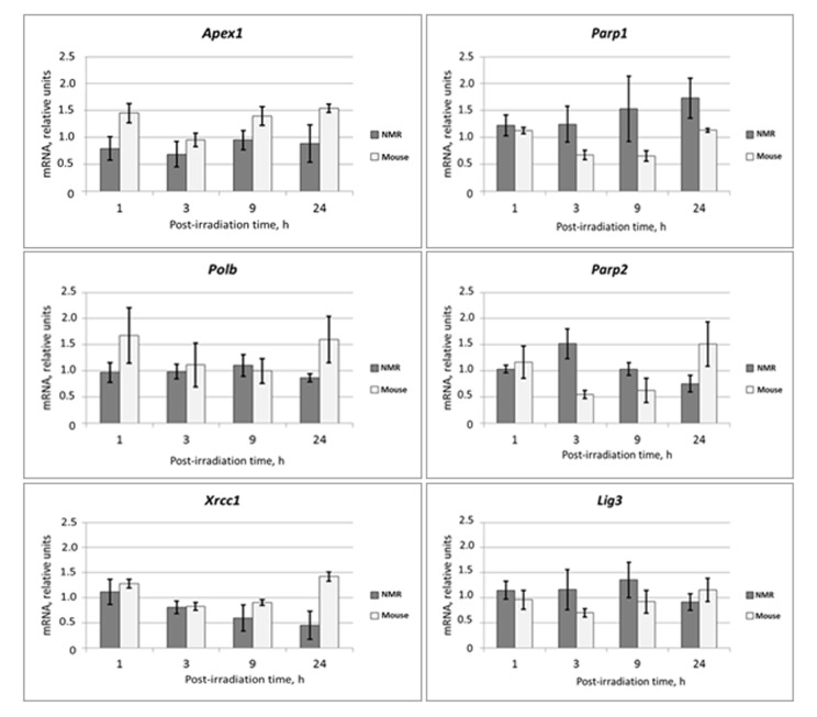
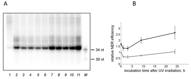
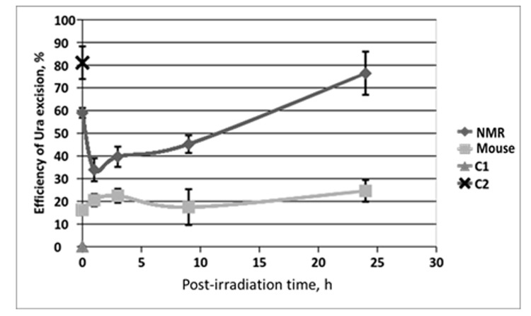
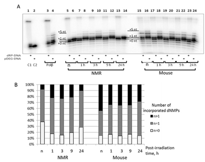
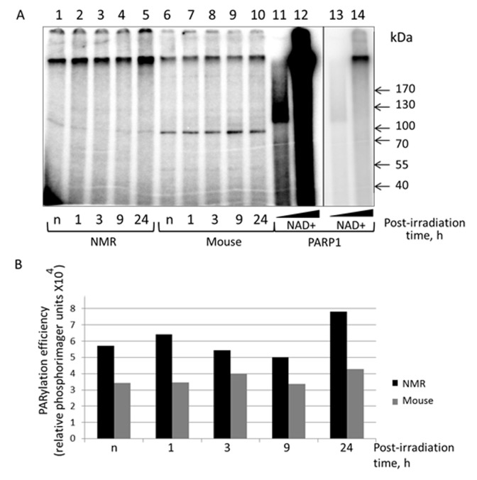
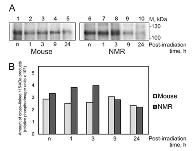
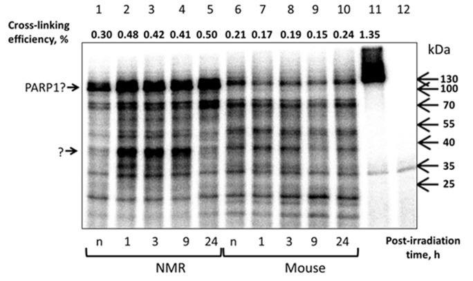
Naked mole rat (NMR) is the long-lived and tumor-resistant rodent. NMRs possess multiple adaptations that may contribute to longevity and cancer-resistance. However, whether NMRs have more efficient DNA repair have not been directly tested. Here we compared base excision repair (BER) and nucleotide excision repair (NER) systems in extracts from NMR and mouse fibroblasts after UVC irradiation. Transcript levels of the key repair enzymes demonstrated in most cases higher inducibility in the mouse vs the NMR cells. Ratios of repair enzymes activities in the extracts somewhat varied depending on post-irradiation time. NMR cell extracts were 2-3-fold more efficient at removing the bulky lesions, 1.5-3-fold more efficient at removing uracil, and about 1.4-fold more efficient at cleaving the AP-site than the mouse cells, while DNA polymerase activities being as a whole higher in the mouse demonstrate different patterns of product distribution. The level of poly(ADP-ribose) synthesis was 1.4-1.8-fold higher in the NMR cells. Furthermore, NMR cell extracts displayed higher binding of PARP1 to DNA probes containing apurinic/apyrimidinic site or photo-reactive DNA lesions. Cumulatively, our results suggest that the NMR has more efficient excision repair systems than the mouse, which may contribute to longevity and cancer resistance of this species.

Keywords: Heterochephalus glaber; Mus musculus; base excision repair; nucleotide excision repair; poly(ADP-ribose) polymerase 1.


Conflict of interest statement

CONFLICTS OF INTEREST: None declared.

Figures

Figure 1
Figure 1
Time dependent levels of mRNA encoding NER proteins in NMR and mouse cells after UVC-light irradiation. Time of cells incubation after UVC-light irradiation is presented on the X-axis. For each gene, the level of its expression in UVC-light irradiated cells was normalized to that in untreated cells with Tubb2a, Tubb1, Polr1b, and Actb genes used for reference. Experiments were repeated at least three times; mean values ± SD are shown.
Figure 2
Figure 2
Time dependent levels of mRNA encoding BER proteins in NMR and mouse cells after UVC-light irradiation. The data are the mean of three independent experiments made in triplicates ±SD. For each gene the level of its expression in UVC-irradiated cells was normalized to that of non-irradiated cells. Three housekeeping genes: Tubb, Actb and Gapdh were used as a reference.
Figure 3
Figure 3
The NER excision activity of NMR and mouse cell extracts. (A) Representative phospho-image of product analysis for mouse (lanes 2-6) and NMR (lanes 7-11) cell extracts. The substrate DNA was incubated for 45 min at 30°C with cell extracts (15 nM substrate DNA, 0.3 mg/ml extract proteins). The excision products were detected by annealing to a specific template containing 5ʹ-GpGpGpGpG overhang, which was then end-labeled using α-[32P]-dCTP and Taq DNA polymerase. The reaction products were resolved on a 10% denaturing polyacrylamide gel. [32P]-5ʹ end-labelled oligonucleotides were used as length markers (lane M). nFlu-DNA without cell extract was used as a negative control (lane 1). (B) Quantification of the levels of excision products based on three independent experiments. The activity in the extract of mouse non-irradiated cells was taken as 1. The data are the mean ±SD, n=3. Grey and black lines correspond to mouse and NMR cell extracts, respectively.
Figure 4
Figure 4
The efficiency of uracil excision from DNA by extracts from NMR and mouse cells with different post-irradiation time. Reaction mixtures contained 100 nM 5'-[32P] 32-mer U-DNA, 50 mM Tris-HCl (pH 8.0), 40 mM NaCl, 10 mM EDTA, 1 mM DTT, 0.1 mg/ml BSA and 0.5 mg/ml of cell extract proteins. The reaction mixtures were incubated at 370C for 10 min. C1 – control reaction mixture contained only U-DNA. C2 – control reaction mixture corresponds to U-DNA incubated with excess of E. coli UDG. Then reaction mixtures were treated and analyzed as described in the section “Uracil excision activity of cell extracts”. Efficiency of Ura removal is determined as percent of cleaved Ura containing DNA chain.
Figure 5
Figure 5
The efficiency of AP site cleavage by extracts from NMR and mouse cells with different post-irradiation time. Reaction mixtures contained 100 nM 5'-[32P] 32-mer AP DNA, 0.5 mg/ml of cell extract and buffer components. The reaction proceeded at 37ºC for 10 min and then was quenched by addition of EDTA (20 mM final) followed by stabilization of intact AP sites with NaBH4 treatment. DNA was then analyzed as described in the section ‘AP site cleavage activity of cell extracts’. C1 – incubation of AP DNA in the absence of cell extracts reflects the level of spontaneous AP site cleavage. C2 – incubation of AP DNA with 100 µM NaOH that brings about full AP site cleavage.
Figure 6
Figure 6
The efficiency of DNA synthesis by extracts from NMR and mouse cells with different post-irradiation time. (A) The 100 nM 5'-[32P] 32-mer DNA with 5'-dRP-nick (odd lanes) or with 5'-pDEG-nick (even lanes) were incubated with 0.1 µM Polβ (lanes 3 and 4), 0.5 mg/ml of cell extract protein of NMR (lanes 5 – 14) or mouse (lanes 15 – 24) fibroblasts and buffer components. The reaction mixtures were incubated at 37ºC for 10 min. The control probes C1 and C2 contained only substrate DNAs and buffer components. The mixtures were supplemented with loading buffer, heated at 970C for 10 min, and the products were then analyzed as described in ‘DNA polymerase activity of cell extracts’. (B) Quantification of the DNA synthesis products for 5'-pDEG-nick DNA shown in A. White parts of bars correspond to non-elongated primer, grey parts reflect the amount of primer elongated by one dNMP and black parts correspond to the products of strand-displacement DNA synthesis. ‘n’ designates non-irradiated cells.
Figure 7
Figure 7
PARylation of cell extract proteins of NMR and mouse cells in presence of [32P]NAD+. (A) The activated DNA was incubated with 0.5 mg/ml of cell extracts of NMR cells (lanes 1 – 5) or mouse cell (lanes 6 – 10). The control probes (lanes 11–14) contained 70 nM PARP1 instead of cell extracts and 40 µM [32P]NAD+ (lanes 11 and 13) or 400 µM [32P]NAD+ (lanes 12 and 14). Lanes 13 and 14 correspond to lanes 11 and 12 but with low exposure. The reaction mixtures were incubated at 370C for 4 min. The mixtures were supplemented with loading buffer, heated at 970C for 10 min and then analyzed as described in the section ‘PARylation of cell extract proteins’. (B) Quantification showing the yield of PARylation products (A). Post-irradiation time is the time the cells were cultivated after irradiation before the cells were harvested for the following extract preparation. “n” designates non-irradiated cells.
Figure 8
Figure 8
Cross-linking of NMR and mouse cell extract proteins to dCFAP-DNA. (A) The 25 nM [32P]dCFAP-DNA (54-mer) was incubated with 1.2 mg/ml of mouse (lanes 1-5) or NMR (lanes 6–10) cell extract proteins and buffer components. The mixtures were exposed to UV-light irradiation at 312 nm, 1.5 J/cm2·min for 10 min. After the UV-light induced cross-linking the reaction mixtures were treated with benzonase (0.1 unit per 1 μl of the reaction mixture for 30 min at 37ºC). Then the products were analyzed as described in ‘Photocross-linking of cell extract proteins with dCFAP-DNA’. ‘n’ denotes the extract of non-irradiated cells; 1–24 designate the extracts of cells cultivated for definite time (in hours) after UVC-irradiation. (B) Quantification showing the yield of the 115-kDa cross-linking products.
Figure 9
Figure 9
Cross-linking of NMR and mouse cell extract proteins with AP DNA. 100 nM 5'-[32P] 32-mer AP DNA was incubated with 0.5 mg/ml of NMR (lanes 1–5) or mouse (lanes 6–10) cell extract proteins. The control probes contained buffer components and AP DNA (lane 12) or buffer components, AP DNA and 70 nM human recombinant PARP1 (lane 11). Samples represented in lanes 1 and 6 correspond to the extracts of non-irradiated cells. The reaction mixtures were incubated at 37ºC for 10 min. Then the probes were treated with 20 mM NaBH4. The products were then separated and analyzed as described in the section ‘Cross-linking of cell extract proteins with AP DNA’. The intensity of the 120-kDa product estimated as a part of cross-linked DNA (%) is shown at the top of the image.

Similar articles

Cited by

References

    1. Vijg J. Somatic mutations, genome mosaicism, cancer and aging. Curr Opin Genet Dev. 2014; 26:141–49. 10.1016/j.gde.2014.04.002 - DOI - PMC - PubMed
    1. Hanawalt PC. Revisiting the rodent repairadox. Environ Mol Mutagen. 2001; 38:89–96. 10.1002/em.1057 - DOI - PubMed
    1. Page MM, Stuart JA. Activities of DNA base excision repair enzymes in liver and brain correlate with body mass, but not lifespan. Age (Dordr). 2012; 34:1195–209. 10.1007/s11357-011-9302-9 - DOI - PMC - PubMed
    1. Lewis KN, Andziak B, Yang T, Buffenstein R. The naked mole-rat response to oxidative stress: just deal with it. Antioxid Redox Signal. 2013; 19:1388–99. 10.1089/ars.2012.4911 - DOI - PMC - PubMed
    1. Buffenstein R. Negligible senescence in the longest living rodent, the naked mole-rat: insights from a successfully aging species. J Comp Physiol B. 2008; 178:439–45. 10.1007/s00360-007-0237-5 - DOI - PubMed

LinkOut - more resources