18F-FDG PET/CT characteristics of pulmonary sclerosing hemangioma vs. pulmonary hamartoma
- PMID: 29930720
- PMCID: PMC6006501
- DOI: 10.3892/ol.2018.8660
18F-FDG PET/CT characteristics of pulmonary sclerosing hemangioma vs. pulmonary hamartoma
Abstract
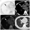
The radiological features of pulmonary sclerosing hemangioma (PSH) and pulmonary hamartoma are poorly specified. Thus, the present study aimed to compare and analyze the characteristics of fluorodeoxyglucose positron emission tomography/computed tomography (18F-FDG PET/CT) in PSH versus pulmonary hamartoma. 18F-FDG PET/CT characteristic findings of 12 patients with PSH and 14 patients with pulmonary hamartoma were retrospectively reviewed. A total of 12 lesions were detected from the 12 patients with PSH, of which 3 masses exhibited calcification. The mean diameter and standardized maximum uptake value (SUVmax) were 1.9±0.7 cm and 2.6±1.0, respectively, and there was no significant correlation between the lesion size and SUVmax (P>0.05). For the 14 patients with pulmonary hamartoma, 14 lesions were found, of which 4 exhibited calcification. The mean diameter and SUVmax were 1.7±0.8 cm and 1.5±0.6, respectively, and there was a significant correlation between the size and SUVmax (r=0.625, r2=0.391, P<0.05). Although there was no significant difference between the size of PSH and pulmonary hamartoma (P>0.05), the SUVmax of PSH was significantly higher than that of pulmonary hamartoma (P<0.05). Moreover, the SUVmax of 1.95 was applied as a cutoff for the diagnosis of PSH, and the resulting sensitivity and specificity for PET/CT to differentiate PSH from pulmonary hamartoma were 83.3 and 78.6%, respectively. Although the morphological features were not specific, PSH showed significantly higher FDG accumulation than pulmonary hamartoma on PET/CT imaging, which may aid the differential diagnosis. Further studies with larger populations are warranted to confirm these study results.
Keywords: fluorodeoxyglucose; positron emission tomography/computed tomography; pulmonary hamartoma; pulmonary sclerosing hemangioma.
Figures




Similar articles
-
Diagnosis value of 18F-Fluoro-D-glucose positron emission tomography-computed tomography in pulmonary hamartoma: a retrospective study and systematic review.BMC Med Imaging. 2023 Feb 7;23(1):28. doi: 10.1186/s12880-023-00981-z. BMC Med Imaging. 2023. PMID: 36747135 Free PMC article.
-
F-18 FDG PET/CT in evaluation of pulmonary sclerosing hemangioma.Clin Nucl Med. 2011 May;36(5):341-3. doi: 10.1097/RLU.0b013e31820aa00c. Clin Nucl Med. 2011. PMID: 21467848
-
18F-FDG PET/CT features of pulmonary sclerosing hemangioma.Acta Radiol. 2013 Feb 1;54(1):24-9. doi: 10.1258/ar.2011.110474. Epub 2012 Jan 30. Acta Radiol. 2013. PMID: 22291338
-
Differential diagnostic value of 18F-FDG PET/CT in pulmonary carcinoids versus hamartomas.Acad Radiol. 2022 Feb;29 Suppl 2:S41-S46. doi: 10.1016/j.acra.2020.10.022. Epub 2020 Nov 9. Acad Radiol. 2022. PMID: 33183953
-
Hyperaccumulation of (18)F-FDG in order to differentiate solid pseudopapillary tumors from adenocarcinomas and from neuroendocrine pancreatic tumors and review of the literature.Hell J Nucl Med. 2013 May-Aug;16(2):97-102. doi: 10.1967/s002449910084. Epub 2013 May 20. Hell J Nucl Med. 2013. PMID: 23687644 Review.
Cited by
-
Contribution of 18F-FDG PET/CT in the Differential Diagnosis of Pulmonary Hamartomas and Pulmonary Carcinoids.Mol Imaging Radionucl Ther. 2021 Jun 3;30(2):101-106. doi: 10.4274/mirt.galenos.2021.20633. Mol Imaging Radionucl Ther. 2021. PMID: 34082511 Free PMC article.
-
Diagnosis value of 18F-Fluoro-D-glucose positron emission tomography-computed tomography in pulmonary hamartoma: a retrospective study and systematic review.BMC Med Imaging. 2023 Feb 7;23(1):28. doi: 10.1186/s12880-023-00981-z. BMC Med Imaging. 2023. PMID: 36747135 Free PMC article.
-
A case of pulmonary sclerosing pneumocytoma diagnosed preoperatively using transbronchial cryobiopsy.Respir Med Case Rep. 2021 Aug 11;34:101494. doi: 10.1016/j.rmcr.2021.101494. eCollection 2021. Respir Med Case Rep. 2021. PMID: 34430194 Free PMC article.
-
Case Report: Pulmonary sclerosing pneumocytoma mimicking as a neuroendocrine tumor on 18F-FDG and 68Ga-DOTATATE PET/CT: a case presentation.Front Oncol. 2025 Feb 21;15:1511595. doi: 10.3389/fonc.2025.1511595. eCollection 2025. Front Oncol. 2025. PMID: 40061895 Free PMC article.
-
Pulmonary sclerosing pneumocytoma: A potential pitfall mimicking lung adenocarcinoma.GHM Open. 2023 Aug 31;3(1):51-55. doi: 10.35772/ghmo.2023.01009. GHM Open. 2023. PMID: 40143840 Free PMC article.
References
LinkOut - more resources
Full Text Sources
Other Literature Sources