. 2019 Feb;94(1):102-115.
doi: 10.1111/brv.12439. Epub 2018 Jun 21.

The evolution of molluscs

Affiliations

The evolution of molluscs

Andreas Wanninger et al. Biol Rev Camb Philos Soc. 2019 Feb.

Abstract

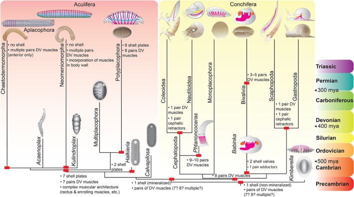
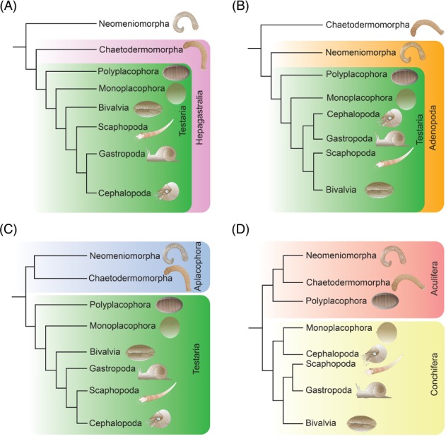
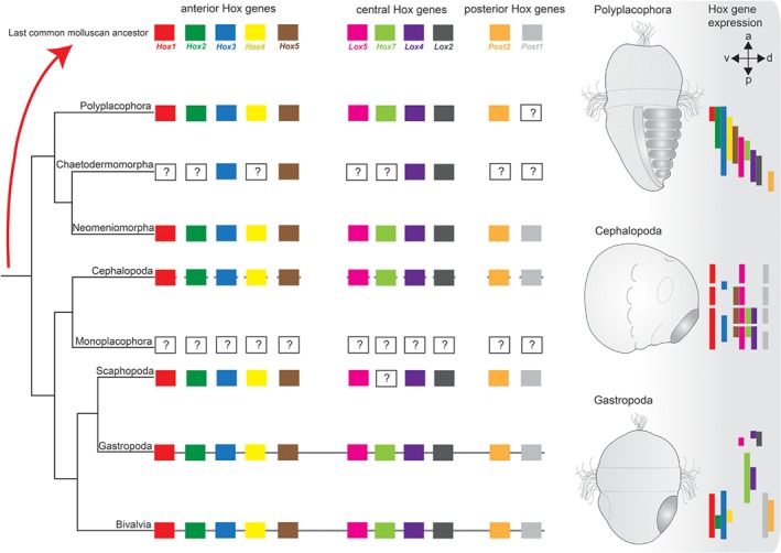
Molluscs are extremely diverse invertebrate animals with a rich fossil record, highly divergent life cycles, and considerable economical and ecological importance. Key representatives include worm-like aplacophorans, armoured groups (e.g. polyplacophorans, gastropods, bivalves) and the highly complex cephalopods. Molluscan origins and evolution of their different phenotypes have largely remained unresolved, but significant progress has been made over recent years. Phylogenomic studies revealed a dichotomy of the phylum, resulting in Aculifera (shell-less aplacophorans and multi-shelled polyplacophorans) and Conchifera (all other, primarily uni-shelled groups). This challenged traditional hypotheses that proposed that molluscs gradually evolved complex phenotypes from simple, worm-like animals, a view that is corroborated by developmental studies that showed that aplacophorans are secondarily simplified. Gene expression data indicate that key regulators involved in anterior-posterior patterning (the homeobox-containing Hox genes) lost this function and were co-opted into the evolution of taxon-specific novelties in conchiferans. While the bone morphogenetic protein (BMP)/decapentaplegic (Dpp) signalling pathway, that mediates dorso-ventral axis formation, and molecular components that establish chirality appear to be more conserved between molluscs and other metazoans, variations from the common scheme occur within molluscan sublineages. The deviation of various molluscs from developmental pathways that otherwise appear widely conserved among metazoans provides novel hypotheses on molluscan evolution that can be tested with genome editing tools such as the CRISPR/Cas9 (clustered regularly interspaced short palindromic repeats/clustered regularly interspaced short palindromic repeats-associated protein9) system.

Keywords: BMP; Bilateria; Cambrian; EvoDevo; Hox; Lophotrochozoa; Mollusca; body axis; development; morphology.


Figures

Figure 1
Figure 1
Molluscan intraphyletic relationships and hypothesized evolutionary pathways of major exoskeletal and muscular subsets. Only changes in character states are indicated (red rectangles). Phylogeny based on Smith et al. (2011) and Vinther et al. (2017). Myoanatomical condition is indicated where known. ‘?’ in Kimberella indicates that it is still debated whether or not the respective serial indentations represent fossilized muscle strands. Assuming that Kimberella is a stem‐group mollusc, the Aculifera–Conchifera concept favours a single‐shelled ancestor to all molluscs, most likely with serially repeated dorso‐ventral (DV) musculature (magenta). After the split from stem‐group aculiferans, a body plan with seven shell plates and corresponding DV muscles as well as multiple subsets of additional muscles evolved on the line leading to Aculifera. Vermification along with incorporation of distinct muscular units [e.g. ventro‐lateral muscle (green), enrolling muscle (light blue), rectus muscle (red), ring muscles (dark blue)] into the adult body wall occurred in the aplacophorans (potentially several times independently), whereby some extinct taxa maintained the shell armour. Within Polyplacophora, the sevenfold seriality was retained in the extinct multiplacophorans; recent polyplacophorans have secondarily acquired an eighth plate with an additional set of DV muscles. The conchiferans retained the single‐shelled condition and muscular seriality from the molluscan ancestor and probably had eight sets of DV muscles as exhibited by recent monoplacophorans and stem‐group bivalves [that evolved a second shell and two adductor muscles (orange)]. Scaphopods, gastropods, and cephalopods have reduced their DV musculature to one pair and have evolved distinct cephalic retractors (yellow). Following the phylogenetic scenario depicted here, this occurred twice independently. Recent bivalves have retained muscular seriality to a certain degree, with most representatives having 3–5 DV muscles.
Figure 2
Figure 2
Major competing phylogenetic scenarios for Mollusca. Note that Conchifera is monophyletic in all cases but its internal relationships are highly debated. (A) Hepagastralia–Testaria concept with Neomeniomorpha as the earliest offshoot; (B) Adenopoda–Testaria concept with Chaetodermomorpha as the earliest offshoot; (C) Aplacophora–Testaria concept with a monophyletic assemblage of Polyplacophora and Conchifera (Testaria) as sister to Aplacophora; and (D) Aculifera–Conchifera concept, according to which Polyplacophora together with Aplacophora form the Aculifera that is the sister clade to Conchifera. Note that scenarios A and B suggest a gradual increase in body plan complexity from the vermiform neomeniomorphs and chaetodermomorphs, a situation that is not supported by paleontological and developmental data. By contrast, the basal dichotomy in C and D and the unresolved molluscan sister group relationship does not allow for an unambiguous reconstruction of molluscan ancestry without the addition of fossil and developmental data.
Figure 3
Figure 3
Molluscan Hox genes and their expression patterns in polyplacophorans, cephalopods, and gastropods. Except for Gastropoda, Bivalvia, and Cephalopoda, for which genomes are available, Hox sequences have only been identified from transcriptomic data sets. Thus, uncertainties exist concerning the definite lack of individual Hox genes in chaetodermomorphs, polyplacophorans, and scaphopods, indicated by ‘?’. No monoplacophoran Hox genes are known to date. Note that for most taxa data were combined from different species. A continuous line between Hox gene symbols indicates an intact Hox cluster for the respective taxon (note that for bivalves, both intact and disrupted Hox gene arrangements are known). The discontinuous line in Cephalopoda indicates a disrupted Hox cluster. The absence of a line in Polyplacophora, Chaetodermomorpha, Neomeniomorpha, and Scaphopoda indicates that the Hox gene arrangement is unknown. Colour‐coded bars show gene expression along the anterior–posterior axis of a mid‐stage polyplacophoran trochophore larva, an early cephalopod embryo, and a late‐stage gastropod trochophore larva. For clarity, expression in specific morphological features is not shown. Note that a staggered expression of Hox genes, as predicted by the concept of colinearity, is found only in polyplacophorans. Shell field(s) in the polyplacophoran and gastropod larvae as well as the mantle anlage in the cephalopod embryo are shaded in dark grey. Abbreviations: a, anterior; d, dorsal; p, posterior; v, ventral.

References

    1. Adema, C. M. , Hillier, L. W. , Jones, C. S. , Loker, E. S. , Knight, M. , Minx, P. , Oliveira, G. , Raghavan, N. , Shedlock, A. , Rodrigues do Amaral, L. , Arican‐Goktas, H. D. , Assis, J. G. , Hideo Baba, E. , Baron, O. L. , Bayne, C. J. , et al. (2017). Whole genome analysis of a schistosomiasis‐transmitting freshwater snail. Nature Communications 8, 15451. - PMC - PubMed
    1. Aguilera, F. , McDougall, C. & Degnan, B. M. (2017). Co‐option and de novo gene evolution underlie molluscan shell diversity. Molecular Biology and Evolution 34, 779–792. - PMC - PubMed
    1. Albertin, C. B. , Simakov, O. , Mitros, T. , Wang, Z. Y. , Pungor, J. R. , Edsinger‐Gonzales, E. , Brenner, S. & Ragsdale, C. W. (2015). The octopus genome and the evolution of cephalopod neural and morphological novelties. Nature 524, 220–224. - PMC - PubMed
    1. Anderson, P. D. & Bokor, G. (2012). Conotoxins: potential weapons from the sea. Journal of Bioterrorism and Biodefence 3, 120.
    1. Anderson, G. J. & Darshan, D. (2008). Small‐molecule dissection of BMP signaling. Nature Chemical Biology 4, 15–16. - PubMed